Evaluation of setup errors and CTV–PTV margin assessment in pelvic cancer patients undergoing image-guided radiotherapy
Vishwas M Shetty1 and S Shylvea Shalom Pandian1,2a
1Department of Radiotherapy Technology, Father Muller College of Allied Health Sciences, Mangaluru, Karnataka 575001, India
2Department of Radiation Oncology, Father Muller Medical College, Mangaluru, Karnataka 575001, India
a https://orcid.org/0009-0006-1744-8313
Abstract
In pelvic radiotherapy, accurate patient positioning is essential to ensure the target area receives sufficient coverage and minimal radiation to the surrounding organs at risk. Although image-guided radiotherapy (IGRT) has significant advances, there are still some setup errors that happen because of patient positioning variability and their anatomical change between treatments, such as bladder filling or rectum stretches. Quantifying these uncertainties is essential for determining appropriate clinical target volume (CTV) to planning target volume (PTV) margins. This study aims to assess setup errors in pelvic cancer patients receiving IGRT and to determine suitable CTV–PTV margins through a population-based methodology. A retrospective study was conducted on 20 patients with pelvic malignancies, who received intensity-modulated radiotherapy or volumetric-modulated arc therapy, in a Halcyon IGRT-based treatment system with daily kilovoltage cone-beam computed tomography (KV-CBCT) imaging. Setup deviation was recorded and analysed in the lateral, longitudinal and vertical directions for each treatment fraction. Population mean errors, along with systematic (S) and random errors (s), were calculated, followed by the estimation of CTV–PTV margins using the Van Herk formula. The results showed minimal setup errors in the overall population mean. Systematic errors were relatively low across all directions, indicating good reproducibility of patient positioning, whereas random errors were comparatively greater, particularly in the longitudinal and lateral directions, indicating daily variation. The calculated CTV–PTV margins were 0.36, 0.78 and 0.59 cm in the vertical, lateral and longitudinal directions, respectively. The largest margin was observed in the lateral direction, indicating greater residual uncertainty despite daily image guidance. These findings indicate that although daily KV-CBCT-based IGRT effectively reduces setup uncertainties, residual variation still influences margin requirements. Institution-specific evaluation of setup errors remains important for optimising CTV–PTV margins and maintaining accurate target coverage while minimising radiation exposure to surrounding normal tissues.
Keywords: pelvic cancer, image-guided radiotherapy setup error, CTV–PTV margin, KV-CBCT, halcyon, IMRT, VMAT
Correspondence to: S Shylvea Shalom Pandian
Email: shylvea@gmail.com
Published: 08/09/2026
Received: 05/04/2026
Publication costs for this article were supported by ecancer (UK Charity number 1176307).
Copyright: © the authors; licensee ecancermedicalscience. This is an Open Access article distributed under the terms of the Creative Commons Attribution License (http://creativecommons.org/licenses/by/4.0), which permits unrestricted use, distribution, and reproduction in any medium, provided the original work is properly cited.
Background
Radiation therapy plays an important role in the management of pelvic malignancies such as carcinoma of the cervix, rectum, endometrium, vagina and prostate. The introduction of advanced radiotherapy treatment techniques such as intensity-modulated radiotherapy (IMRT) and volumetric-modulated arc therapy (VMAT) aims to deliver radiation dose accurately to the target volume while minimising the radiation exposure to surrounding normal tissues and organs at risk (OAR), such as bladder, rectum, bowel, femur head. Despite these technological advancements, achieving this precise radiation delivery is challenging due to various uncertainties that arise throughout the radiotherapy treatment process, including patient immobilisation, treatment planning and treatment delivery. Among the uncertainties, patient setup error remains one of the most common contributors to treatment uncertainty.
These setup errors are defined as deviations between the patient’s planned position during the simulation and the actual treatment position at each treatment fraction delivery. These errors are broadly classified as systematic errors (S), which are consistent and continued throughout the treatment courses, and random errors (s), which vary on a day-to-day basis. Both types of errors can lead to compromise target coverage and increase the risk of unintended radiation to the normal tissues if not adequately accounts it during the radiotherapy treatment process [1].
The introduction of image-guided radiotherapy (IGRT) has significantly improved treatment accuracy. Techniques such as cone-beam computed tomography (CBCT), integrated into linear accelerators, enable three-dimensional visualisation of patient anatomy prior to treatment, allowing verification and correction of translational setup deviations [2]. Although technology advances, some residual setup uncertainties still exist, particularly in pelvic radiotherapy. The uncertainty is mainly due to interfractional anatomical variations caused by changes in bladder filling, rectal stretches, patient movement, which can lead to shifts in both target volumes and surrounding OAR.
Several studies have evaluated setup uncertainties in pelvic radiotherapy with different treatment techniques and imaging modalities, reporting variability in the magnitude of systematic and random errors [3, 4]. In a recent study [5], CBCT-based IGRT was used on a Halcyon machine and identified axis-dependent variations in planning target volume (PTV) margins, underscoring the necessity of precise error quantification in IGRT-based treatment delivery. By analysing the literature article, it is evident that the setup error variation highlights the importance of institution-specific assessment, as factors such as patient positioning [6], imaging protocols and treatment delivery systems can influence setup accuracy.
Accurate evaluation of setup errors is essential for determining appropriate clinical target volume (CTV)–PTV margins, which ensures adequate target coverage while minimising unnecessary radiation to surrounding normal tissues [7]. Margin calculation methods, such as Van Herk formula, account for both systematic and random errors, thereby providing a statistically reliable estimation of the margins required to achieve optimal treatment accuracy. Therefore, with the increasing use of advanced treatment techniques such as IMRT, VMAT and IGRT platforms such as Halcyon system, it is essential to reassess setup uncertainties within clinical settings.
Therefore, the present study aimed to evaluate translational setup errors in pelvic cancer patients treated at our Father Muller Medical College, Mangaluru ,using IGRT-based radiotherapy on the Halcyon system. This aim will be addressed by assessing both population-based systematic and random setup errors and by determining appropriate CTV–PTV margins using the Van Herk margin recipe.
Methods
Study design and setting
This retrospective, observational study was conducted in the Department of Radiation Oncology, Father Muller Medical College, Mangaluru, India. The study evaluated translational setup errors in pelvic cancer patients treated with IMRT/VMAT using IGRT-based radiotherapy on the Halcyon treatment platform over a period of 6 months. Data collection and analysis were carried out in accordance with institutional ethical guidelines.
Study population
A total of 20 patients with histologically confirmed pelvic malignancies were included in this study. The study population included patients with Ca cervix (n = 6), Ca endometrium (n = 4), Ca rectum (n = 5), Ca Vagina (n = 3) and Ca prostate (n = 2). All patients underwent definitive radiotherapy using either IMRT or VMAT techniques on the Halcyon treatment system.
Inclusion and exclusion criteria
Patients receiving definitive radiotherapy (2 Gy per fraction) using IMRT/VMAT techniques with daily image guidance were included. Patients treated with palliative radiotherapy (3 Gy per fraction), those with para-aortic involvement, and patients treated using 3D conformal radiotherapy were excluded.
Patient immobilisation and simulation
Patients were instructed to follow a bladder protocol by emptying the bladder, drinking a fixed amount of water (500–700 mL) and waiting a set time to achieve a comfortably full bladder, ensuring reproducible treatment and reduced bowel and bladder toxicity.
Then each patient was positioned supine on a flat couch with a base plate. A knee rest was used to maintain comfort and reproducibility of patient position. A thermoplastic pelvic mask was softened in a hot water bath and moulded over the patient’s pelvic region from umbilicus to mid-thigh. After cooling, the mask was inspected for fit and reproducibility. The reference alignment marker was placed on the mask to ensure reproducible setup during each treatment fraction.
Computed tomography (CT) simulation
CT images were acquired with a slice thickness of 5 mm from diaphragmatic surface to mid-thigh. Reference marks were aligned with room lasers to establish the treatment isocentre. The CT images were transferred to the Eclipse treatment planning system.
Contouring and treatment planning
Target volumes and OAR were contoured according to institutional protocols and international radiotherapy guidelines. A uniform 5-mm margin was applied from CTV to PTV. Treatment plans were generated using IMRT/VMAT techniques and delivered on a Varian Halcyon Elite V3.1 with 6 MV FFF energy. All treatment plans underwent routine quality assurance procedures prior to treatment delivery.
Treatment delivery
Patient setup
Patients were positioned on the treatment couch with immobilisation device and aligned the reference markers with lasers (Figure 1).
Image acquisition and setup verification
Daily image guidance was performed using integrated kilovoltage cone-beam computed tomography (KV-CBCT) before each treatment fraction. Acquired CBCT images were registered with the planning CT using bony anatomy matching (Figure 2). Translational couch shifts were applied in the vertical (X), lateral (Y) and longitudinal (Z) directions to correct patient positioning before radiation delivery.
The translational setup shifts recorded after image registration were documented for every treatment fraction and subsequently analysed.
Recording of setup errors
The applied translational shifts in the X (vertical), Y (lateral) and Z (longitudinal) directions were recorded in centimetres (cm) for each fraction (Figure 2) and were used for analysis.
Setup error analysis
Statistical analysis
Descriptive statistics, including mean and standard deviation (SD), were used to summarise translational setup errors in all three directions (lateral, longitudinal and vertical). Based on translational shift, setup errors were analysed on both individual and population levels.

Figure 1. Patient setup.
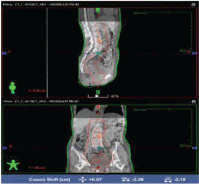
Figure 2. KV-CBCT and planning CT image registration for patient setup verification.
At individual level,
For each patient, the mean setup error was calculated and considered as systematic error, while the SD of repeated measurements represented the individual random error.
At population level,
Systematic error (S) was calculated as the SD of the individual mean setup errors, which indicates the variation of systematic deviations across the population.
Random error (s) was calculated as the mean of the individual random errors, representing the average random variation within the population.
CTV–PTV margin calculation
After the calculation of systematic and random error, population-based CTV to PTV margins were calculated for all patients using the Van Herk margin recipe, which ensure adequate dose coverage for 90%–95% of patients:
Margin = 2.5Σ + 0.7σ
Margins were computed separately for each translational direction–lateral, longitudinal and vertical to account for directional variations in setup uncertainties.
Results
A total of 20 pelvic cancer patients undergoing IGRT treatment with IMRT and VMAT techniques were used to analyse the magnitude of setup uncertainties. All patients underwent daily setup verification using KV-CBCT prior to treatment and translational shift was recorded in all three orthogonal directions – vertical (X), lateral (Y) and longitudinal (Z). Baseline patient characteristics are presented in Table 1.
Individual patient setup errors
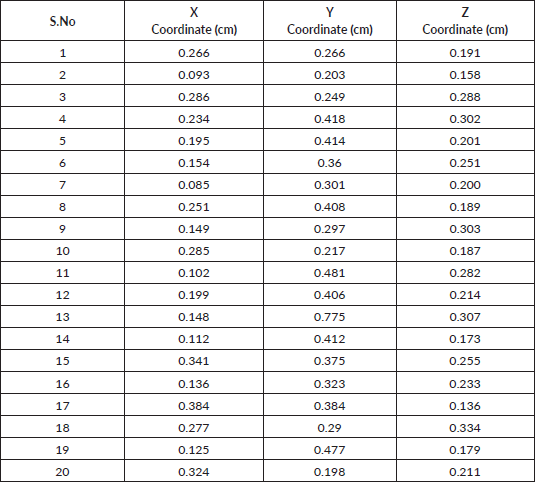
The mean translational setup errors for each patient with respect to the planned treatment isocentre were calculated in the vertical (X), lateral (Y) and longitudinal (Z) directions. The calculated individual patient mean setup errors are summarised in Table 2.
The mean setup errors ranged from 0.085 to 0.0384 cm in the vertical direction, 0.198 to 0.775 cm in the lateral direction and 0.136 to 0.334 cm in the longitudinal direction.
Table 1. Baseline characteristics of study population.
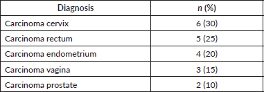
Table 2. Mean individual setup errors in the vertical (X), lateral (Y) and longitudinal (Z) directions.
Population systematic and random errors
To evaluate consistent deviations occurring throughout the treatment process (systematic errors) and day-to-day variations in patient positioning (random errors), systematic and random errors were analysed at the population level. The calculated values for each translational direction are presented in Table 3.
The highest systematic and random errors were observed in the lateral direction.
CTV to PTV margin calculation
The population-based CTV–PTV margins were calculated using the Van Herk margin formula. Calculated CTV–PTV margins for each translational direction are presented in Table 4.
The largest CTV–PTV margin was observed in the lateral direction, followed by the longitudinal and vertical directions.
Table 3. Population systematic error (S) and random error (s).

Table 4. Calculated CTV–PTV margins.

Discussion
Analysis of setup errors
Accurate patient positioning is essential in pelvic radiotherapy to ensure adequate target coverage while minimising radiation exposure to surrounding normal tissues. Despite the routine use of daily image guidance, residual setup uncertainties remain unavoidable because of interfraction anatomical variations and patient positioning differences. The present study evaluated translational setup errors in pelvic cancer patients treated with daily KV-CBCT-guided IMRT/VMAT on the Halcyon system and estimated institution-specific CTV–PTV margins using the Van Herk Margin formula.
The results demonstrated relatively small population systematic errors in all three translational directions, indicating good reproducibility of patient positioning throughout the treatment. In contrast, random errors were greater, particularly in lateral and longitudinal directions, indicating day-to-day variations despite daily image guidance. Among the three translational directions, the lateral direction shows the greatest setup uncertainty, resulting in the largest calculated CTV–PTV margin (0.78 cm). Similar findings have been reported in previous studies evaluating setup errors in pelvic radiotherapy, where lateral positioning errors were frequently greater than vertical error because of variations in patient anatomy, immobilisation reproducibility and interfraction changes in bladder and rectum filling [10, 11]. These findings highlight the importance of maintaining consistent patient preparation protocols throughout the treatment [8].
The calculated CTV–PTV margins in the present study were comparable to those reported in previous investigations using daily CBCT-based image guidance [9]. Although a uniform 5-mm institutional margin was used during treatment planning, the calculated lateral margin exceeded. This does not necessarily indicate that the current institutional margin is inadequate but rather highlights that institution-specific evaluation of setup uncertainties can provide valuable information for optimising treatment accuracy and margin selection.
Daily KV-CBCT considerably improves treatment precision by allowing correction of translational setup deviations before each treatment fraction. Nevertheless, image guidance cannot completely eliminate residual uncertainties caused by internal organ motion, patient relaxation and anatomical changes occurring after image acquisition. Therefore, an appropriate CTV–PTV margin remains essential even in the era of daily IGRT.
The findings of this study support the continued use of routine image guidance and emphasise the importance of periodic institutional audits of setup accuracy. Such evaluations can help to ensure that treatment margins remain appropriate for clinical practice.
Limitation
Despite the valuable observations obtained from this study, certain limitations must be recognised. Firstly, it was conducted at a single institution with a relatively small sample size, which may limit the broader coverage of the findings. Secondly, only translational setup errors were evaluated, whereas rotational errors were not assessed [11]. Finally, because this was a retrospective study, detailed clinicopathological data, including tumour stage and history, were not included. Therefore, subgroup analyses evaluating the influences of these factors on setup errors could not be performed.
Future study
In future, research should focus on larger populations with multi-institutional studies to validate these findings across different clinical settings. Additionally, involving rotational setup errors along with translational errors would provide a more comprehensive setup uncertainties assessment.
The use of advanced motion management techniques and adaptive radiotherapy approaches may further help in minimising setup variations and improving treatment accuracy.
Conclusion
Pelvic cancer radiotherapy delivered using treatment techniques such as IMRT and VMAT in combination with daily KV-CBCT image guidance demonstrates improved setup verification comparable to other studies. However, measurable residual translational setup variation was observed in all three orthogonal directions, with higher deviation in the lateral direction. Population-based analysis demonstrated low systematic errors and comparatively higher random errors, resulting in calculated CTV–PTV margins of 0.36, 0.78 and 0.59 cm in the vertical, lateral and longitudinal directions, respectively.
These findings highlight the importance of institution-specific assessment of setup uncertainties to support appropriate margin selection and ensure optimal target coverage while minimising radiation exposure to surrounding normal tissues. Further studies involving larger patient population are required to validate these observations.
Acknowledgment
The author(s) thank the Department of Radiation Oncology, Father Muller Medical College Hospital, for providing facilities and support for this study.
Conflicts of interest
The author(s) declare no conflicts of interest.
Funding
The authors declare that no financial support was received for this study.
Author contributions
This work was carried out in collaboration between both authors. Author Vishwas M Shetty contributed to data collection, analysis and interpretation and preparation of the manuscript. Author Shylvea Shalom Pandian S contributed to study design, supervision, critical review and final approval of the manuscript.
References
1. Li Z, Cheng Y, and Dong J, et al (2024) The impact of setup errors on dose distribution in cervical cancer radiotherapy and the margin from CTV to PTV J Cancer Res Clin Oncol 150(12) 516 https://doi.org/10.1007/s00432-024-06032-6 PMID: 39621121 PMCID: 11611962
2. Webster A, Appelt AL, and Eminowicz G (2020) Image-guided radiotherapy for pelvic cancers: a review of current evidence and clinical utilisation Clin Oncol (R Coll Radiol) 32(12) 805–816 https://doi.org/10.1016/j.clon.2020.09.010 PMID: 33071029
3. Amaoui B, Hadaoui A, and Mouhssine D, et al (2020) Evaluation of setup errors in conformal radiotherapy for pelvic tumours: case of the Regional Center of Oncology Radiat Med Prot 1(2) 99–102 https://doi.org/10.1016/j.radmp.2020.05.003
4. Badajena A, Raturi VP, and Sirvastava K, et al (2020) Prospective evaluation of the setup errors and its impact on safety margin for cervical cancer pelvic conformal radiotherapy Rep Pract Oncol Radiother 25(2) 260–265 https://doi.org/10.1016/j.rpor.2020.02.006 PMID: 32140083 PMCID: 7049598
5. Xiao H, Han Q, and Wei S, et al (2024) Setup errors analysis in iterative KV CBCT: a clinical study of cervical cancer treated with volumetric modulated arc therapy J Appl Clin Med Phys 25(10) e14480 https://doi.org/10.1002/acm2.14480 PMID: 39120606 PMCID: 11466492
6. Matsubayashi F, Kamima T, and Ito Y, et al (2024) Air inflow into vacuum-type immobilization devices impacts setup errors Radiol Phys Technol 17(3) 697–702 https://doi.org/10.1007/s12194-024-00822-w PMID: 38890268
7. Tong Y, Gong G, and Chen J, et al (2017) The heterogeneous CTV-PTV margins should be given for different parts of tumors during tomotherapy Oncotarget 8(51) 89086–89094 https://doi.org/10.18632/oncotarget.21631 PMID: 29179501 PMCID: 5687671
8. Lnu A, Kumar P, and Chauhan AK, et al (2023) Assessment of setup errors in gynecological malignancies treated with radiotherapy using onboard imaging Cureus 15(4) e37435 [https://doi.org/10.7759/cureus.37435.] PMID: 37056218 PMCID: 10088567
9. Elshahat K, El-Sheshtawy W, and Al-Agamawi A, et al (2020) Evaluation of setup uncertainty in radiotherapy of pelvic cancer Al-Azhar Int Med J 2020 [https://doi.org/10.21608/aimj.2020.28802.1209]
10. Nigam J, Kumar P, and Balan U (2016) Set up errors and recommended safety margins in the pelvic radiotherapy fields of cancer cervix patients: an institutional experience SRMS J Med Sci 1 https://doi.org/10.21761/jms.v1i2.7125
11. Hwang C (2023) Impact of dose distribution on rotational setup errors in radiotherapy for prostate cancer Med Dosim 48(3) 181–186 https://doi.org/10.1016/j.meddos.2023.03.007 PMID: 37085402