Patterns of treatment failure and survival outcomes following definitive intensity-modulated radiotherapy in locally advanced head and neck squamous cell carcinoma: a single centre experience
Abhishek Chakravarty1, Sweety Gupta2, Sumit Singh3, Aviral Rastogi4, Atokali Chophy4, Ravindra Babu Shylaja Namitha2 , Ravi Roushan Kumar5, Mayank Soni2, Deepa M Joseph2 and Manoj Gupta6
1Department of Radiation Oncology, NIMSR Jaipur, Jaipur, Rajasthan 303121, India
2Department of Radiation Oncology, AIIMS Rishikesh, Rishikesh, Uttarakhand 249203, India
3Department of Clinical Oncology, NMC, Biratnagar, Koshi Pradesh 56700, Nepal
4Department of Radiation Oncology, AIIMS Delhi, New Delhi 110029, India
5Department of Radiation Oncology, TMC, Muzaffarpur, Bihar 842004, India
6Department of Radiation Oncology, SGRR Indresh Hospital, Dehradun, Uttarakhand 248001, India
Abstract
Background: Locally advanced head and neck squamous cell carcinoma (LA-HNSCC) continues to have suboptimal outcomes, with locoregional and distant failures remaining a significant cause of morbidity and mortality despite advances in radiotherapy techniques. This study evaluated failure patterns and survival outcomes following definitive intensity-modulated radiotherapy (IMRT) in LA-HNSCC.
Materials and methods: An ambispective analysis was conducted on LA-HNSCC patients who received definitive IMRT with weekly concurrent chemotherapy. Locoregional failures (both residual disease and new loco-regional recurrences) as well as distant metastases were identified. Patterns of failure were categorised as local, regional or distant. Kaplan-Meier method was used to assess survival outcomes and univariate and multivariate Cox regression models were utilised to identify factors associated with failures and survival.
Results: A total of 332 patients were analysed, with a median age of 58 years; 94% had stage III–IVB disease and 83.1% received concurrent chemotherapy. At a median follow-up of 10 months, failure was observed in 44.9% of patients. Local control rates at 3, 6, 12 and 24 months were 97.9%, 81.6%, 69.9% and 52.5%, respectively. Local failure was the most common failure pattern (33.4%), followed by regional (15.6%) and distant failure (9.3%). Primary tumour volume greater than 30 cc and an oral cavity primary site were independent predictors of local failure. Median disease-free survival and overall survival were 8.3 and 13.8 months, respectively.
Conclusion: Despite the use of modern IMRT techniques, locoregional failure - particularly local failure - remains the predominant pattern of treatment failure in LA-HNSCC. Primary tumour volume is a critical determinant of outcome, highlighting the need for volumetric risk stratification and individualised treatment strategies to improve disease control in high-risk patients. However, the interpretation of survival outcomes should be considered in light of the wide range of follow-up duration in this cohort, which represents a limitation of the present study.
Keywords: radiotherapy, head and neck cancer, chemotherapy, failure
Correspondence to: Sweety Gupta
Email: drsg2411@yahoo.co.in
Published: 26/05/2026
Received: 06/01/2026
Publication costs for this article were supported by ecancer (UK Charity number 1176307).
Copyright: © the authors; licensee ecancermedicalscience. This is an Open Access article distributed under the terms of the Creative Commons Attribution License (http://creativecommons.org/licenses/by/4.0), which permits unrestricted use, distribution, and reproduction in any medium, provided the original work is properly cited.
Introduction
Head and neck cancers (HNC) globally account for 947,221 new cases and 450,000 deaths per year as per GLOBOCAN 2022 estimates [1]. The incidence of Head and Neck Squamous cell carcinoma (HNSCC) has been increasing in developing countries, particularly in the younger population, with a projected 30% increase in incidence by 2030. The highest incidence comes from India, which is 17% and the mortality of 9.6% worldwide [2]. This trend is partly attributed to changes in lifestyle factors, such as increased alcohol consumption and tobacco use in developing nations. The most attributable factor for the higher incidence of HNSCC in India is tobacco consumption, i.e., 80% [3]. Other factors, such as human papillomavirus (HPV), contribute to oropharyngeal cancer arising from the tonsil [4]. The current treatment strategy aims to achieve locoregional disease control with an improved quality of life. Definitive concurrent chemoradiation is the treatment of choice for locally advanced disease. This allows for organ preservation, enabling salvage surgery for residual or relapsed cancers [5, 6]. Evidence from the meta-analysis of chemotherapy in squamous cell head and neck cancer (MACH-NC) showed that concurrent radiotherapy (RT) and chemotherapy reduced mortality risk by 19% and improved 5-year survival overall (OS) by 6.5%. This benefit was primarily due to a 13.5% increase in locoregional control. The 2.9% reduction in the risk of distant metastasis (DM) was not statistically significant. An update of the MACH-NC meta-analysis confirmed the superiority of concurrent chemoradiotherapy (CRT) compared with RT alone across all HNC primary sites [7, 8].
Intensity-modulated radiotherapy (IMRT) has replaced conventional RT techniques as the standard of care in the definitive and adjuvant treatment of HNSCC [9]. IMRT enables the delivery of conformal doses and the sparing of normal tissues, thereby reducing toxicity while maintaining similar control rates [10]. However, the possibility of geographical miss remains a potential contributor to post-treatment failures [11, 12]. In the present study, we analysed the outcomes after definitive IMRT, types of failures and the factors associated with failure in patients with HNSCC.
Materials and methods
Study design
This ambispective study included histopathologically diagnosed HNSCC undergoing definitive RT with IMRT treated from February 2018 to December 2022. Institutional Ethics Committee approval was obtained before the commencement of the study.
Patient selection
All patients were histologically proven localised HNSCC, previously untreated (RT naive) stage I-IVB American Joint Committee of Cancer (AJCC) Classification System 2018, with epicentre in oropharynx, larynx, hypopharynx and unresectable oral cavity. Patients undergoing induction chemotherapy and/or concurrent chemotherapy were included. All patients were above 18 years and had an Eastern Cooperative Oncology Group Performance Score (ECOG PS) 0–2 or Karnofsky Performance Score ≥60. Nasopharyngeal, para nasal sinus, salivary gland, T1-T2 N0 glottic malignancies, dual malignancies (synchronous and metachronous), post operative HNSCC, de novo metastatic disease, prior history of RT and patients not undergoing full course of planned treatment were excluded from the study.
Study protocol
In this ambispective study, we analysed medical records of patients undergoing definitive IMRT for HNSCC. The data included comprehensive assessments, such as medical history, clinical examination, endoscopy and additional imaging studies, including contrast-enhanced (CE) computed tomography of the face, neck and thorax/CE magnetic resonance imaging (MRI) of the face, neck or whole body positron emission tomography–computed tomography (PET-CT). Staging was done according to 8th edition of the AJCC.
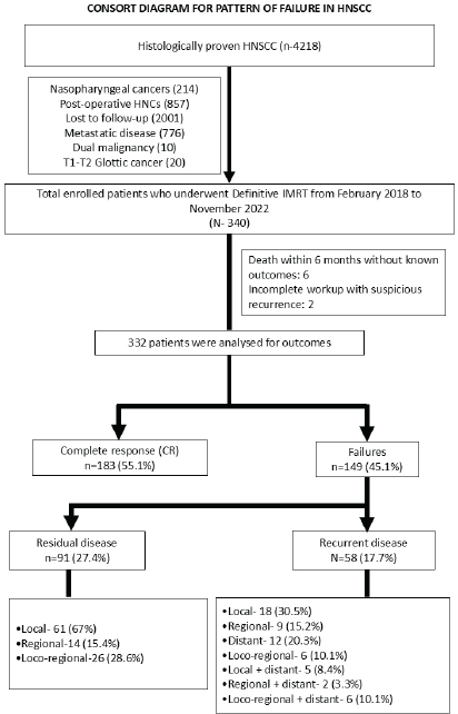
Figure 1. Consort diagram of patients included in the study.
Treatment details
Target volume (TV) definitions
TVs were contoured on axial computed tomography (CT) slices of the treatment planning system (TPS). The gross tumour volume (GTV) of the primary and nodes was contoured using relevant clinical examination findings, imaging (CT/MRI/PET CT), with the best possible image registration by the treating physician. In patients undergoing induction chemotherapy, pre-chemotherapy images and clinical findings were utilised to delineate the primary and nodal GTVs. Clinical target volume primary delineation was based on consensus guidelines provided by Grégoire et al [13] while clinical target volume nodal delineation were based on consensus guidelines from Grégoire et al [14] and selection of nodal stations were provided by the 2019 update from Biau et al [15]. The choice of level of dose prescription (either 2 volumes or 3 volumes) were based on primary and nodal GTV size and clinicians’ choice. A clinical target volume to planning target volume (PTV) expansion of 5 mm was used, which was based on institutional protocol [16].
IMRT plans were generated on Monaco v5.0 for Elekta and Varian TPS. Treatment was delivered using either sequential IMRT (50 Gy in 25 fractions followed by two boost phases of 10 Gy in 5 fractions each) or simultaneously integrated boost (SIB)-IMRT, with two regimens prescribing 69.96/59.4/54 Gy in 33 fractions or 66/60/54 Gy in 30 fractions to high-, intermediate- and low-risk PTVs, respectively. All plans were generated using volumetric arc therapy (VMAT) or RapidArc technique. During the COVID-19 period, hypofractionated schedule was employed, delivering 63/55.6/49.14 Gy in 21 fractions to the respective PTVs in accordance with institutional practice to ensure treatment continuity during the pandemic. However, concurrent chemotherapy was never used during this treatment protocol. Treatment was delivered using daily image guided radiotherapy using mega voltage cone beam computed tomography.
Concurrent chemotherapy with weekly cisplatin at 40 mg/m2 was administered in patients with CrCl >60 mg/ml/min and normal to mild sensory neural hearing loss. Concurrent carboplatin at an area under the curve of two was administered once weekly to patients who were ineligible for cisplatin. Patients who had received concurrent oxaliplatin in a previous study were also followed up and analysed for locoregional and distant control.
Follow-up
Patients were evaluated weekly during IMRT for acute toxicities. During each follow-up, patients were assessed using a history, clinical examination, 70-degree endoscopy and/or imaging with CT/MRI/PET-CT at 3 months and later, if suspicious for residual/recurrent disease. The follow-up duration was calculated from the time of initiation of RT to the last date of follow-up. Patients with persistent disease up to 6 months or with persistent symptomatic disease (either as Partial response, Stable disease or Progressive disease) at last follow-up were considered to have residual disease and deemed failures. Patients who achieved a complete response (CR) following treatment and later developed new lesions identified on imaging and confirmed histopathologically were considered to have experienced recurrence. Recurrences were categorised as local, regional, locoregional or distant. Disease-free survival (DFS) was calculated from initiation of RT to last follow-up in case of patients with CR or until patients labelled failed after treatment. OS was defined as the time from histopathological diagnosis to death, which could be due to malignancy or other non-malignant causes. Patients alive at the last follow-up were censored beyond that time period for the survival analysis.
Statistical analysis
Statistical analysis was performed using SPSS version 23.0 (IBM Corp., Armonk, NY). The demographic details were presented as frequencies, accompanied by histograms or pie charts as appropriate. Chi-square tests were used to measure the strength of association between recurrence patterns and tumour, patient and treatment parameters, including tumor, node, metastasis (TNM) stage, age group, ECOG score, radiation dose, use of concurrent chemotherapy and acute treatment-related toxicities. Mann-Whitney U test was used to measure the strength of association between the GTV volumes of the primary and node. Univariate and multivariate analyses for factors affecting outcome were analysed using the Cox proportional hazards model. Kaplan-Meier curves were used to illustrate DFS and OS.
Results
A total of 332 locally advanced head and neck squamous cell carcinoma (LA-HNSCC) treated between February 2018 and November 2022 were analysed as per the inclusion criteria in the present study (Figure 1).
Table 1. Patient and treatment details.
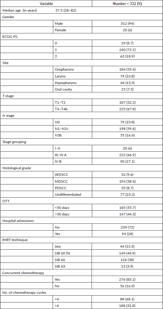
Table 2. Univariate analysis of factors associated with local and regional recurrence.
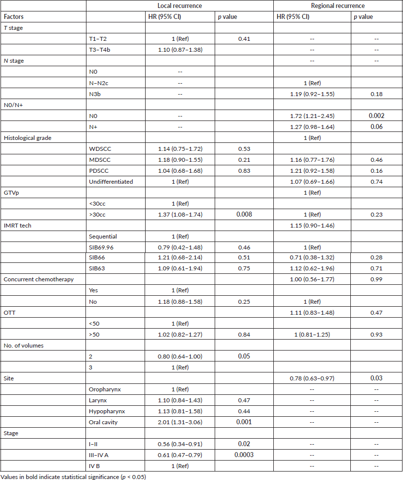
Table 3. Multivariate analysis of factors associated with local and regional recurrence.
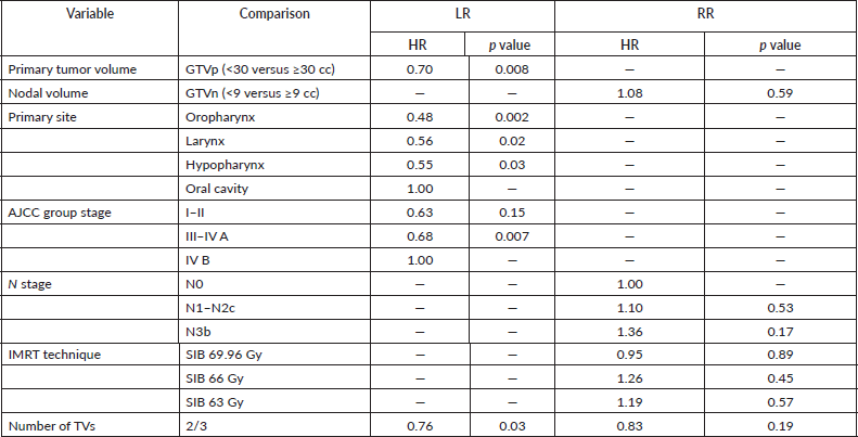
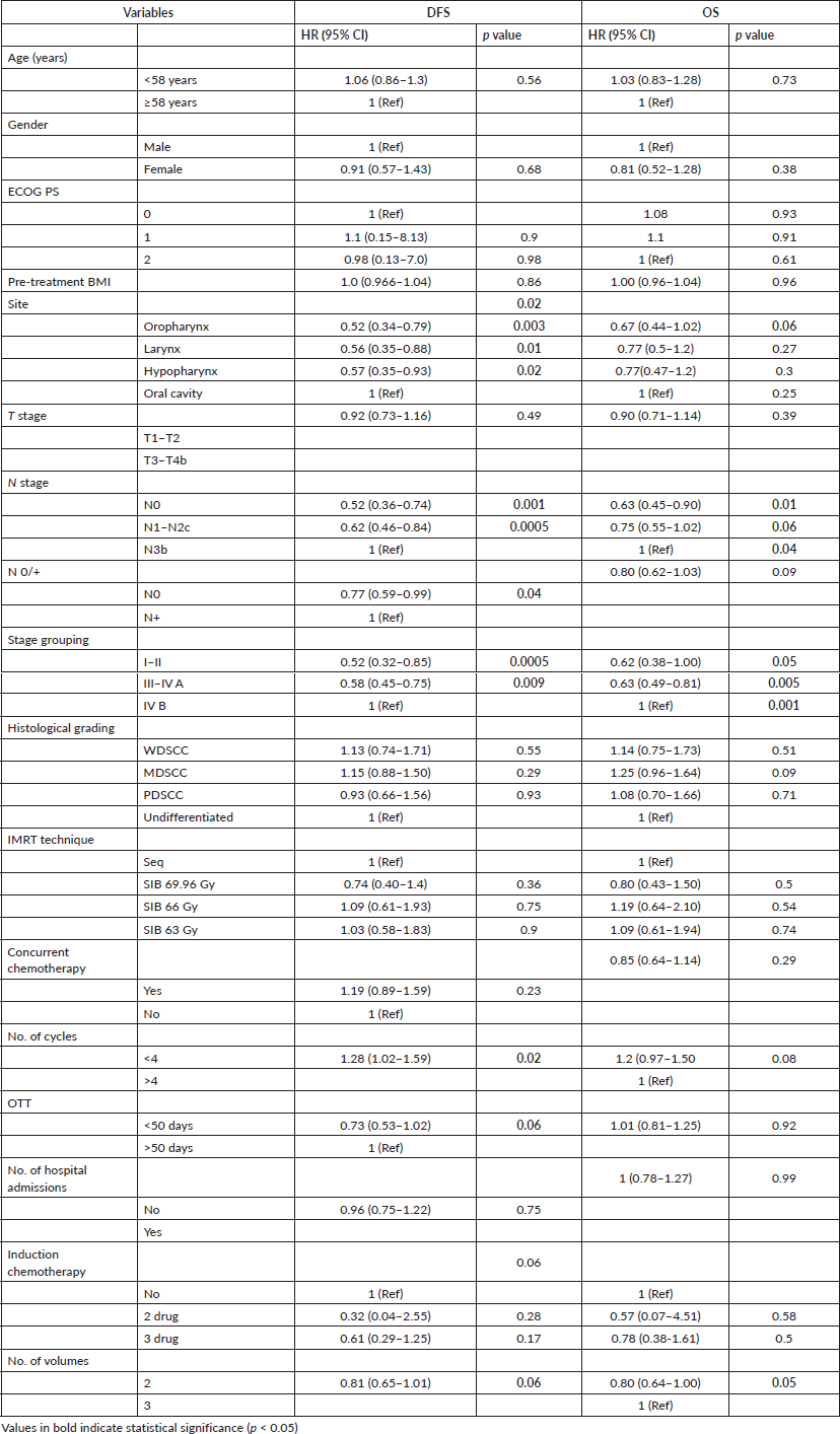
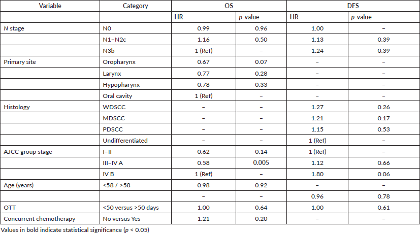
Table 4. Univariate analysis of factors associated with DFS and OS.
Patient characteristics and treatment details
The median age at diagnosis was 57.5 years (28–82 years). A majority of patients were males (94%). The most common primary tumour site was the oropharynx (55.4%; 184). 94% of patients were stage III–IV B. The median GTVp and GTVn volumes were 29.4 cm³ (IQR-15.65–52.15) and 8.7 cm³ (IQR-2.6–24.6), respectively. A total of 47.3% and 51.5% of patients were planned with 2 and 3 volumes, respectively. A total of 86.5% of patients received RT by SIB technique, with dose schedules of 69.96 Gy/33 fractions in 44.9% and 66 Gy/30 fractions in 38%. Concurrent chemotherapy was administered to 83.1% of patients, with 75.7% receiving concurrent cisplatin, 3.6% receiving concurrent oxaliplatin and 20.6% receiving concurrent carboplatin. The median number of chemotherapy cycles was 5 (range 1–7).
Induction chemotherapy was administered to 18.2% of patients, with 46 patients receiving two drugs (Paclitaxel/Cisplatin + 5-FU) and eight patients receiving three drugs (Paclitaxel/Cisplatin + 5-FU). The median treatment completion duration was 50 days (range, 39–127 days). Patients, treatment and details are summarised in Table 1.
Treatment outcomes
The median follow-up period was 10 months (range: 3 to 57.8 months). Treatment failure was observed in 44.9% (149/332). Local control rates (LCRs) at 3, 6, 12 and 24 months were 97.9%, 81.6%, 69.9% and 52.5%, respectively. The predominant site of failure was in oropharyngeal primaries (61.0%; 91/149), followed by laryngeal (19.4%; 29/149), hypopharyngeal (10.0%; 15/149) and oral cavity cancers (9.4%; 14/149). The cumulative incidence of failure included local failure in (33.4%; 111/332), regional failure in (15.6%; 52/332) and DM in (9.3%; 31/332). Among DM, lungs were the most common site (83.8%), followed by bones (38.7%). One patient developed compressive myelopathy and died within 1.5 months after completion of treatment; this patient was included in the analysis.
The median time to failure was:
- Local failure: 9.6 months (95% CI: 8.6–10.6 months)
- Regional failure: 9.9 months (95% CI: 9.0–10.7 months)
- Distant failure: 10.2 months (95% CI: 9.4–11.0 months)
Table 5. Multivariate analysis of factors associated with DFS and OS.
Factors associated with treatment failure
Local failure showed a statistically significant result in patients with a GTVp volume greater than 30 cc and a tumour site, which was consistent in both univariate and multivariate analyses. GTVp volumes >30 cc, oral cavity primaries and Stage IV B were associated with worse local failure patterns.
The nodal stage (N-stage) and GTV of nodal disease (GTVn > 9 cc) were predictive of regional failure on univariate analysis but did not retain significance in multivariate analysis.
N stage and AJCC stage grouping were significant in univariate analysis but not in multivariate analysis. Volumetric impact on outcomes showed that two-volume plans showed improved outcomes compared to three-volume plans (HR 0.80, 95% CI 0.64–1.00, p = 0.05) in terms of OS benefit. Larger PTV high-risk volumes (> 228.5 cc) were correlated with an increased risk of treatment failure. Tables 2 and 3 show univariate and multivariate analyses of factors associated with local and regional failure.
These findings suggest that while modern IMRT techniques achieve excellent target coverage, primary tumour volume remains a critical determinant of treatment outcomes. The strong correlation between GTVp >30 cc and local failure risk highlights the need for potential dose escalation strategies or alternative treatment approaches for larger tumour volumes.
Survival analysis
At a median follow-up of 10 months, 78.9% (262/332) of patients were alive, while 21.1% (70/332) had died. Among these deaths, 70% (49/70) were disease-related, while the status of 8 patients was unknown.
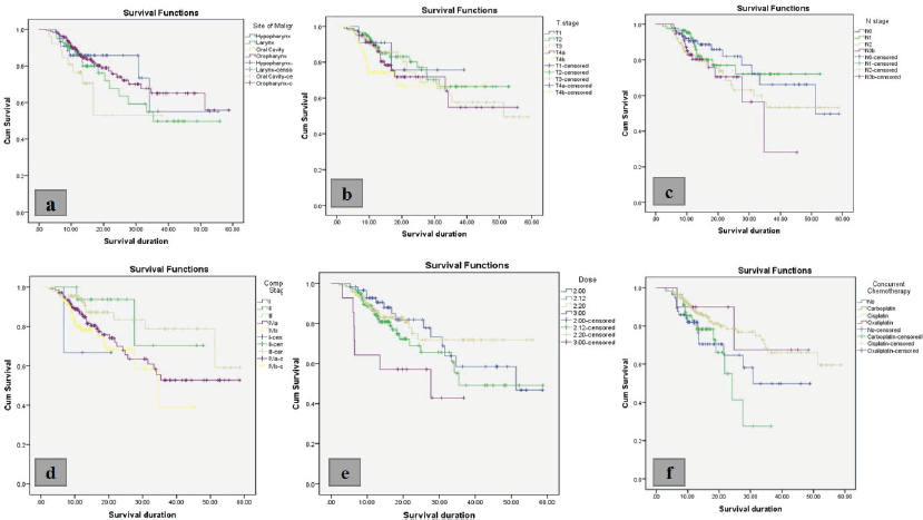
Figure 2. (a): Kaplan-Meier curve for association of OS with (a): site of malignancy, p = 0.583; (b): T stage, p = 0.083; (c): N stage, p = 0.004; (d): concurrent chemotherapy, p = 0.853.
The medial OS for the entire cohort was 13.8 months (3–58.7 months).
In the subgroup of patients achieving CR, the median OS was 16.4 months (8–62.3 months), while the median DFS was 13.8 months (6–57.8 months). Second primary malignancies were identified in 8/183 (4%) patients. Among these, six occurred within the upper aerodigestive tract (two head and neck primaries, three lung primaries and one esophageal primary), while one case each of endometrial carcinoma and nuclear protein in testis midline carcinoma was observed. Furthermore, 33 patients (18%) died due to non-malignant causes, most commonly lower respiratory tract infections or unknown causes.
Among patients who developed recurrence after achieving CR, the median time to any recurrence (local, regional or distant) was 10.2 months (6–41.2 months). The median time to local recurrence was 8.7 months (6–41.2 months), while the median time to regional recurrence was 9.3 months (6–20.5 months).
Among patients who experienced treatment failure, the median DFS was 6.9 months (3–41.2 months) and the median OS was 13.8 months (5.3–60.8 months).
On univariate and multivariate analyses, age, gender, ECOG performance status, pre-treatment BMI, histological grade, IMRT technique, concurrent chemotherapy, overall treatment time (OTT), hospital admissions and T stage were not significantly associated with OS or DFS. However, N stage and AJCC stage grouping were significantly associated with DFS, while only AJCC stage grouping was significantly associated with OS. The number of treatment volumes was significantly associated with OS and showed a trend toward significance for DFS. Detailed results of the univariate and multivariate analyses are presented in Tables 4 and 5.
For the entire cohort, the median DFS was 11.1 months (IQR: 8.8–15.5 months). There was no statistically significant association between site of malignancy, T stage or concurrent chemotherapy and OS, while N stage and composite stage showed a statistically significant association with OS. Figure 2 shows the Kaplan-Meier curves for factors associated with OS.
A statistically significant association was observed between site of malignancy, T stage, N stage and composite stage with DFS. The 3- and 6-month LCRs for the study cohort were 91.2% and 64.4%, respectively. There was no association between dose or concurrent chemotherapy and DFS. Figure 3 shows the Kaplan-Meier curves for DFS.
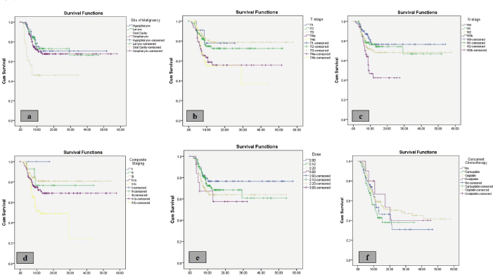
Figure 3. (a): Kaplan-Meier curve for association of DFS with (a): site of malignancy, p = 0.002; (b): T stage, p = 0.006; (c): N stage, p = 0.003; (d): composite stage, p = 0.002; (e): dose, p = 0.266; (f): concurrent chemotherapy, p = 0.281.
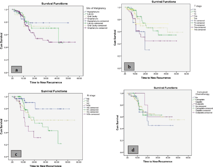
Figure 4. (a): Kaplan-Meier curve for association of time to recurrence with (a): site of malignancy, p = 0.232; (b): T stage, p = 0.262; (c): N stage, p = 0.209; (d): composite stage, p = 0.05; (e): dose, p = 0.08; (f): concurrent chemotherapy, p = 0.03.
Figure 4 shows the Kaplan-Meier curves for time to new recurrences. N stage and composite stage were significantly associated with new recurrences, while concurrent chemotherapy demonstrated a statistically significant association with time to new recurrences.
These findings suggest that patients achieving CR demonstrated relatively improved survival outcomes, although recurrences predominantly occurred within the first year of follow-up.
Discussion
It has been observed that about 35%–40% patients fail loco-regionally after definitive chemoradiation in locally advanced head and neck cancers. With the introduction of IMRT and VMAT, more conformal RT techniques have been developed, offering improved dose delivery to TVs while sparing normal tissues. However, a significant concern arises with geographical miss associated with this treatment method. Various institutions have reported their patterns of failure, highlighting that the majority of failures occur within the first year after completion of treatment and their prognosis remains poor [17, 18]. To guide future attempts at improving the therapeutic ratio after IMRT/VMAT, it is essential to discuss the pattern of failure. The present study reports the failure pattern of 332 patients with LA HNCs at our institute from February 2018 to December 2022. The predominant pattern of failure was locoregional (40.6%), which was higher compared to other contemporary studies [19]. This could be because a large proportion (27%) of patients in our study were classified as Stage IVB. These patients were deemed suitable for definitive treatment due to good response to induction therapy; however, they failed at their index sites either locally or regionally, which might be a result of the chemoresistant niche being the primary target during RT. It was predictable to find that these patients also had a significantly poorer overall and DFS. The OTT of less than 50 days had an impact on survival (HR 0.73, p = 0.06) in our study. Evidence consistently shows that prolonging OTT for definitive CRT in HNSCC worsens local control and survival, with detriment becoming clear once OTT exceeds 7 weeks [20]. Classic radiobiological estimates suggest a loss of roughly 0.5–1.0 Gy of ‘effective dose’ per extra day of OTT beyond a threshold, explaining why interruptions and OTT exceeding 50 days correlate with poorer locoregional control. It was also interesting to note that larger GTV in primary tumours >30 cc were associated with a significantly poorer LCR (10 versus 8.8 months, p = 0.008). Ahmed et al [21] also reported similar findings with a primary GTV <30 cc as an important prognostic indicator with improved CR rates (82.6% versus 51.9%) and a significantly improved OS (59.2 versus 21.4 months). The role of GTV primary volume also holds its importance in predicting the risk of DM, which is often incurable once diagnosed. The DAHANCA group evaluated this in a multivariate model and identified that GTV >50 cc had a significant association with DM, with hazard ratios of 7.6 (2.5–23.4) for p16-positive OPSCC and 4.1 (2.3–7.2) in other HNSCC [22]. The efficacy of intensified RT to GTV-T showed improvement in local failure, DFS and OS as used in ARTSCAN and ARTSCAN III with doses of 68–73.1 Gy to GTV-T. However, this effect seemed to plateau at a primary tumour volume of 40 cm³ [23]. The majority of regional recurrences were observed at the index site with in-field recurrence. Marginal recurrence was observed in two patients. These were followed in the contralateral neck at Level Ib (right tonsil primary) and Level 3 (laryngeal primary), which were included in PTV_IR. One patient with T2N1 laryngeal primary failed marginally both loco-regionally, which was diagnosed at 11 months. The other patient was diagnosed with nodal recurrence 12 months after treatment completion. Nodal burden was predictive of both regional and distant failure. Patients with N3b disease were shown to have higher regional and distant failure and significantly poorer DFS and OS, while the opposite was true for N0 disease. However, in the case of N1-N2c, there did not seem to be a significant impact on the pattern of failure. The predominant pattern of recurrence in our cohort was local recurrence (33.4%), followed by regional (15.7%) and distant recurrence (9.3%). This pattern aligns with previous studies by Studer et al [24] who reported similar recurrence patterns in patients with HNSCC treated with IMRT. This finding suggests that while IMRT provides excellent conformality, local tumour control remains a significant challenge, particularly in advanced-stage disease. Our analysis revealed that stage and nodal status were independent predictors of both OS and DFS. Stage IVB disease showed significantly worse outcomes compared to earlier stages (HR 1.0 versus 0.63 for Stage III–IVA, p = 0.005), consistent with volumetric staging studies that have demonstrated the prognostic value of traditional staging parameters in the era of IMRT [25]. The significantly worse outcomes observed in N3b disease (HR 1.72, 95% CI 1.21–2.45, p = 0.002) underscore the continuing challenge of managing advanced nodal disease. Our analysis revealed that TNM staging and nodal status had a significant impact on both OS and DFS. This finding aligns with previous studies, including the work of Takes et al [26] which demonstrated the fundamental importance of TNM classification in predicting treatment outcomes. Patients with advanced nodal disease (N2-3) showed notably inferior survival rates compared to those with limited nodal involvement (N0-1). The implementation of different IMRT techniques (sequential versus SIB) did not significantly impact treatment outcomes, suggesting that both approaches can be effectively utilised when appropriate. The high proportion of patients receiving concurrent chemotherapy (83.1%) reflects current standard practice; however, our analysis revealed varying impacts of different chemotherapy agents on outcomes. The limited number of patients who were eligible for salvage treatment (15 out of 149 failures) highlights the challenging nature of recurrent disease and the need for improved initial local control. The distribution of salvage approaches between reirradiation (2 patients) and surgery (12 patients) reflects the complex decision-making process in managing recurrences and the limited options available for this patient population.
Future research should focus on identifying novel strategies to improve local control, particularly for patients with larger tumour volumes and oral cavity primaries. Additionally, studies targeting biological markers such as epidermal growth factor receptor and other chemotherapeutic options, such as Docetaxel, as well as dose escalation strategies with PET-based planning techniques, may help identify patients at higher risk of treatment failure who might benefit from treatment intensification or alternative approaches [27, 28].
Strengths of this study include a large single-institution cohort, uniform IMRT planning and comprehensive assessment of failure patterns in a real-world population, whereas lack of HPV/p16 testing, especially given the high number of oropharyngeal cancers (55.4%), a wide range of follow up and the ambispective nature of the study, are some of the limitations.
Conclusion
Definitive IMRT provides reasonable disease control in LA-HNSCC; however, locoregional failure remains the predominant pattern of relapse. Primary tumour volume is a key independent predictor of failure, emphasising the need for volumetric risk stratification and tailored treatment intensification to improve outcomes in high-risk patients.
List of abbreviations
AJCC, American Joint Committee on Cancer; BMI, Body mass index; CECT, Contrast-enhanced computed tomography; CI, Confidence interval; CRT, Chemoradiotherapy; CT, Computed tomography; DFS, Disease-free survival; DM, Distant metastasis; ECOG, Eastern Cooperative Oncology Group; GTV, Gross tumour volume; GTVn, Gross tumour volume (Nodal); GTVp, Gross tumour volume (Primary); HNSCC, Head and neck squamous cell carcinoma; HPV, Human Papillomavirus; HR, Hazard ratio; IMRT, Intensity-modulated radiotherapy; IQR, Interquartile range; KM, Kaplan-Meier; LA-HNSCC, Locally advanced head and neck squamous cell carcinoma; LC, Local control; LCR, Local control rate; LF, Local failure; LR, Local recurrence; LRR, Locoregional recurrence; MRI, Magnetic resonance imaging; OS, Overall survival; OTT, Overall treatment time; OPSCC, Oropharyngeal squamous cell carcinoma; PDSCC, Poorly differentiated squamous cell carcinoma; PET-CT, Positron emission tomography-computed tomography; PFS, Progression-free survival; PTV, Planning target volume; RR, Regional recurrence; RT, Radiotherapy; SIB, Simultaneous integrated boost; TPS, Treatment planning system; VMAT, Volumetric modulated arc therapy; WDSCC, Well-differentiated squamous cell carcinoma.
Conflicts of interest
The authors declare that they have no conflicts of interest.
Funding
This research did not receive any specific grant from funding agencies in the public, commercial or not-for-profit sectors.
Author contributions
Abhishek Chakravarty (AC); Sweety Gupta (SG); Sumit Singh (SS); Aviral Rastogi (AR); Atokali Chophy (AC); Namitha RS(NRS); Ravi Roushan Kumar (RRK); Mayank Soni (MS); Deepa M Joseph (DMJ); Manoj Gupta (MG).
Conception and design: AC, SG, DMJ, MG; Administrative support: SG, DMJ, MG; Collection and assembly of data: AC, SS, AR, AC, NRS. Data analysis and interpretation: AC, RRK, MS, SG. Technical inputs and manuscript review: AC, SG, DMJ, MG. Proofreading: AC, SG, DMJ, MG. Manuscript writing: All authors. Final approval of manuscript: All authors. Accountable for all aspects of the work: AC, SG, DMJ, MG.
References
1. Bray F, Laversanne M, and Sung H, et al (2022) Global cancer statistics 2022: GLOBOCAN estimates of incidence and mortality worldwide for 36 cancers in 185 countries CA Cancer J Clin 74 229–263 [https://doi.org/10.3322/caac.21794]
2. Barsouk A, Aluru JS, and Rawla P, et al (2023) Epidemiology, risk factors, and prevention of head and neck squamous cell carcinoma Med Sci (Basel) 11 42 [https://doi.org/10.3390/medsci11020042] PMID: 37367741 PMCID: 10304137
3. Basu R, Mandal S, and Ghosh A, et al (2008) Role of tobacco in the development of head and neck squamous cell carcinoma in an eastern Indian population Asian Pac J Cancer Prev 9 381–386 [PMID: 19256752] PMID: 18990006
4. Johnson DE, Burtness B, and Leemans CR, et al (2020) Head and neck squamous cell carcinoma Nat Rev Dis Prim 6 92 [10.1038/s41572-020-00224-3] https://doi.org/10.1038/s41572-020-00224-3 PMID: 33243986 PMCID: 7944998
5. De Roest RH, van der Heijden M, and Wesseling FWR, et al (2022) Disease outcome and associated factors after definitive platinum-based chemoradiotherapy for advanced-stage HPV-negative head and neck cancer Radiother Oncol 175 112–121 https://doi.org/10.1016/j.radonc.2022.08.013
6. Iyer NG, Tan DSW, and Tan VK, et al (2015) Randomized trial comparing surgery and adjuvant radiotherapy versus concurrent chemoradiotherapy in patients with advanced, nonmetastatic squamous cell carcinoma of the head and neck: 10-year update and subset analysis Cancer 121 1599–1607 https://doi.org/10.1002/cncr.29251 PMID: 25639864
7. Pignon JP, Le Maître A, and Bourhis J (2007) Meta-analyses of chemotherapy in head and neck cancer (MACH-NC): an update Int J Radiat Oncol Biol Phys 69(2) S112–S114 https://doi.org/10.1016/j.ijrobp.2007.04.088
8. Lacas B, Carmel A, and Landais C, et al (2021) Meta-analysis of chemotherapy in head and neck cancer (MACH- NC): an update on 107 randomized trials and 19,805 patients, on behalf of MACH-NC group Radiotherapy Oncol 156 281–293 https://doi.org/10.1016/j.radonc.2021.01.013
9. Marta GN, Silva V, and de Andrade Carvalho H, et al (2014) Intensity-modulated radiation therapy for head and neck cancer: systematic review and meta-analysis Radiother Oncol 110(9) 9–15 https://doi.org/10.1016/j.radonc.2013.11.010
10. Gupta T, Kannan S, and Ghosh-Laskar S, et al (2018) Systematic review and meta-analyses of intensity-modulated radiation therapy versus conventional radiotherapy in head and neck squamous cell carcinoma PLoS One 13(0200137) e0200137 https://doi.org/10.1371/journal.pone.0200137
11. Chao KS, Ozyigit G, and Tran BN, et al (2003) Patterns of failure in patients receiving definitive and postoperative IMRT for head-and neck cancer Int J Radiat Oncol Biol Phys 55(2) 312–321 https://doi.org/10.1016/S0360-3016(02)03940-8
12. Dawson LA, Anzai Y, and Marsh L, et al (2000) Patterns of local-regional recurrence following parotid-sparing conformal and segmental intensity-modulated radiotherapy for head and neck cancer Int J Radiat Oncol Biol Phys 46(5) 1117–1126 https://doi.org/10.1016/S0360-3016(99)00550-7 PMID: 10725621
13. Grégoire V, Evans M, and Le QT, et al (2018) Delineation of the primary tumour clinical target volumes (CTV-P) in laryngeal, hypopharyngeal, oropharyngeal and oral cavity squamous cell carcinoma: AIRO, CACA, DAHANCA, EORTC, GEORCC, GORTEC, HKNPCSG, HNCIG, IAG-KHT, LPRHHT, NCIC CTG, NCRI, NRG Oncology, PHNS, SBRT, SOMERA, SRO, SSHNO, TROG consensus guidelines Radiotherapy Oncol 126(1) 3–24 https://doi.org/10.1016/j.radonc.2017.10.016
14. Grégoire V, Ang K, and Budach W, et al (2014) Delineation of the neck node levels for head and neck tumors: a 2013 update Radiotherapy Oncol 110(1) 172–181 https://doi.org/10.1016/j.radonc.2013.10.010
15. Biau J, Lapeyre M, and Troussier I, et al (2019) Selection of lymph node target volumes for definitive head and neck radiation therapy: a 2019 update Radiotherapy Oncol 134 1–9 https://doi.org/10.1016/j.radonc.2019.01.018
16. Sikdar D, Krishnan AS, and Namitha RS, et al (2024) MV CBCT based assessment of setup uncertainties and planning target volume margin in head and neck cancer Rep Pract Oncol Radiother 29(2) 141–147 [https://doi.org/10.5603/rpor.99361]
17. Bollen H, van der Veen J, and Laenen A, et al (2021) Recurrence patterns after IMRT/VMAT in head and neck cancer Front Oncol 11 720052 https://doi.org/10.3389/fonc.2021.720052 PMID: 34604056 PMCID: 8483718