Longitudinal changes and predictors of early chemotherapy-induced cancer-related fatigue in women with breast cancer: findings from a prospective Nigerian Cohort
Gbenro E. Olukiran1, Adewumi O. Alabi2,3, Anthonia C. Sowunmi2,3, Babatunde Fadipe4,5, Gbenga O. Popoola6, Sunday A. Okinbaloye7, Hnin L. Nwe Thein7 and Ololade Kehinde2
1Department of Radiation & Clinical Oncology, Lagos University Teaching Hospital, Lagos 100023, Nigeria
2Department of Radiation Biology, Radiotherapy & Radiodiagnosis, College of Medicine, University of Lagos, Lagos 100023, Nigeria
3Department of Radiation & Clinical Oncology, MEDSERVE-LUTH Cancer Centre, Lagos University Teaching Hospital, Lagos 100023, Nigeria
4Department of Psychiatry, Lagos University Teaching Hospital, Idi-Araba, Lagos 100023, Nigeria
5Arnold Lodge Medium Secure Unit, Nottinghamshire Healthcare NHS Foundation Trust, Leicester LE5 0LE, UK
6Psychiatric Intensive Care Unit, Lincolnshire Partnership NHS Foundation Trust, Lincoln LN1 1FS, UK
7United Lincolnshire Teaching Hospitals NHS Trust, Lincoln LN2 5QY, UK
Abstract
Background: Cancer-related fatigue is a common and disabling symptom among women receiving chemotherapy for breast cancer, yet its early trajectory and associated factors remain insufficiently characterised in African settings. This study examined longitudinal changes in fatigue and baseline factors associated with fatigue deterioration among women with breast cancer receiving chemotherapy at a Nigerian tertiary oncology centre.
Methods: This prospective cohort study included 79 chemotherapy-naïve women with histologically confirmed breast cancer who were assessed before chemotherapy and after completion of the third cycle. Fatigue was measured using the Functional Assessment of Chronic Illness Therapy–Fatigue (FACIT-Fatigue) scale. The primary outcome was the within-participant change in FACIT-Fatigue score, calculated as the cycle 3 score minus the baseline score. Baseline demographic, clinical, socioeconomic, haematological and study-specific patient-reported variables were evaluated as potential predictors of fatigue deterioration.
Results: At baseline, the mean FACIT-Fatigue score was 34.0 ± 8.4, and 36 participants (45.6%) had clinically significant fatigue. By cycle 3, the mean score had declined to 24.8 ± 11.7, representing significant fatigue deterioration (mean change −9.23 points; 95% CI −11.82 to −6.64; p < 0.001; Cohen’s dz = 0.80). Seventy participants (88.6%) experienced a clinically meaningful decline of at least three FACIT-Fatigue points. Surgical status was associated with fatigue change in the bivariate analysis, while age and appetite demonstrated weak or borderline associations. However, none of the evaluated baseline factors were independently associated with fatigue deterioration in the multivariable analysis. The adjusted model had virtually no explanatory value (adjusted R² = 0.001).
Conclusion: Women with breast cancer began chemotherapy with a substantial burden of fatigue, which worsened markedly during the first three treatment cycles. Although no evaluated baseline factor independently predicted this deterioration, the prospective within-participant findings demonstrate a substantial early symptom burden. These findings support fatigue assessment before chemotherapy and repeated monitoring during treatment, together with timely, guideline-directed supportive care when clinically indicated.
Keywords: breast neoplasms, cancer-related fatigue, chemotherapy, haemoglobins, prospective studies
Correspondence to: Adewumi O. Alabi
Email: adewumialabi@gmail.com
Published: 21/10/2026
Received: 03/06/2026
Publication costs for this article were supported by ecancer (UK Charity number 1176307).
Copyright: © the authors; licensee ecancermedicalscience. This is an Open Access article distributed under the terms of the Creative Commons Attribution License (http://creativecommons.org/licenses/by/4.0), which permits unrestricted use, distribution, and reproduction in any medium, provided the original work is properly cited.
Introduction
Breast cancer is the most frequently diagnosed malignancy among women worldwide and remains a major cause of cancer mortality. In 2022, approximately 2.3 million women were diagnosed with breast cancer, and 670,000 died from the disease. This burden is projected to increase substantially by 2050, with the greatest proportional rise expected in countries with a low Human Development Index [1,2]. These global estimates conceal marked geographical inequalities. Although breast cancer incidence is generally lower in sub-Saharan Africa than in many high-income regions, mortality is disproportionately high because of delayed diagnosis, advanced-stage presentation, limited diagnostic and treatment capacity, treatment interruption and restricted access to comprehensive oncology services [2,3].
Cancer-related fatigue is a persistent and distressing sense of physical, emotional or cognitive exhaustion attributable to cancer or its treatment. It is disproportionate to recent activity, is not reliably relieved by rest and interferes with usual functioning [4]. Its clinical significance extends beyond the subjective experience of tiredness. Cancer-related fatigue can impair mobility, cognition, occupational and social functioning, treatment tolerance and health-related quality of life. It may also reduce a patient’s capacity to participate consistently in cancer care [5,6]. The symptom may be present at diagnosis, intensify during active treatment and persist for months or years after treatment completion [5,6].
Fatigue is particularly common during systemic anticancer treatment. A meta-analysis of 129 studies involving more than 71,000 patients estimated an overall fatigue prevalence of 49%, increasing to approximately 62% among patients receiving active cancer treatment [7]. In breast cancer, fatigue may be present before chemotherapy and commonly worsens after treatment begins. In a prospective study of women assessed before and during the first three cycles of adjuvant chemotherapy, fatigue was more severe than in age-matched women without cancer and increased after chemotherapy commenced [8]. Another prospective cohort demonstrated that fatigue prevalence and severity changed across chemotherapy cycles, with the pattern varying according to chemotherapy regimen and with partial improvement following treatment completion [9]. These average trends nevertheless conceal substantial interindividual variation. Longitudinal studies have identified patients with persistently low fatigue, persistently high fatigue, treatment-reactive fatigue, progressive fatigue and fatigue that improves after an initially high pretreatment burden [10]. Furthermore, a meta-analysis involving 12,327 breast cancer survivors estimated that approximately one-quarter experienced severe post-treatment fatigue, demonstrating that the symptom may remain clinically important beyond active therapy [11].
Cancer-related fatigue has a multifactorial pathogenesis. Proposed determinants include age, socioeconomic circumstances, comorbidity, body composition, frailty, baseline performance status, tumour stage, pain, cachexia, sleep disturbance, psychological distress, nutritional compromise, reduced physical activity and limited social support [5,6,11]. Treatment-related contributors may include chemotherapy regimen, cumulative exposure, dose intensity, concurrent systemic therapy and the burden of other treatment toxicities. Anaemia may contribute through impaired tissue oxygen delivery, while inflammatory signalling, neuroendocrine dysregulation, altered energy metabolism and mitochondrial dysfunction have also been implicated [5,6]. The relevance of these factors may vary between patients and across treatment phases, making it unlikely that any single baseline characteristic will adequately explain the full fatigue trajectory.
Evidence from African oncology populations confirms that fatigue is common, although much of the available literature is cross-sectional and includes heterogeneous cancer diagnoses. In an Ethiopian mixed-methods study, 77.4% of adult oncology patients reported fatigue; late-stage disease, anaemia, comorbidity and uninsured medical expenditure were independently associated with its occurrence [12]. These findings illustrate the potential contribution of clinical, biological and structural determinants in African settings. However, evidence from mixed-cancer populations cannot be assumed to describe the specific experience of women receiving chemotherapy for breast cancer, particularly given regional differences in stage at presentation, treatment availability, chemotherapy exposure, supportive-care provision and socioeconomic circumstances [3,12].
Emerging Nigerian evidence demonstrates a substantial burden of fatigue during active treatment and survivorship. In a prospective study of 120 women receiving chemotherapy at University College Hospital, Ibadan, 94.2% reported fatigue during the first three treatment courses, making it one of the most frequently reported adverse effects [13]. However, fatigue was recorded as a treatment adverse effect rather than measured serially from a pretreatment baseline using a validated multidimensional fatigue instrument. In a subsequent case-control study from southwestern Nigeria, clinically meaningful fatigue was reported by 24.3% of breast cancer survivors, compared with 10.0% of age-matched controls. Survivors also reported greater interference with concentration and interpersonal functioning [14]. A further Nigerian study found fatigue in 22.5% of breast cancer survivors; advanced stage at presentation, employment status and more recent chemotherapy were independently associated with its occurrence [15]. More recently, a cross-sectional study of 139 women treated in two tertiary hospitals in Delta State found that fatigue affected 65% of participants after chemotherapy and commonly co-occurred with pain and sleep disturbance [16]. Differences between these estimates probably reflect variation in patient populations, treatment phases, fatigue definitions, measurement instruments and assessment timing.
Collectively, studies from Nigeria establish that fatigue is common during chemotherapy and can persist into survivorship. Nevertheless, the available evidence is predominantly cross-sectional or based on adverse-effect reporting and therefore provides limited information about within-patient change from the pretreatment period through the early chemotherapy cycles [13–16]. There is also limited prospective Nigerian evidence on whether routinely available demographic, clinical, psychosocial, socioeconomic and haematological characteristics can identify women at increased risk of early fatigue deterioration. Addressing this gap is clinically important because early recognition of worsening fatigue may facilitate timely evaluation of contributory factors and appropriate supportive care. This prospective study therefore evaluated longitudinal change in cancer-related fatigue from pretreatment baseline to completion of the third chemotherapy cycle and examined potential baseline factors associated with fatigue deterioration among women with breast cancer receiving care at a Nigerian tertiary oncology centre.
Methods
Study design and setting
This prospective cohort study was conducted at the Radiotherapy and Oncology Department of Lagos University Teaching Hospital (LUTH), Lagos, Nigeria. LUTH is a major tertiary referral centre providing cancer care to patients from Lagos State and other parts of southwestern Nigeria.
Participants were recruited consecutively between January and June 2022 and followed from immediately before their first chemotherapy cycle until completion of the third cycle. Recruitment continued throughout the study period. Fatigue was assessed at baseline, before chemotherapy initiation and after completion of the third cycle, approximately 9 weeks later under the predominantly three-weekly chemotherapy schedules.
The third cycle was selected a priori to capture early cumulative toxicity following repeated chemotherapy exposure while limiting attrition associated with later treatment cycles. It also represented an early clinical reassessment point and was consistent with previous longitudinal studies evaluating symptom changes during the first three chemotherapy cycles [8,13].
Study population and recruitment
Eligible participants were women aged 18 years or older with histologically confirmed breast cancer who were chemotherapy-naïve at enrolment, scheduled to commence systemic chemotherapy and willing to provide written informed consent. Patients with recurrent breast cancer, previous chemotherapy for the current malignancy, severe uncontrolled medical comorbidities or cognitive or functional impairment that precluded reliable participation were excluded.
Consecutive sampling was used. All potentially eligible women presenting during the study period were screened sequentially and invited to participate. Seventy-nine women with paired baseline and cycle 3 fatigue assessments constituted the analytic cohort.
Inability to read or write was not an explicit exclusion criterion. Questionnaires were administered through face-to-face interviews by study personnel in a designated private room. The same English-language questionnaire, item wording and response options were used for all participants to standardise administration and minimise interviewer influence. No translated or culturally adapted non-English version was used.
Sample-size considerations
Sample-size adequacy was evaluated in relation to the primary outcome of within-participant change in FACIT-Fatigue score rather than fatigue prevalence. With 79 paired observations, a two-sided paired t-test at a 5% significance level provided 80% power to detect a standardised paired mean difference, Cohen’s dz, of approximately 0.32.
The multivariable model estimated approximately 10 predictor coefficients using 78 complete records. The predictor analysis was therefore considered exploratory, and interpretation emphasised regression coefficients and confidence intervals rather than statistical significance alone.
Data collection and study variables
Baseline demographic, socioeconomic, clinical and patient-reported data were collected using a structured interviewer-administered questionnaire and review of participants’ clinical records. Follow-up fatigue assessment was conducted after completion of the third chemotherapy cycle. Baseline haemoglobin concentration was obtained from participants’ clinical records and examined as a potential biological predictor of fatigue change.
Clinical variables included age at treatment, menopausal status, disease stage, previous breast surgery, chemotherapy regimen and Karnofsky Performance Status. The Karnofsky Performance Status scale ranges from 0 to 100, with higher scores indicating better functional capacity [21].
The study did not employ a validated multidimensional psychosocial instrument. Instead, selected patient-reported characteristics were assessed at baseline using individual study-specific questions. Social isolation was assessed using a single yes-or-no question asking whether the participant had experienced feelings of isolation during the preceding 7 days. Loss of emotional control and feelings of dejection were similarly assessed using separate yes-or-no questions referring to the preceding 7 days. Appetite was assessed using a single question referring to the preceding month, with responses categorised as good, fair or poor. Sleep quality during the preceding week was classified as good, fair or poor.
Living arrangement was assessed by asking whether the participant lived alone, with responses recorded as yes or no, and by recording the type of residence. Functional participation was assessed using four separate questions asking participants to rate their involvement in preparing food, cleaning the house, visiting family or friends and light lifting on a five-point scale. These study-specific questions were not combined into a composite score, and no clinical cut-off was applied. They were therefore reported individually and were not presented as validated psychosocial constructs. Missing responses were retained as missing and were not imputed.
Socioeconomic variables included marital status, educational attainment, employment status, work disruption and average monthly income. Monthly income was classified as less than ₦30,000, ₦30,000–₦100,000, ₦100,001–₦200,000 or more than ₦200,000.
Assessment of cancer-related fatigue
Cancer-related fatigue was assessed using the 13-item Functional Assessment of Chronic Illness Therapy–Fatigue Scale, version 4 (FACIT-Fatigue). The instrument evaluates fatigue severity and its effects on daily activities and functioning during the preceding 7 days. Each item is rated on a five-point Likert scale ranging from 0, ‘not at all’, to 4, ‘very much’ [17].
Items were reverse-scored where required and summed according to the standard FACIT scoring procedure. Total scores range from 0 to 52, with higher scores indicating less fatigue and better fatigue-related functioning.
A published Nigerian validation of the FACIT-Fatigue scale was not identified. The instrument was nevertheless selected because it is brief, designed specifically for fatigue assessment and has established measurement properties in oncology. It has demonstrated satisfactory validity and reliability among women with breast cancer in Iran and acceptable internal consistency in an Indian oncology population, supporting its use in comparable low- and middle-income settings [18,19].
Before the main study, the instrument was piloted in 15 women who were not included in the principal cohort. Two research assistants independently administered the questionnaire. Administration required approximately 5–8 minutes. Inter-rater agreement was 93.3%, with Cohen’s kappa of 0.815. Cronbach’s alpha values were 0.844 and 0.828 for the two research assistants.
Internal consistency was reassessed in the analytic cohort. Cronbach’s alpha and McDonald’s omega were 0.949 and 0.952, respectively, at baseline and 0.942 and 0.943 at cycle 3, demonstrating excellent internal consistency.
FACIT-Fatigue scoring and data quality
All FACIT-Fatigue item responses were checked against the permissible range of 0–4. One baseline An14 response had a recorded value of 23 and was treated as missing because the original response could not be recovered reliably. One baseline HI7 response was also missing. No replacement value was inferred for either response.
A total FACIT-Fatigue score was calculated when at least 7 of the 13 items had valid responses. Eligible incomplete questionnaires were prorated by multiplying the observed item sum by 13 and dividing by the number of completed items. This resulted in prorated baseline scores for two participants. All cycle 3 questionnaires contained 13 valid responses.
Outcome measures
The primary outcome was within-participant change in FACIT-Fatigue score from baseline to cycle 3, calculated as follows:
Change in FACIT-Fatigue score = cycle 3 FACIT-Fatigue score − baseline FACIT-Fatigue score
Because higher FACIT-Fatigue scores indicate less fatigue, negative change scores represented worsening fatigue, whereas positive change scores represented improvement.
Secondary outcomes were the mean change in FACIT-Fatigue score, clinically meaningful fatigue worsening and internal consistency of the FACIT-Fatigue scale.
Clinically meaningful worsening was defined as a reduction of at least three FACIT-Fatigue points. This threshold was based on anchor- and distribution-based oncology studies identifying approximately 3–4 points as the minimally important difference for the FACIT-Fatigue scale [20]. The binary outcome complemented the continuous change analysis by providing a clinically interpretable participant-level measure.
Data management
Data were checked for duplicate records, missing observations, inconsistent coding, impossible values and values outside prespecified ranges. The original workbook was retained without overwriting recorded observations. Analyses of the primary outcome were restricted to participants with paired baseline and cycle 3 fatigue assessments.
Statistical analysis
Continuous variables were summarised using means and standard deviations and, where appropriate, medians and ranges. Categorical variables were summarised using frequencies and percentages.
The distribution of within-participant change in FACIT-Fatigue score was assessed using a histogram, normal quantile–quantile plot, skewness, outlier inspection and the Shapiro–Wilk test. The paired differences were approximately normally distributed. Baseline and cycle 3 FACIT-Fatigue scores were therefore compared using a paired-samples t-test. The mean change, 95% confidence interval and two-sided p-value were reported. The magnitude of the within-participant change was quantified using Cohen’s dz, calculated as the mean paired difference divided by the standard deviation of the paired differences.
In addition to the continuous change analysis, clinically meaningful fatigue deterioration was defined as a reduction of at least three FACIT-Fatigue points from baseline to cycle 3. This threshold was selected on the basis of oncology studies identifying an approximately 3–4-point change as the minimally important difference for the FACIT-Fatigue scale [20]. The number and percentage of participants meeting this criterion were reported to provide a clinically interpretable participant-level estimate of fatigue deterioration.
Bivariate associations between baseline participant characteristics and change in FACIT-Fatigue score were examined according to variable type. Pearson correlation analysis was used for continuous variables. Binary categorical variables were compared using independent-samples t-tests, while variables with more than two categories were evaluated using one-way analysis of variance. The study-specific patient-reported variables, including appetite and social isolation, were analysed separately and were not combined into an overall psychosocial score.
A multivariable ordinary least-squares linear regression model was fitted with change in FACIT-Fatigue score as the dependent variable. Prespecified predictors were age at treatment, disease stage, income category, social isolation, appetite status, Karnofsky Performance Status and baseline haemoglobin concentration. Disease stage, income category and appetite status were entered as categorical variables, while age, Karnofsky Performance Status and baseline haemoglobin were entered as continuous variables. Regression coefficients, standard errors, 95% confidence intervals and p-values were reported. Overall model performance was assessed using the F-statistic, R² and adjusted R². Automated stepwise variable selection was not used.
Regression assumptions were evaluated using residual-versus-fitted and quantile–quantile plots, the Shapiro–Wilk test, the Breusch–Pagan test, variance inflation factors, leverage and Cook’s distance. All statistical tests were two-sided, and p < 0.05 was considered statistically significant. Analyses were performed using R statistical software (R Foundation for Statistical Computing, Vienna, Austria).
Ethical considerations
Ethical approval was obtained from the Lagos University Teaching Hospital Health Research Ethics Committee (approval number ADM/DCST/HREC/APP/2862). Written informed consent was obtained before enrolment. Participation was voluntary, and participants could withdraw without any effect on their clinical care.
Study records were coded to protect participants’ identities. Paper documents were stored securely, while electronic data were password-protected and accessible only to authorised study personnel.
Results
Participant characteristics at baseline
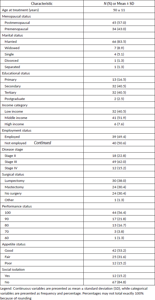
A total of 79 women with histologically confirmed breast cancer were enrolled and completed baseline assessments prior to initiation of chemotherapy. The mean age at treatment was 50 ± 11 years. Forty-five participants (57.0%) were postmenopausal, while 34 (43.0%) were premenopausal. Most participants were married (66, 83.5%), whereas 7 (8.9%) were widowed, 4 (5.1%) were single and one participant each was divorced (1.3%) or separated (1.3%). Educational attainment was predominantly secondary or tertiary level, each reported by 32 participants (40.5%), while 13 (16.5%) had primary education and 2 (2.5%) had postgraduate education.
With respect to socioeconomic status, 41 participants (51.9%) belonged to the middle-income group, 32 (40.5%) to the low-income group and 6 (7.6%) to the high-income group. Thirty-nine participants (49.4%) were employed.
Most patients presented with locally advanced disease. Stage III disease was observed in 49 participants (62.0%), while 18 (22.8%) had stage II disease and 12 (15.2%) had stage IV disease. Regarding surgical management, 30 participants (38.0%) had undergone lumpectomy, 24 (30.4%) had undergone mastectomy and 24 (30.4%) had not undergone surgery at the time of enrolment.
Performance status was generally favourable, with 44 participants (56.4%) having a performance status score of 100 and 17 (21.8%) having a score of 90. Lower performance status scores of 80, 70 and 60 were observed in 13 (16.7%), 3 (3.8%) and 1 (1.3%) participants, respectively.
Appetite was reported as good by 42 participants (53.2%), fair by 25 (31.6%) and poor by 12 (15.2%). Twelve participants (15.2%) reported social isolation at baseline. Baseline demographic, clinical, psychosocial and socioeconomic characteristics are summarised in Table 1.
Table 1. Baseline demographic, clinical, psychosocial and socioeconomic characteristics of study participants (N = 79).
Baseline burden of cancer-related fatigue
At baseline, prior to initiation of chemotherapy, the mean FACIT-F score was 34.0 ± 8.4, with a median score of 35.0 and a range of 13–48. Using the predefined FACIT-F threshold for clinically significant fatigue (≤34), 36 participants (45.6%) met criteria for clinically significant fatigue, while 43 (54.4%) did not.
The baseline fatigue profile of the study cohort is summarised in Table 2.
Longitudinal changes in cancer-related fatigue during early chemotherapy
Cancer-related fatigue worsened significantly during the first three cycles of chemotherapy. The mean FACIT-F score declined from 34.0 ± 8.4 at baseline to 24.8 ± 11.7 following completion of the third chemotherapy cycle.
The mean change in FACIT-F score was −9.23 points (95% CI: −11.82 to −6.64), indicating a substantial deterioration in fatigue status over time. Paired-samples t-test analysis demonstrated a statistically significant difference between baseline and post-cycle 3 fatigue scores (t = 7.09, df = 78, p < 0.001).
The magnitude of change corresponded to a large within-participant effect size (Cohen’s dz = 0.80). Longitudinal changes in FACIT-F scores are summarised in Table 3 and illustrated in Figure 1.
Error bars represent 95% confidence intervals. Higher FACIT-F scores indicate lower fatigue burden and better fatigue-related quality of life
Bivariate predictors of fatigue deterioration
Bivariate analyses were performed to identify baseline demographic, clinical, psychosocial, socioeconomic and haematological factors associated with longitudinal changes in fatigue during chemotherapy. More negative ΔFACIT-F values indicated greater fatigue deterioration. Age demonstrated a weak negative correlation with ΔFACIT-F (r = −0.221, 95% CI: −0.422 to −0.0002; p = 0.050), indicating greater fatigue deterioration among older participants. In contrast, baseline haemoglobin level (r = 0.002, p = 0.988) and performance status (r = 0.024, p = 0.838) were not associated with fatigue change.
Postmenopausal women experienced greater fatigue deterioration than premenopausal women (mean ΔFACIT-F: −11.2 versus −6.6), although this association did not reach statistical significance (p = 0.072). No significant associations were observed for disease stage (p = 0.515), social isolation (p = 0.704) or income category (p = 0.490). Surgical status was significantly associated with fatigue deterioration (p = 0.026). Appetite status also demonstrated a borderline association with fatigue change (p = 0.079), with participants reporting fair appetite experiencing the greatest decline in FACIT-F scores. Detailed results of the bivariate analyses are presented in Table 4.
Table 2. Baseline fatigue profile prior to chemotherapy initiation (N = 79).
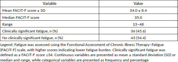
Table 3. Longitudinal change in FACIT-Fatigue scores from baseline to completion of the third chemotherapy cycle (N = 79).
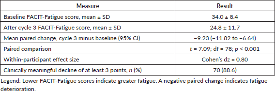
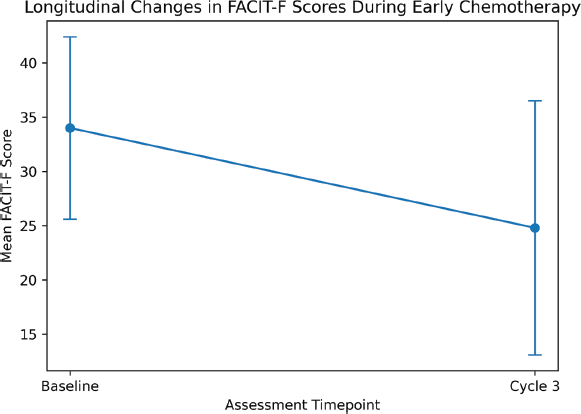
Figure 1. Longitudinal changes in mean Functional Assessment of Chronic Illness Therapy–Fatigue (FACIT-F) scores from baseline to completion of the third chemotherapy cycle among women with breast cancer (N = 79).
Table 4. Bivariate associations between baseline characteristics and longitudinal change in FACIT-F score (ΔFACIT-F).
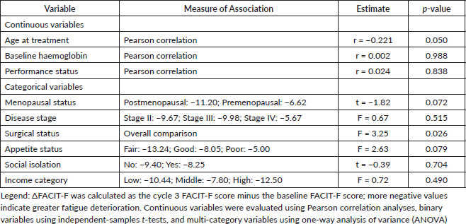
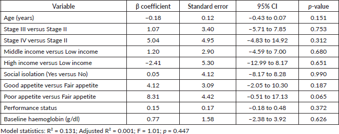
Table 5. Multivariable linear regression model identifying predictors of fatigue deterioration during early chemotherapy.
Multivariable predictors of fatigue deterioration
A multivariable linear regression model was fitted to identify independent predictors of longitudinal fatigue deterioration during chemotherapy. Variables entered into the model included age at treatment, disease stage, income category, social isolation, appetite status, performance status and baseline haemoglobin level. After adjustment for potential confounders, none of the evaluated variables demonstrated a statistically significant independent association with change in FACIT-F score. Increasing age was associated with greater fatigue deterioration, although the association was not statistically significant (β = −0.18, 95% CI: −0.43 to 0.07; p = 0.151).
Compared with participants reporting fair appetite, those reporting poor appetite demonstrated a trend towards less negative FACIT-F change scores (β = 8.31, 95% CI: −0.51 to 17.13; p = 0.065), representing the strongest association observed in the adjusted model. Appetite classified as good was not significantly associated with fatigue change (β = 4.12, 95% CI: −2.05 to 10.30; p = 0.187).
Disease stage, income category, social isolation, performance status and baseline haemoglobin level were not independently associated with fatigue deterioration during chemotherapy. The overall model explained 13.1% of the variance in ΔFACIT-F scores (R² = 0.131; adjusted R² = 0.001) and was not statistically significant overall (F = 1.01, p = 0.447). Results of the multivariable analysis are presented in Table 5.
Discussion
This prospective cohort study demonstrates that women with breast cancer in this Nigerian tertiary oncology setting had a substantial burden of fatigue before chemotherapy and experienced pronounced worsening during the first three treatment cycles. At baseline, 45.6% of participants met the predefined criterion for clinically significant fatigue. By the completion of cycle 3, the mean FACIT-F score had declined by 9.23 points, representing a statistically significant and clinically important deterioration with a large within-participant effect size. Although age, surgical status, menopausal status and appetite produced signals in the bivariate analyses, none of the baseline variables included in the multivariable model independently predicted fatigue deterioration. These findings establish the early burden and trajectory of chemotherapy-associated fatigue in this cohort but do not demonstrate that chemotherapy alone caused the observed change.
The prevalence of clinically significant fatigue before chemotherapy indicates that fatigue was already an important component of the participants’ symptom burden before treatment exposure. Nigerian evidence supports the relevance of this observation, although differences in study populations, assessment instruments and measurement periods limit direct numerical comparison. A prospective study of Nigerian women receiving chemotherapy identified fatigue among the commonly reported treatment-related adverse effects, while a case-control study from southwestern Nigeria found that breast cancer survivors had a higher prevalence of fatigue than apparently healthy controls [13,14]. A subsequent Nigerian study reported clinically meaningful fatigue in 22.5% of breast cancer survivors, whereas a study conducted after chemotherapy in Delta State reported fatigue in 65% of participants [15,16]. The 45.6% baseline prevalence in the present study is higher than the prevalence reported among survivors in southwestern Nigeria but lower than that reported in the post-chemotherapy Delta State cohort. These differences are plausible because fatigue varies according to the point in the cancer trajectory, treatment exposure, disease status, case definition and measurement instrument. Importantly, previous Nigerian studies have been predominantly cross-sectional or conducted during survivorship. The present study complements this literature by documenting fatigue before chemotherapy and prospectively measuring within-patient change during early treatment.
The decline in FACIT-F score was the principal finding. Because higher FACIT-F scores indicate less fatigue and better fatigue-related functioning, the mean change of −9.23 points indicates substantial worsening between baseline and cycle 3. The 95% confidence interval, from −11.82 to −6.64 points, excludes both no change and the commonly accepted 3–4-point threshold for a minimally important difference on the FACIT-F scale [20]. The standardised within-participant effect size was also large (Cohen’s dz = 0.80), indicating that the deterioration was substantial relative to the variability in individual change. Using the predefined threshold of a reduction of at least three FACIT-Fatigue points, 70 participants (88.6%) experienced clinically meaningful fatigue deterioration between baseline and cycle 3. Nevertheless, nine participants did not reach this threshold, demonstrating that individual responses to treatment was not uniform.
The early deterioration observed in this cohort is consistent with previous longitudinal studies of women receiving chemotherapy for breast cancer. Fatigue has been shown to worsen after chemotherapy initiation, with changes occurring alongside treatment-related symptoms such as nausea, oral toxicity and sleep disturbance [8]. Another prospective study similarly reported increased fatigue during adjuvant chemotherapy, although the timing and magnitude of peak fatigue varied according to chemotherapy regimen [9]. Longer-term longitudinal evidence has identified distinct fatigue trajectories from diagnosis through survivorship, including persistently high, increasing, recovering and consistently low-fatigue patterns [10]. The present findings therefore confirm that clinically important fatigue deterioration may occur within the first three chemotherapy cycles while also demonstrating that individual responses are heterogeneous.
The limited follow-up period does not establish whether the observed deterioration resolved, persisted or worsened after cycle 3. Longitudinal research indicates that some women recover after treatment, whereas others develop persistent or delayed fatigue [10,11]. Nigerian survivorship studies have similarly shown that fatigue may remain clinically relevant after completion of breast cancer treatment [14,15]. Consequently, the present study should be interpreted as describing early chemotherapy-associated deterioration that complements, rather than contradicts, evidence of persistent fatigue among survivors. Moreover, because the study did not include an untreated breast cancer comparison group, the observed change cannot be attributed exclusively to chemotherapy. Disease evolution, repeated hospital attendance, concurrent symptoms and other time-varying patient or treatment factors may also have contributed.
Approximately three-fifths of the participants had stage III disease, reflecting the advanced-stage presentation commonly encountered in sub-Saharan African oncology practice [6]. However, disease stage was not associated with fatigue change in either the bivariate or adjusted analysis. This differs from the Nigerian survivor study in which advanced stage was associated with fatigue and from a meta-analysis showing a modestly greater risk of severe fatigue among survivors with stage II or III disease than among those with stage 0 or I disease [11,15]. These differences may reflect the present study’s assessment of short-term change rather than fatigue prevalence, the predominance of stage III disease, the small stage-specific subgroups and differences in treatment phase. The findings therefore do not support the conclusion that advanced disease stage caused the early deterioration observed in this cohort.
Surgical status was associated with FACIT-F change in the unadjusted analysis. This is broadly consistent with de Jong et al [9], who reported greater fatigue among women who had undergone mastectomy than among those treated with breast-conserving surgery. Nevertheless, surgical status may also represent differences in disease extent, treatment sequencing, postoperative recovery or the interval between surgery and chemotherapy. Because it was not included in the reported adjusted model, its association should be regarded as unadjusted and potentially confounded rather than as evidence of an independent effect of surgical status.
Baseline Karnofsky Performance Status was not associated with fatigue deterioration. This contrasts with a systematic review and meta-analysis in which poor performance status was strongly associated with cancer-related fatigue [22]. The apparent difference may be explained by the generally favourable and restricted performance-status distribution in the present cohort: most participants had scores of 90 or 100, leaving relatively few women with substantial functional impairment. This restricted range reduces the ability to detect an association. It may also indicate that baseline functional capacity does not necessarily predict the magnitude of fatigue change during the first three chemotherapy cycles.
Age showed a weak association with fatigue deterioration in the bivariate analysis, with older participants experiencing greater decline, but the association was no longer statistically significant after adjustment. Age should therefore be considered a tentative signal rather than a confirmed predictor. Published evidence on age is inconsistent. The Nigerian survivor study found greater fatigue among younger women in its univariate analysis, while other longitudinal and meta-analytic studies have similarly identified younger age as a potential risk factor for persistent fatigue [10,11,15]. A large prospective prediction study also identified younger age, together with baseline fatigue, body mass index, anxiety, insomnia and pain, as a predictor of severe post-treatment fatigue [23]. The direction observed in the present cohort may reflect differences in comorbidity, physiological reserve, menopausal status, treatment selection, dose intensity or symptom reporting. The loss of statistical significance after adjustment, together with the modest sample size, prevents a firm age-related inference.
Postmenopausal women experienced a numerically greater decline than premenopausal women, but the difference did not reach statistical significance. This finding should not be interpreted as evidence that menopausal status independently determines fatigue worsening. It also differs from larger survivorship studies in which younger or premenopausal women have sometimes demonstrated greater long-term fatigue risk [11,23]. Menopausal status and chronological age are closely related, and their effects may be further modified by endocrine symptoms, treatment-induced ovarian dysfunction, comorbidity and treatment selection. These factors were not measured sufficiently to clarify the relationship in the present cohort.
Appetite showed a borderline association with fatigue change, but participants reporting fair appetite, not poor appetite, experienced the greatest mean deterioration. In the adjusted analysis, poor appetite was associated with a less negative change relative to fair appetite, although this estimate also failed to reach statistical significance. This unexpected direction may reflect the small number of participants reporting poor appetite, imprecision in a single baseline appetite question, regression to the mean or unmeasured differences in disease and supportive care. Previous research has linked chemotherapy-related symptoms, including nausea and oral toxicity, with fatigue worsening, while contemporary models recognise nutritional compromise as one of several possible contributors to fatigue [4,5,8]. The present results do not demonstrate that appetite caused fatigue deterioration; they indicate only that more detailed and repeated nutritional assessment may be informative in future studies.
Neither income category nor the single study-specific measure of social isolation was associated with fatigue change. These null findings differ from Nigerian survivorship research in which employment status was associated with fatigue and from longitudinal studies identifying psychological distress, depression and sleep disturbance as important correlates of persistent fatigue [10,15,23]. However, income and social isolation are not equivalent to employment, depression, distress or validated social-support constructs. The use of a single question to assess social isolation may have lacked the sensitivity required to capture the quality, availability and adequacy of social support. These results should therefore not be interpreted as evidence that socioeconomic and psychosocial circumstances are unimportant.
Baseline haemoglobin was not correlated with FACIT-F change and was not significant in the adjusted model. This result addresses whether haemoglobin before treatment predicted subsequent fatigue deterioration; it does not test whether a change in haemoglobin was associated with a concurrent change in fatigue. Baseline haemoglobin was not associated with fatigue deterioration in the present study. In contrast, longitudinal evidence from anaemic patients with cancer indicates that improvement in haemoglobin may be accompanied by clinically meaningful improvement in fatigue [24]. These findings are not directly contradictory because the present analysis examined whether baseline haemoglobin predicted subsequent changes in fatigue, rather than whether changes in haemoglobin corresponded with changes in fatigue. The predictive value of a baseline measurement may be limited by a restricted haemoglobin range, modest subsequent reductions or measurement timing. Moreover, fatigue may be influenced by pain, sleep disturbance, emotional distress, inflammation, nutritional status and other treatment-related toxicities. The relationship may also be nonlinear, becoming clinically important only below a particular haemoglobin threshold, and may be modified by chemotherapy regimen, transfusion or supportive medication. Because paired changes in haemoglobin and fatigue were not analysed, the present study cannot determine whether their longitudinal trajectories were associated.
The multivariable model had an adjusted R² of 0.001 and was not statistically significant, indicating virtually no adjusted explanatory value for the included predictors. This should not be interpreted as evidence that baseline patient characteristics have no influence on fatigue. The model included ten regression coefficients in only 79 participants, resulting in limited power and imprecise estimates, particularly for small categories. Other possible explanations include restricted predictor ranges, single-item measurement of appetite and social isolation, measurement error in change scores, influential observations, nonlinear associations, interactions and possible overadjustment. Although the reported variance inflation factors did not indicate important multicollinearity, the model did not include several potentially influential treatment-related or time-varying factors. Chemotherapy regimen, cumulative dose, relative dose intensity, dose delays, concurrent targeted or endocrine therapy, corticosteroid exposure, transfusion and other treatment toxicities were not evaluated. The absence of these variables may have contributed substantially to the model’s limited explanatory performance.
For practice, the findings directly support assessment of fatigue before chemotherapy and repeated assessment during the early treatment period. Screening alone is insufficient when clinically important fatigue is identified; it should be followed by evaluation for reversible or treatable contributors. Recommendations to assess pain, sleep, emotional distress, nutritional status, anaemia, comorbidity and treatment toxicity are derived from established guidelines and the wider literature, rather than from intervention testing in this study [3,25]. Evidence-based guidelines recommend appropriately tailored exercise, cognitive behavioural therapy and mindfulness-based programmes for fatigue during and after cancer treatment [25]. A meta-analysis of randomised trials similarly found that exercise and psychological interventions produced greater reductions in fatigue than the pharmaceutical interventions evaluated [26]. The present study did not test any of these interventions. Their feasibility, accessibility and cultural acceptability should therefore be evaluated specifically within Nigerian oncology services.
Limitations
This study has several limitations. It was conducted at a single tertiary centre with a modest sample, which limits generalisability and statistical power. Some disease, income, appetite and surgical subgroups contained few participants, reducing the precision of subgroup comparisons. Follow-up ended after cycle 3; consequently, the study did not determine fatigue trajectories across the remaining chemotherapy cycles or after treatment. The absence of an untreated comparison group also prevents causal attribution of the observed deterioration specifically to chemotherapy. Several potentially important patient-related factors were not comprehensively measured, including body mass index, frailty, comorbidity, baseline physical activity, sleep disturbance, depression, anxiety and nutritional status. Unmeasured disease-related factors included pain, cachexia, tumour burden, metastatic burden and inflammatory activity. Treatment-related information on regimen, individual drugs, cumulative dose, relative dose intensity, cycle delays, concurrent systemic treatment, corticosteroids, transfusions and other adverse effects was unavailable for analysis. Inflammatory biomarkers, renal and thyroid function and haematological indices other than baseline haemoglobin were also not evaluated. Appetite and social isolation were assessed using single study-specific questions rather than validated nutritional or psychosocial instruments, which may have resulted in measurement imprecision. These omissions may partly explain the low explanatory performance of the multivariable model and further limit causal interpretation.
Notwithstanding these limitations, the study makes an important contribution by prospectively quantifying both pretreatment fatigue burden and early within-patient deterioration using a validated fatigue measure. Future research should extend this work through multicentre Nigerian cohorts with larger samples and prespecified, parsimonious predictor models. Fatigue and relevant laboratory parameters should be assessed repeatedly across each phase of chemotherapy and after treatment. Future studies should record chemotherapy regimen, dose, dose intensity, treatment delays, concurrent therapies and supportive medications, alongside patient-, disease- and toxicity-related variables. Validated instruments should be used to assess depression, anxiety, social support, sleep, physical activity and nutritional status, while appropriately selected inflammatory, endocrine and metabolic biomarkers may help clarify the underlying biological pathways. Intervention studies should then evaluate feasible, culturally appropriate approaches to fatigue management within Nigerian oncology services.
Conclusion
Women with breast cancer in this prospective Nigerian cohort entered chemotherapy with a substantial burden of fatigue, which worsened markedly during the first three treatment cycles. The magnitude of deterioration was clinically important and large at the individual level, although responses varied and no baseline factor included in the adjusted model independently predicted the change. The study’s prospective design, complete paired assessments, use of a validated fatigue instrument and focus on an underrepresented Nigerian population are important strengths despite its single-centre setting, modest sample size and limited follow-up. These findings provide robust evidence of early chemotherapy-associated fatigue burden and support fatigue assessment before treatment and repeated surveillance during chemotherapy. Larger multicentre studies incorporating detailed treatment exposure, validated psychosocial measures, relevant clinical variables and repeated biological assessments are required to identify patients at greatest risk and test effective, locally feasible supportive-care interventions.
Conflicts of interest
The authors declared no potential conflicts of interest with respect to the research, authorship and/or publication of this article.
Funding
The authors received no financial support for the research, authorship and/or publication of this article.
References
1. Bray F, Laversanne M, and Sung H, et al (2024) Global cancer statistics 2022: GLOBOCAN estimates of incidence and mortality worldwide for 36 cancers in 185 countries CA Cancer J Clin 74(3) 229–263 [https://doi.org/10.3322/caac.21834] PMID: 38572751
2. International Agency for Research on Cancer 2025 Breast cancer cases and deaths are projected to rise globally [Internet] (Lyon: International Agency for Research on Cancer) [https://www.iarc.who.int/wp-content/uploads/2025/02/pr361_E.pdf] Date accessed: 17/08/26
3. National Comprehensive Cancer Network 2025 NCCN Clinical Practice Guidelines in Oncology: Cancer-Related Fatigue. Version 1.2026 [Internet] (Plymouth Meeting: National Comprehensive Cancer Network) [https://www.nccn.org/professionals/physician_gls/pdf/fatigue.pdf] Date accessed: 17/08/26
4. Bower JE (2014) Cancer-related fatigue—mechanisms, risk factors, and treatments Nat Rev Clin Oncol 11(10) 597–609 https://doi.org/10.1038/nrclinonc.2014.127 PMID: 25113839 PMCID: 4664449
5. Thong MSY, Van Noorden CJF, and Steindorf K, et al (2020) Cancer-related fatigue: causes and current treatment options Curr Treat Options Oncol 21(2) 17 https://doi.org/10.1007/s11864-020-0707-5 PMID: 32025928 PMCID: 8660748
6. Anyigba CA, Awandare GA, and Paemka L (2021) Breast cancer in sub-Saharan Africa: the current state and uncertain future Exp Biol Med (Maywood) 246(12) 1377–1387 https://doi.org/10.1177/15353702211006047 PMID: 33926257 PMCID: 8243219
7. Al Maqbali M, Al Sinani M, and Al Naamani Z, et al (2021) Prevalence of fatigue in patients with cancer: a systematic review and meta-analysis J Pain Symptom Manage 61(1) 167–189 https://doi.org/10.1016/j.jpainsymman.2020.07.037
8. Jacobsen PB, Hann DM, and Azzarello LM, et al (1999) Fatigue in women receiving adjuvant chemotherapy for breast cancer: characteristics, course, and correlates J Pain Symptom Manage 18(4) 233–242 https://doi.org/10.1016/S0885-3924(99)00082-2 PMID: 10534963
9. De Jong N, Candel MJJM, and Schouten HC, et al (2004) Prevalence and course of fatigue in breast cancer patients receiving adjuvant chemotherapy Ann Oncol 15(6) 896–905 https://doi.org/10.1093/annonc/mdh229 PMID: 15151946
10. Bower JE, Ganz PA, and Irwin MR, et al (2021) Do all cancer patients experience fatigue? A longitudinal study of fatigue trajectories in women with breast cancer Cancer 127(8) 1334–1344 https://doi.org/10.1002/cncr.33327 PMID: 33606273 PMCID: 8562726
11. Abrahams HJG, Gielissen MFM, and Schmits IC, et al (2016) Risk factors, prevalence, and course of severe fatigue after breast cancer treatment: a meta-analysis involving 12,327 breast cancer survivors Ann Oncol 27(6) 965–974 https://doi.org/10.1093/annonc/mdw099 PMID: 26940687
12. Asefa T, Bitew G, and Tezera H, et al (2024) Prevalence of cancer-related fatigue, associated factors and adult cancer patients’ experiences at Hawassa University Comprehensive Specialized Hospital in Ethiopia: a mixed methods study Front Oncol 14 1480246 https://doi.org/10.3389/fonc.2024.1480246
13. Folorunso SA, Abiodun OO, and Abdus-Salam AA, et al (2023) Evaluation of side effects and compliance to chemotherapy in breast cancer patients at a Nigerian tertiary hospital Ecancermedicalscience 17 1537 [https://doi.org/10.3332/ecancer.2023.1537] PMID: 37138960 PMCID: 10151076
14. Folorunso SA, Ntekim A, and Abdus-Salam AA, et al (2022) Fatigue as a complicating factor in the recovery of breast cancer survivors treated at an oncology clinic in South West Nigeria: a case-control study Ecancermedicalscience 16 1420 [https://doi.org/10.3332/ecancer.2022.1420] PMID: 36158984 PMCID: 9458267
15. Folorunso SA, Abdus-Salam AA, and Ntekim AI, et al (2024) Sociodemographic and treatment-related correlates of fatigue in breast cancer survivors at an oncology clinic in Nigeria Ecancermedicalscience 18 1659 [https://doi.org/10.3332/ecancer.2024.1659] PMID: 38425762 PMCID: 10901630
16. Brotobor D, Nwozichi C, and Edeawe OI, et al (2026) Symptom burden and its effect on the quality of life in a selected group of breast cancer patients in Delta State, Nigeria, after chemotherapy: a cross-sectional study Pan Afr Med J 53(7) 7 [https://doi.org/10.11604/pamj.2026.53.7.46948]
17. Yellen SB, Cella DF, and Webster K, et al (1997) Measuring fatigue and other anaemia-related symptoms with the Functional Assessment of Cancer Therapy measurement system J Pain Symptom Manage 13(2) 63–74 https://doi.org/10.1016/S0885-3924(96)00274-6 PMID: 9095563
18. Meysami A, Yari H, and Haghighat S, et al (2017) The Functional Assessment of Chronic Illness Therapy–Fatigue: validity and reliability of the Iranian version Oncol Res Treat 40(12) 789–793 https://doi.org/10.1159/000479588 PMID: 29183033
19. Kapoor A, Singhal MK, and Bagri PK, et al (2015) Cancer-related fatigue: a ubiquitous problem yet so underreported, under-recognized and undertreated South Asian J Cancer 4(1) 21–23 https://doi.org/10.4103/2278-330X.149942 PMID: 25839015 PMCID: 4382777
20. Cella D, Eton DT, and Lai JS, et al (2002) Combining anchor- and distribution-based methods to derive minimal clinically important differences on the Functional Assessment of Cancer Therapy anaemia and fatigue scales J Pain Symptom Manage 24(6) 547–561 https://doi.org/10.1016/S0885-3924(02)00529-8
21. Karnofsky DA, and Burchenal JH 1979 The clinical evaluation of chemotherapeutic agents in cancer Evaluation of chemotherapeutic agents ed MacLeod CM (New York: Columbia University Press) pp 191–205 [https://ndlsearch.ndl.go.jp/en/books/R100000136-I1570291225027627008]
22. Ma Y, He B, and Jiang M, et al (2020) Prevalence and risk factors of cancer-related fatigue: a systematic review and meta-analysis Int J Nurs Stud 111 103707 https://doi.org/10.1016/j.ijnurstu.2020.103707 PMID: 32920423
23. Di Meglio A, Havas J, and Soldato D, et al (2022) Development and validation of a predictive model of severe fatigue after breast cancer diagnosis: toward a personalized framework in survivorship care J Clin Oncol 40(10) 1111–1123 https://doi.org/10.1200/JCO.21.01252 PMID: 35061509 PMCID: 8966972
24. Cella D, Kallich J, and McDermott A, et al (2004) The longitudinal relationship of haemoglobin, fatigue and quality of life in anaemic cancer patients: results from five randomized clinical trials Ann Oncol 15(6) 979–986 https://doi.org/10.1093/annonc/mdh235 PMID: 15151958
25. Bower JE, Lacchetti C, and Alici Y, et al (2024) Management of fatigue in adult survivors of cancer: ASCO–Society for Integrative Oncology guideline update J Clin Oncol 42(20) 2456–2487 https://doi.org/10.1200/JCO.24.00541
26. Mustian KM, Alfano CM, and Heckler C, et al (2017) Comparison of pharmaceutical, psychological, and exercise treatments for cancer-related fatigue: a meta-analysis JAMA Oncol 3(7) 961–968 https://doi.org/10.1001/jamaoncol.2016.6914 PMCID: 5557289