Actinic keratosis underlying cutaneous horn at an unusual site—a case report
Pragya A Nair, Arvind H Chaudhary and Malay J Mehta
Department of Dermatology, Pramukhswami Medical College, Karamsad, Gujarat 388 325, India
Correspondence to: Pragya A Nair. Email: drpragash2000@yahoo.com
Abstract
Cutaneous horns are usually found on chronic sun-damaged skin. A cutaneous horn is a rare tumour, often conical, circumscribed, and composed of dead keratin usually derived from base keratinocytes. It occurs mainly in association with underlying benign, premalignant, and malignant cutaneous diseases. The commonest malignancy is squamous cell carcinoma. Thus, to accurately ascertain the nature of the condition at the base of the lesion and to rule out malignancy, an excision biopsy is indicated. Here, we report a case of cutaneous horn over the palmar aspect of the left middle finger in a 45-year-old man whose histopathology showed actinic keratosis.
Keywords: cutaneous horn, cornucutaneum, actinic keratosis
Introduction
A cutaneous horn is a conical projection of hard hyperkeratotic excrescence of cohesive keratin projecting over skin. Although it grossly resembles an animal horn, it lacks a bony core. Cutaneous horns are found in the upper parts of the body, such as the face, neck, and shoulders, and are frequently related to actinic damage [1]. Cutaneous horns are thought to result from underlying benign, premalignant, or malignant in 61.1%, 23.2%, and 15.7% of cases, respectively [2]. Cutaneous horns have been noted on top of many clinical conditions such as keratoacanthoma, actinic keratosis, warts, molluscum contagiosum, seborrheic keratosis, Bowen’s disease, malignant melanoma, basal cell carcinoma, or squamous cell carcinoma [3].
We hereby report a case of a cutaneous horn at an unusual site, that is, the palmar aspect of the middle finger, with underlying histopathologic changes of actinic keratosis.
Case report
A 45-year-old man presented with a single lesion over the palmar aspect of the middle phalanx of the left middle finger since 12 months prior. There was no history of trauma or surgery at that site in the past. No itching, pain, or bleeding was present. The patient was a farmer by occupation. No significant family history was present. On examination, a firm, horny, and curved growth of around 2 cm in length with a broad base was present over the palmar aspect of middle phalanx of the left middle finger with no erythema at the base (Figure 1). No lymphadenopathy was present. Routine blood investigations, urine examination, and chest X-ray were normal. An excision biopsy was done.
Histopathology showed extensive hyperkeratosis and focal parakeratosis. The epidermis was thickened and showed irregular downward proliferation in dermis (Figure 2a). A varying proportion of keratinocyte in the stratum malphigian showed a loss of polarity and disordered arrangement with part of the normal dermis showing collagen bundles horizontally placed (Figure 2b). A diagnosis of actinic keratosis was given.
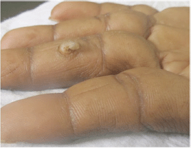
Figure 1: A horny growth of around 2 cm in length with a broad base over the palmar aspect of the middle phalanx of the left middle finger.
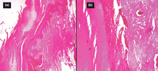
Figure 2: (a) Hyperkeratosis and focal parakeratosis, Hematoxylin & Eosin (H & E) stain 4x; (b) loss of polarity and disordered arrangement of keratinocyte in stratum malphigian with part of normal dermis, H & E stain 10x.
Discussion
Cutaneous horns, or cornucutaneum, are benign, elongated, keratinous projections from the skin, ranging in size from a few millimetres to many centimetres and resembling a miniature horn. The base of the horn may be flat, nodular, or crateriform. All animal horns except those of rhinoceroses contain bone casts that are not seen in cutaneous horns in human beings, which are simply composed of compact keratin. Cutaneous horns may be considered a common entity in the Caucasian population; a study reports 643 patients over a ten-year period with 32 new patients annually [2]. It is usually seen over the face, pina, nose, forearm, and dorsal aspect of the hand [3]. Cases over the areas not exposed to sunlight, such as the penis, mucosal part of the lower lip, and nasal vestibule, have also been reported [4, 5]. Clinically, it is a hard, yellowish-brown horn, often curved, having circumferential ridges surrounded by normal epidermis or acanthotic collarette. Weeson’s defined criteria (1987) for a horn are that it should be straight or curved and 2–2.5 cm long [6].
Classical cutaneous horns have dysplastic epidermal changes similar to solar keratosis with no atypicality or loss of polarity.
The pathogenesis of this abnormal formation of keratinised material has not been fully elucidated. It may be of clinical importance because the underlying condition may be a malignant lesion. It is difficult to define the underlying lesion, especially in superficial biopsies; therefore, deep biopsies or the total excision of small lesions is recommended [7]. Malignancy is present in 16–20% of cases, with squamous cell carcinoma being the most common type [8] in 94% of horns with a malignant base. Among the predisposing factors are advanced age, male sex, and sun exposure [7]. Tenderness at the base of the lesion, large size, wide base, or low height-to-base ratio horns are more likely to display a malignant base [9]. Multiple horns may occur in some patients [10]. Actinic keratoses have been reported as the most common horn base entity (37.4%) in a study of 230 horns [11], but they may also result from seborrheic keratoses, warts, keratoacanthoma, squamous cell carcinoma, basal cell carcinoma. Extremely rare cases associated with metastatic renal cell carcinoma [12], lymphoma [13], dermatofibroma [14], and pyogenic granuloma [15] have been reported. The base of the horn will display the characteristic feature of the pathologic process responsible for the development of the horn [16].
Actinic keratosis is a premalignant condition. The lesion starts as a flat scaly lesion and later grows into a large wart-like area. It commonly ranges from between 2 and 6 mm in size, may be dark, light, tan, pink, red, or a combination of all these shades. It occurs on sun-exposed areas of the body such as face, ears, neck, scalp, chest, back of hands, forearms, and lips. Up to 20% of untreated actinic keratosis can progress to malignancy. Histologically there is thickened stratum corneum with scattered areas of parakeratosis, associated loss of the granular layer and thickening of the epidermis. The normal ordered maturation of the keratinocytes is disordered to varying degrees, there may be widening of the intracellular spaces, cytologic atypia, such as abnormally large nuclei. The underlying dermis often shows severe actinic elastosis and a mild chronic inflammatory infiltrate. Cutaneous horn that represents actinic keratosis demonstrates similar but more exaggerated changes with massive tiers of hyperkeratosis and parakeratosis.
The unique feature of this case is that the cutaneous horn was located on the palmar aspect of the middle finger, which is an uncommon site. Only one such case has been reported at an unusual site over the right index finger tip, but it also did not show any underlying premalignant or malignant changes [9]. It suggests that trauma might be a predisposing factor for the development of cutaneous horn in areas that are not exposed to actinic damage.
Excision biopsy of the lesion and histopathological examination to rule out malignancy is mandatory. As malignancies should be excised with appropriate margins and evaluated for metastasis, a careful physical examination of the lymph nodes draining the area of lesion is required [17]. Treatment options include wide surgical excision, electrocautry, cryotherapy, carbon dioxide, or NdYAG laser [18].
Conflicts of interest
The authors have no conflicts of interest to declare.
Acknowledgments
The authors acknowledge Dr Keyuri Patel, Professor, Department of Pathology and Central Diagnostic Laboratory, for histopathology.
References
1. Bondeson J (2001) Everard Home, John Hunter Cutaneous horn: a historical review Am J Dermatopathol 23 362–9 DOI: 10.1097/00000372-200108000-00014 PMID: 11481530
2. Yu RC, Pryce DW, Macfarlane AW and Stewart TW (1991) A histopathological study of 643 cutaneous horns Br J Dermatol 124(5) 449–52 DOI: 10.1111/j.1365-2133.1991.tb00624.x PMID: 2039721
3. Copcu E, Sivrioglu N and Culhaci N (2004) Cutaneous horns: are these lesions as innocent as they seem to be? World J Surg Oncol 2 18 DOI: 10.1186/1477-7819-2-18 PMID: 15176977 PMCID: 421749
4. Rekha A and Ravi A (2004) Cornucutaneum-cutaneous horn on the penis Indian J Surg 66 296–7
5. Mehmet Mutaf (2007) A rare perioral lesion: cutaneous horn of the lower lip Eur J Plast Surg 29 339–41 DOI: 10.1007/s00238-006-0101-y
6. Gupta A and Jain AK (1997) A cutaneous horn over seborrhoeic keratosis of pinna Ind J Otolaryngol Head Neck Surg 49 140–1
7. Schwartz RA, Bridges TM, Butani AK and Ehrlich A (2008) Actinic keratosis: an occupational and environmental disorder J Eur Acad Dermatol Venereol 22 606–15 DOI: 10.1111/j.1468-3083.2008.02579.x PMID: 18410618
8. Solivan GA, Smith KJ and James WD (1990) Cutaneous horn of the penis: its association with squamous cell carcinoma and HPV-16 infections J Am Acad Dermatol 23 969–72 DOI: 10.1016/0190-9622(90)70315-9 PMID: 2172337
9. Tauro LF, Martis JS, John SK and Kumar KP (2006) Cornucutaneum at an unusual site Ind J Plast Surg 39 76–8 DOI: 10.4103/0970-0358.26911
10. Pyne J, Sapkota D and Wong JC (2013) Cutaneous horns: clues to invasive squamous cell carcinoma being present in the horn base Dermatol Pract Conc 3 2
11. Schosser RH, Hodge SJ, Gaba CR and Owen LG (1979) Cutaneous horns: a histopathologic study South Med J 72 1129–31 DOI: 10.1097/00007611-197909000-00014 PMID: 472837
12. Ozturk S, Cil Y, Sengezer M, Yigit T, Eski M and Ozcan A (2006) Squamous cell carcinoma arising in the giant cutaneous horns accompanied with renal cell carcinoma Eur J Plast Surg 28 483–5 DOI: 10.1007/s00238-005-0004-3
13. Dasgupta S, Mitra D, Bhattacharya A and Sur P (2006) B cell lymphoma with unusual clinical cutaneaous presentation J Cancer Res Ther 2 203–5 DOI: 10.4103/0973-1482.29834
14. Kim YJ, Jeon J, Son SW, Kim AR, Oh CH and Song HJ (2006) Dermatofibroma: unusual lesion with underlying cutaneous horn Kor J Dermatol 44 754–6
15. Findlay RF and Lapins NA (1983) Pyogenic granuloma simulating a cutaneous horn Cutis 31 610–2 PMID: 6861513
16. Mencia-Gutierrez E, Gutierrez-Diaz E, Redondo-Marcos I, Ricky JR and Garcia-Torre JP (2004) Cutaneous horns of the eyelid: a clinicopathological study of 48 cases J Cutan Pathol 31 539–43 DOI: 10.1111/j.0303-6987.2004.00226.x PMID: 15268708
17. Kumaresan M, Kumar P and Pai MV (2008) Giant cutaneous horn Ind J Dermatol 53 199–200 DOI: 10.4103/0019-5154.44800
18. Lowe FC and McCullough AR (1985) Cutaneous horn of the penis: An approach to management: case report and review of literature J Am Acad Dermatol 13 369–73 DOI: 10.1016/S0190-9622(85)70177-6 PMID: 4031162


