Socio-demographic determinants and comparative diagnostic performance of VIA, Pap smear and HPV DNA testing for cervical cancer screening in North Indian women
Pallavi Rajput1, Vandita Pahwa1, Charu Singh2, Sankalp Sancheti3, Ravikiran Pawar4, Shikha Jain1, Gurwinder Kaur5, Kundan Rao6, Navin Bansal7, Rahat Brar8, Tapas Dora9 and Ashish Gulia10
1Department of Preventive Oncology, Homi Bhabha Cancer Hospital & Research Centre; Tata Memorial Centre, Homi Bhabha National Institute (HBNI), New Chandigarh 140901, India
2Department of Microbiology, Homi Bhabha Cancer Hospital & Research Centre; Tata Memorial Centre, Homi Bhabha National Institute (HBNI), New-Chandigarh 140901, India
3Department of Oncopathology, Homi Bhabha Cancer Hospital & Research Centre; Tata Memorial Centre, Homi Bhabha National Institute (HBNI), New-Chandigarh 140901, India
4Department of Homi Bhabha Cancer Hospital & Research Centre; Tata Memorial Centre, Homi Bhabha National Institute (HBNI), New-Chandigarh 140901, India
5Department of Preventive Oncology, Homi Bhabha Cancer Hospital, Sangrur, India
6Clinical Research Secretariat, Homi Bhabha Cancer Hospital & Research Centre; Tata Memorial Centre, Homi Bhabha National Institute (HBNI), New Chandigarh 140901, India
7Department of Transfusion Medicine, Homi Bhabha Cancer Hospital & Research Centre; Tata Memorial Centre, Homi Bhabha National Institute (HBNI), New-Chandigarh 140901, India
8Department of Radiology, Homi Bhabha Cancer Hospital & Research Centre; Tata Memorial Centre, Homi Bhabha National Institute (HBNI), New-Chandigarh 140901, India
9Department of Radiation Oncology, Homi Bhabha Cancer Hospital & Research Centre; Tata Memorial Centre, Homi Bhabha National Institute (HBNI), New-Chandigarh 140901, India
10Department of Surgical Oncology, Homi Bhabha Cancer Hospital & Research Centre; Tata Memorial Centre, Homi Bhabha National Institute (HBNI), New-Chandigarh 140901, India
Abstract
Background: Cervical cancer is the second most common cancer among Indian women, with low screening coverage, especially in rural areas. Persistent infection with high-risk human papillomavirus (HPV), particularly types 16 and 18, is the main cause. This study evaluated socio-demographic factors associated with visual inspection with acetic acid (VIA), Pap smear and HPV positivity and compared the diagnostic performance of these screening methods.
Methods: This retrospective study included 641 women aged 30 to 55 years screened at two preventive oncology centres in Mullanpur and Sangrur between October 2024 and April 2025. Participants underwent VIA, Pap smear and HPV DNA testing. VIA findings were interpreted according to the World Health Organization guidelines, and Pap smear results were assessed using standard criteria. HPV results were categorised as HPV 16/18 or other high-risk types. Women positive on any screening test underwent colposcopy, with cervical punch biopsy performed when indicated.
Results: Among the women screened, VIA positivity was 31.9%, HPV positivity was 18.8% and Pap smear positivity was 1.7%. VIA positivity was higher among rural women and those with lower income, whereas HPV positivity was associated with early age at marriage and higher parity. For detecting cervical intraepithelial neoplasia 2+ (CIN2+) lesions, VIA demonstrated 100% sensitivity and lower specificity, Pap smear showed high specificity (99.5%) but lower sensitivity, while HPV DNA testing demonstrated 93.3% sensitivity and 82.8% specificity, with the highest diagnostic accuracy.
Conclusion: HPV DNA testing demonstrated high diagnostic accuracy for detecting CIN II+ lesions, while VIA showed high sensitivity but lower specificity, making it a practical screening tool in low-resource settings. Pap smear remained highly specific but detected fewer high-grade lesions. Early marriage, higher parity and lower socioeconomic status were associated with increased HPV positivity. These findings support risk-based cervical cancer screening strategies that integrate HPV DNA testing and VIA to improve early detection and reduce the burden of cervical cancer in resource-limited settings.
Keywords: cervical cancer screening, human papillomavirus, visual inspection with acetic acid, Pap smear, socio-economic factors
Correspondence to: Vandita Pahwa
Email: drvanditapahwa@gmail.com
Published: 15/10/2026
Received: 10/02/2026
Publication costs for this article were supported by ecancer (UK Charity number 1176307).
Copyright: © the authors; licensee ecancermedicalscience. This is an Open Access article distributed under the terms of the Creative Commons Attribution License (http://creativecommons.org/licenses/by/4.0), which permits unrestricted use, distribution, and reproduction in any medium, provided the original work is properly cited.
Introduction
Despite decades of cancer control programmes in India, cervical cancer screening coverage remains very low. Globally, cervical cancer is the fourth most common cancer among women, with an estimated 660,000 new cases and 350,000 deaths reported in 2022 [1]. In India, it is the second most common cancer among women; however, the screening rate for women aged 30 to 49 years in both urban and rural populations remains below 2.5% [2–4]. According to GLOBOCAN 2020, nearly half a million new cervical cancer cases are diagnosed worldwide each year, with 50% mortality [3]. Most patients present at advanced stages, resulting in substantial morbidity and mortality [2,3]. Low literacy levels, limited awareness and socioeconomic challenges, particularly in rural areas, remain major barriers to effective utilisation of screening services [4]. Persistent infection with high-risk human papillomavirus (HPV), particularly types 16 and 18, is the primary risk factor for cervical cancer [1,5]. Although most HPV infections are transient, persistent infection in the presence of cofactors such as early age at marriage, high parity, smoking, obesity and immuno-suppression can progress to high-grade cervical intraepithelial neoplasia (CIN II/III) and invasive cervical cancer [1,5,7]. Additional socio-demographic and behavioural factors, including age at marriage, parity, genital hygiene, medication use and substance use, have also been shown to influence cervical cancer risk [4,7,8].
Screening methods such as visual inspection with acetic acid or Lugol’s iodine (VIA/VILI), Pap smear cytology and HPV DNA testing are widely used for early detection and prevention [1,6,9]. Evidence from India and other low- and middle-income countries suggests that HPV-based screening, combined with cytology or VIA, represents an effective and feasible approach in low-resource settings [5,8,13]. Socio-demographic determinants strongly influence screening outcomes; women from rural areas, those with low literacy or socioeconomic status, and those married at an early age or with high parity are more likely to be HPV positive and less likely to access screening services [4,7,8]. Targeted interventions for these high-risk groups are therefore essential to improve early detection and reduce the burden of cervical cancer. However, variability in the performance of VIA, Pap smear and HPV testing has been reported across different Indian populations, highlighting the need for locally relevant, evidence-based screening strategies [6,9,10].
This study aimed to explore socio-demographic factors associated with HPV, Pap smear and VIA positivity; examine the strengths and limitations of each screening method and evaluate their sensitivity, specificity and diagnostic accuracy. The findings are intended to inform the development of evidence-based cervical cancer screening programmes in India.
Materials and methods
Study design and setting
This retrospective observational study was conducted at the Preventive Oncology Departments of the Mullanpur and Sangrur centres. Medical records of women who underwent cervical cancer screening using Pap smear cytology, HPV DNA testing, VIA/VILI and colposcopy between October 2024 and April 2025 were systematically reviewed.
Study population
Women aged 30 to 55 years who underwent all three screening modalities (VIA, Pap smear and HPV DNA testing) along with colposcopic examination were included. Records were identified from hospital databases and screening registers. Women with incomplete documentation, a prior hysterectomy, known cervical cancer, or pregnancy were excluded. Only complete records were included in the final analysis.
Data collection
Data on demographic characteristics, reproductive history, contraceptive use, tobacco exposure, prior screening history, per-speculum clinical findings and screening test results were extracted using a standardised data collection format. Data extraction was performed independently by trained personnel, and discrepancies were resolved through cross-verification with source documents to ensure accuracy and reliability.
Screening procedures
Visual inspection with acetic acid (VIA)
VIA was performed using 5% acetic acid. VIA positivity was defined as the presence of sharply demarcated, dense aceto-white areas in the transformation zone or columnar epithelium.
Pap smear collection and interpretation
Cervical cytology samples were collected following appropriate counselling and positioning. Ectocervical and endocervical samples were obtained using cotton swabs, smeared onto labelled glass slides and fixed in 95% ethanol. Cytological interpretation was performed according to the Bethesda system. Results were considered positive if atypical squamous cells of undetermined significance (ASC-US) or higher-grade abnormalities (including low-grade squamous lntraepithelial lesion and high-grade squamous intraepithelial lesion (HSIL)) were detected, and negative if no intraepithelial lesion or malignancy (NILM) was reported.
HPV DNA testing
Cervical samples were collected using a Digene conical brush rotated 360° 3 to 4 times at the cervical OS and placed in 1 mL of Digene transport medium. HPV DNA testing was performed using the Anyplex™ II HPV HR Detection Assay, which detects 14 high-risk HPV genotypes, including HPV-16, HPV-18 and 12 additional types (31, 33, 35, 39, 45, 51, 52, 56, 58, 59, 66 and 68). Results were categorised as positive for HPV 16/18, positive for other high-risk HPV types or negative if no HPV DNA was detected.
Colposcopy and histopathology
Women who tested positive by any screening method underwent colposcopic examination. Cervical biopsies were obtained from suspicious areas and submitted for histopathological evaluation to confirm the presence of CIN or invasive malignancy.
Statistical analysis
Descriptive statistics were used to summarise demographic and clinical characteristics. Age was categorised as <45 years and ≥45 years. Associations between socio-demographic variables and screening outcomes were assessed using the chi-square test or Fisher’s exact test, as appropriate [12].
Univariate logistic regression analysis was performed to estimate odds ratios for associations between demographic and clinical factors and screening test positivity. Variables with an adequate number of events (e.g. VIA positivity = 31.9%) were included in multivariate logistic regression models, with stepwise removal of non-significant predictors [13].
The association between screening test results and histopathological findings was evaluated using the chi-square test or Fisher’s exact test. Diagnostic performance measures including sensitivity, specificity, accuracy, positive predictive value (PPV) and negative predictive value (NPV) were calculated using histopathology as the reference standard. Agreement between HPV DNA testing, Pap smear cytology, and VIA/VILI was assessed using κ statistics.
All statistical analyses were performed using SPSS version 29 and R version 4.5.1 [14,15]. A p-value < 0.05 was considered statistically significant.
Data quality control
Records were reviewed for completeness and duplicate entries were removed. Uniform criteria for test positivity were applied consistently across all records.
Ethical considerations
Ethical approval was obtained from the Institutional Ethics Committee of Homi Bhabha Cancer Hospital and Research Centre (Project ID: 15000064). As this study involved a retrospective review of medical records, no direct patient contact occurred. All patient data were de-identified, and confidentiality was strictly maintained. Informed consent for cervical cancer screening had been obtained from all participants at the time of their initial evaluation.
Results
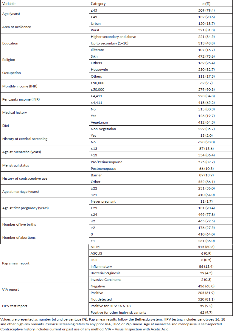
A total of 641 women were included in this retrospective analysis. The majority of participants (79.4%) were <45 years. Most women resided in rural areas (81.3%), while 18.7% were from urban settings. Regarding educational status, 16.7% of participants were illiterate, 48.8% had completed education up to the tenth grade and 34.5% had attained education beyond secondary school. Most participants belonged to the Sikh community (73.6%), with the remaining 26.4% representing other religious groups. The majority of women (82.7%) were housewives. Assessment of per capita income showed that nearly two-thirds of participants (65.2%) had a monthly income below INR 4,411, indicating a predominantly low-income population.
Reproductive characteristics revealed that early marriage was common, with 64.0% of women married at or before 21 years of age and 49.5% of participants had their first pregnancy at an early age. Screening results showed that 205 women (31.9%) were VIA positive, 121 women (18.8%) tested positive for HPV DNA, and 11 women (1.7%) had a positive Pap smear. Baseline demographic, socioeconomic, reproductive characteristics and screening outcomes are summarised in Table 1.
As shown in Figure 1, among the 284 women who tested positive by at least 1 screening method, 204 (31.8% of the total screened population) had abnormal findings on colposcopy, while the remaining screen-positive women did not show visible cervical lesions. Cervical punch biopsy was performed in 188 women with abnormal colposcopy (92.1%), whereas 16 women (7.8%) declined biopsy despite clinical recommendation. Histopathological examination showed benign results in 173 women (92.0%), CIN II in 6 women (3.2%) and CIN III or higher lesions in 9 women (4.8%).
The association of various epidemiological factors including age, socioeconomic status, literacy, gynaecological history0 and other relevant characteristics was evaluated in relation to the outcomes of VIA, Pap smear and HPV testing (Table 2). Some socio-demographic characteristics showed statistically significant associations, particularly with VIA and HPV positivity.
VIA positivity
Among the 641 women screened, 205 (31.9%) tested positive for VIA. The majority of VIA-positive women were <45 years (85.4%), whereas women >45 years were less likely to test positive (odds ratio (OR) = 0.60; 95% CI: 0.38–0.95; p = 0.03). Rural residence was strongly associated with VIA positivity, with women from rural areas having more than twice the odds of testing positive compared to urban residents (OR = 2.13; 95% CI: 1.45–4.10; p < 0.001). Low per capita income (<4,411 INR) was also significantly associated with higher VIA positivity (OR = 1.78; 95% CI: 1.25–2.53; p < 0.001). No significant associations were observed with education level or other socio-demographic factors. These findings suggest that younger age, rural residence and lower income were the main factors influencing VIA positivity in this population.
Table 1. Demographic, reproductive and screening characteristics of study participants (n = 641).
Pap smear positivity
Pap smear positivity was observed in 11 women (1.7%). Most smears were negative (80.3%), followed by inflammatory changes (13.4%) and bacterial vaginosis (4.5%). Among the positive smears, five cases were ASC-US, three were HSIL and two showed invasive carcinoma. Although no socio-demographic factor reached statistical significance, Pap positivity tended to be slightly higher in women from rural areas, those with low income, early marriage (≤21 years), or having >2 live births.
HPV positivity
HPV DNA positivity was observed in 121 women (18.8%), with nearly half (48.7%) positive for high-risk HPV types 16 and 18. Early marriage (≤21 years) was significantly associated with HPV positivity (OR = 1.72; 95% CI: 1.11–2.68; p = 0.02), and women with >2 live births had higher odds of being HPV positive (OR = 1.60; 95% CI: 1.05–2.44; p = 0.03). Other socio-demographic factors, including age, residence and per capita income, were not significantly associated with HPV positivity (Table 2).
Adjusted odds analysis (AOR)
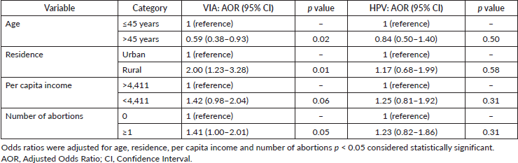
After adjusting for socio-demographic and reproductive factors, VIA positivity was significantly associated with younger age, rural residence, lower income and a history of abortion. Women >45 years had lower odds of VIA positivity compared to those aged ≤45 years (AOR = 0.59; 95% CI: 0.38–0.93; p = 0.02), while rural residents had about twice the odds compared to urban residents (AOR = 2.00; 95% CI: 1.23–3.28; p = 0.01). Lower per capita income showed a borderline increase in odds (AOR = 1.42; 95% CI: 0.98–2.04; p = 0.06) and having at least one abortion was also associated with higher odds of VIA positivity (AOR = 1.41; 95% CI: 1.00–2.01; p = 0.05).
In contrast, HPV positivity was not significantly associated with age, residence, income or history of abortion. Early marriage (≤21 years) (AOR = 1.72; 95% CI: 1.11–2.68; p = 0.02) and having >2 live births (AOR = 1.60; 95% CI: 1.05–2.44; p = 0.03) were the main factors associated with HPV positivity. Adjusted odds were not calculated for Pap smear due to the small number of positive cases (Table 3).
The performance of VIA, Pap smear and HPV testing for detecting high-grade cervical lesions is summarised in Table 4.
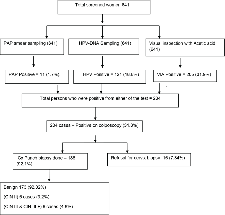
Figure 1. Flow diagram of study participants and screening outcomes. The flow diagram shows detail figures of screening procedures (VIA, PAP, and HPV-DNA), Colposcopic evaluation, and histopathological outcomes among the total screened.
VIA demonstrated high sensitivity but moderate specificity, making it effective for identifying women with CIN2+ and CIN3+ lesions. Pap smear was highly specific but less sensitive, especially when only high-grade lesions were considered positive. HPV testing showed a balanced combination of sensitivity and specificity. These findings indicate that the three screening modalities complement each other, providing an effective approach for detecting high-grade cervical lesions in this population.
Table 2. Prevalence of VIA, Papand HPV positivity according to socio-demographic and reproductive factors (n = 641).
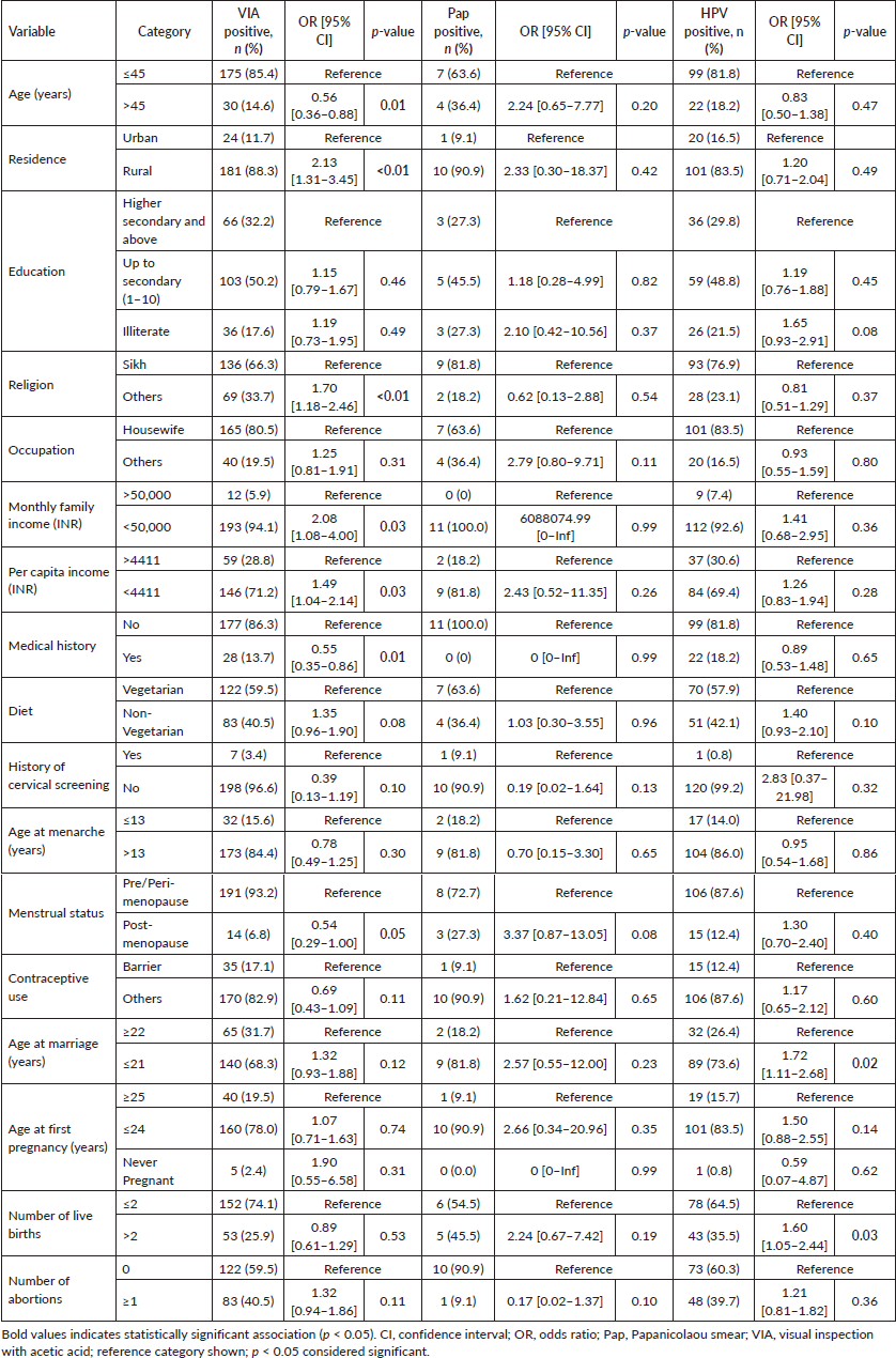
Table 3. Adjusted odds ratios for factors associated with VIA and HPV positivity.
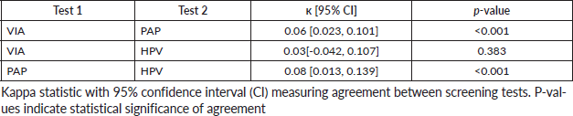
As shown in Table 5. The agreement of test results was slightly better in PAP versus HPV (κ: 0.08 [0.013, 0.139; p-value < 0.001) and VIA versus PAP (κ: 0.06 [0.023, 0.101]; p-value < 0.001) than VIA versus HPV (κ: 0.03 [-0.042, 0.107]; p-value: 0.0383). Overall, the agreement of test results was poor for all the pairs of tests (κ range: 0.03–0.08).
Table 4. Diagnostic performance of VIA, Pap smear, and HPV testing for detection of CIN2+ and CIN3+ lesions.
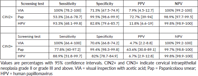
Table 5. Concordance between screening test results.
Discussion
Cervical cancer remains a significant public health challenge in India, particularly among women in rural and socio-economically disadvantaged settings. Effective screening is crucial for early detection of precancerous lesions and reduction of cervical cancer incidence.
In our study, a relatively large proportion of women, 205 (31.9%), tested positive on VIA. This high VIA positivity is likely influenced by several factors. VIA can appear positive in the presence of benign cervical conditions such as inflammation, ectropion or metaplasia, and its interpretation depends on visual assessment of aceto-white changes. These factors are commonly encountered in rural populations and have been reported previously [7,8]. Another contributor to the elevated VIA positivity in our cohort is the recruitment process. Many women were referred from community screening camps after already being screened VIA positive, resulting in an overrepresentation of VIA-positive women and an overall positivity rate approaching 30%. Similar observations have been reported in hospital-based services, highlighting how VIA positivity can be influenced by the healthcare environment. For instance, an opportunistic screening study of 329 women attending a gynaecology out patient department in India found that 18.5% were VIA positive, reflecting higher detection rates in symptomatic or hospital-attending populations [20]. Likewise, a study in rural Uttar Pradesh reported VIA positivity of 21% among women presenting for routine screening [20]. These facility-based positivity rates, notably higher than those reported in population-based programmes, underline the impact of patient selection and recruitment on VIA outcomes, emphasising that VIA positivity is shaped not only by underlying cervical pathology but also by the setting and characteristics of the screened population. This rate is substantially higher than those reported in large population-based screening programmes. For instance, a South Indian screening programme reported VIA positivity of 10.75% among 18,869 women [7]. Similarly, studies from Nepal and Iran reported VIA positivity rates of 7.4% and 12%–15%, respectively [9,10], highlighting variability influenced by examiner experience, training, lighting conditions and population characteristics.
Socio-demographic analysis revealed that rural residence significantly increased the odds of VIA positivity (OR = 2.13; p < 0.01), which is aligned with prior literature suggesting higher prevalence of cervical inflammation, limited access to preventive care and greater rates of symptomatic presentation among women in rural areas [7,8]. Age >45 years was associated with lower VIA positivity (OR = 0.56; p = 0.01), possibly due to regression of the transformation zone into the endocervix with advancing age, making acetowhite lesions less visible. Socioeconomic factors also played an important role: low family income (<Rs. 50,000/month) and lower per capita income (<4,411 INR) were associated with increased VIA positivity (OR = 2.08 and 1.49, respectively; p = 0.03), reflecting the influence of limited access to health services and preventive care. Other variables, including diet, contraceptive use and prior cervical complaints, did not show significant associations.
Reproductive factors further influenced VIA positivity. Early age at marriage (≤21 years) and higher parity (>2 live births) were associated with slightly increased VIA positivity (OR = 1.32 and 0.89, respectively), suggesting that cervical trauma, hormonal influences and prolonged exposure to potential HPV infection may contribute to acetowhite changes [15,16].
Pap smear positivity
Pap smear cytology in our cohort demonstrated only 11 women testing positive (1.7%). This low prevalence aligns with the documented tendency for Pap smear positivity to be lower than VIA or HPV DNA testing, especially in opportunistic screening settings [6,8]. Previous Indian studies have reported Pap positivity rates ranging from 10% to 25%, largely influenced by sample adequacy, presence of inflammation and quality of cytological assessment [7,20].
Our analysis revealed that older age (>45 years) was associated with higher odds of Pap positivity (OR = 2.24), although this was not statistically significant (p = 0.2). Rural residence showed a similar trend (OR = 2.33; p = 0.42). Reproductive factors, particularly early marriage (≤21 years; OR = 2.57) and high parity (>2 live births; OR = 2.24), were associated with increased Pap positivity, consistent with evidence that prolonged sexual activity, cervical trauma and hormonal changes influence cytological outcomes [15,16]. Other determinants, including education, occupation, monthly income, contraceptive use and menstrual status, did not significantly affect Pap results.
HPV DNA test positivity
HPV DNA testing in our cohort revealed positivity in 121 women (18.9%), with HPV 16 and HPV 18 genotypes accounting for nearly 50% of positive cases. This prevalence aligns with other Indian studies, where HPV positivity ranges between 15% and 25% in general populations [5,7,16]. Peri-urban studies in Andhra Pradesh and Mumbai report similar or slightly higher prevalence rates (>20%), highlighting HPV DNA testing as the most sensitive screening modality for detecting high-grade cervical lesions [7,16].
In our study, early age at marriage (≤21 years) and higher parity (>2 live births) were significant predictors of HPV positivity (OR = 1.72; p = 0.02 and OR = 1.6; p = 0.03, respectively). These associations are in concordance with a previously reported hospital-based study from Bihar, which found that women with more than two children were significantly more likely to be HPV positive (OR = 2.49; 95% CI: 1.93–3.21) [18], and those from lower socioeconomic groups had an even higher risk (OR = 3.05; 95% CI: 2.16–4.32) [15]. Similarly, a study conducted among women in North India reported that those married before the age of 20 years had a significantly higher risk of HPV infection (OR = 1.80) [11]. Furthermore, in a cohort of women living with HIV in Mumbai, being married before 20 years of age was associated with increased HPV positivity (OR = 2.02; 95% CI: 1.13–3.58) [19].
Other socio-demographic variables, including rural residence, low education and low family income, displayed non-significant trends toward higher HPV prevalence. These results are in line with previous research demonstrating that socioeconomic disadvantage, limited access to healthcare and higher infection burden contribute to higher HPV prevalence in low-resource settings [7,11].
Diagnostic performance of cervical cancer screening methods
In our study, VIA, Pap smear cytology and high-risk HPV DNA testing were evaluated for their ability to detect CIN II+ and CIN III+ lesions.
VIA demonstrated very high sensitivity but low specificity for detecting CIN II+ and CIN III+ lesions, reflecting its tendency to register some non-neoplastic acetowhite areas as abnormal. Meta-analytic data also showed that VIA’s pooled sensitivity and specificity vary widely, with a sensitivity of around 69% (95% CI: 54%–81%) and a specificity of 87% (95% CI: 79%–92%), reflecting operator dependency and variability in visual interpretation in low-resource settings [23]. In contrast, in a few Indian studies, the International Agency for Research on Cancer rural demonstration project observed VIA sensitivity of 67.9% and specificity of 93.2% for CIN III+ [22], while a study from Dadri, Uttar Pradesh reported sensitivity of ~50% and specificity of 96.7% for CIN III+ lesions [20]. Additionally, a Taiwanese study reported higher sensitivity for VIA compared with Pap smear, with lower specificity, highlighting the risk of false positives [25]. These findings are in line with our study, which also demonstrated 100% sensitivity for VIA, showing that it can detect all high-grade lesions in selective settings, but its accuracy may vary in larger population-based screening programmes due to differences in interpretation and patient characteristics.
Pap smear cytology in our cohort showed high specificity for both CIN II+ (98.8%) and CIN III+ (98.3%) lesions, with moderate sensitivity (53.3% and 77.8%, respectively). These findings are consistent with Indian studies, including the Andhra Pradesh cervical cancer screening study, which reported high specificity (>85%) but relatively lower sensitivity for early high-grade lesions, particularly CIN II [24]. The high NPVs observed in our study indicates the ability of Pap cytology to rule out high-grade disease effectively, even though some precancerous lesions were not detected.
In terms of screening methods, HPV DNA testing showed the highest sensitivity of 93.3% and specificity of 83.8% for CIN II lesions, with similar results for CIN III+ cases [23]. This performance aligns with both meta-analytic and Indian studies, where HPV testing generally shows higher sensitivity than Pap cytology and VIA while maintaining acceptable specificity (~82%–90%) [23,24]. HPV testing retained high sensitivity and very high NPVs (>99%), reflecting its ability to detect clinically significant lesions [23,24].
Inter-test concordance (κ agreement)
Inter-test agreement between VIA, Pap smear and HPV DNA testing in our cohort was low, reflecting limited overlap in the detection of cervical abnormalities across these screening modalities. This pattern is consistent with findings from Andhra Pradesh, where Gravitt et al reported poor concordance among VIA, cytology and HPV assays (κ = 0.04–0.11) [17]. Similar trends have been observed in other low-resource settings: studies in peri-urban Indian communities and rural North India noted only slight to fair agreement between VIA and HPV testing [6,17,24]. Factors such as operator experience, subjective interpretation of acetowhite changes, specimen quality and population prevalence of cervical lesions contribute to these discrepancies. These observations underscore that VIA, Pap and HPV DNA testing often identify different subsets of women at risk, supporting the complementary use of multiple modalities in screening programmes to enhance overall detection coverage.
Limitations of the study
Several factors should be considered when interpreting the findings. The small number of women testing positive on Pap smear (n = 11) restricted the statistical power to identify associations with socio-demographic characteristics. Similarly, the relatively low prevalence of HPV positivity and the small number of CIN2+ and CIN3+ cases resulted in low PPVs across screening tests. The predominantly rural and lower socioeconomic composition of the study population may limit the applicability of these findings to urban or higher-income populations.
Conclusion
Early marriage, higher parity and lower socioeconomic status were associated with increased HPV positivity, reflecting patterns observed in similar Indian cohorts. VIA captured a larger proportion of potential lesions but showed lower specificity, while Pap cytology demonstrated high specificity but moderate detection of high-grade lesions. HPV DNA testing identified high-risk infections, particularly types 16 and 18, which were most frequently associated with CIN II and CIN III+ lesions. These observations highlight the differential performance of screening modalities in a North Indian population and the potential role of HPV testing in identifying women at higher risk, alongside complementary use of VIA testing in resource-limited settings.
Conflicts of interest
The authors declare no conflicts of interest.
Funding
No external funding was received for this study.
Ethical approval
Ethical approval for this study was obtained from the Institutional Ethics Committee of Homi Bhabha Cancer Hospital and Research Centre (Project ID: 15000064). All patient data were de-identified, and confidentiality was strictly maintained.
Author contributions
Conceptualisation, Data collection, manuscript drafting- Dr. Pallavi, Conceptualisation, manuscript review and editing- Dr.Vandita Pahwa, Data collection, manuscript drafting and review- Dr. Shikha, Dr. Gurwinder kaur, Microbiological review of samples- Dr. Charu, Histopathological review of samples- Dr. Sankalp, Cytopathological review of samples- Dr. Ravikiran, Statistical analysis- Kundan rao, Manuscript review and overall supervision- Dr. Ashish Gulia.
References
1. World Health Organization (2024) Cervical Cancer Key Facts (Geneva: WHO)
2. Ministry of Statistics and Programme Implementation (India) Percentage of Women Aged 30–49 Years Ever Undergone a Screening Test for Cervical Cancer (NFHS-5, 2019–21) (New Delhi: MOSPI)
3. Arbyn M, Weiderpass E, and Bruni L, et al (2020) Estimates of incidence and mortality of cervical cancer in 2020: a worldwide analysis Lancet Glob Health 8(2) e191–e203 https://doi.org/10.1016/S2214-109X(19)30482-6 PMCID: 7025157
4. Nigam G, Pandey A, and Singh T, et al (2022) Socioeconomic and regional variation in breast and cervical cancer screening among Indian women: NFHS-5 (2019–21) BMC Cancer 22 1279 https://doi.org/10.1186/s12885-022-10387-9
5. Basu P, Banerjee D, and Niyogi P, et al (2009) Human papillomavirus genotype distribution in cervical cancer in India: a multicentre study Int J Cancer 124(9) 2342–2347
6. Sankaranarayanan R, Esmy PO, and Rajkumar R, et al (2004) Diagnostic accuracy of VIA and HPV detection as primary and sequential screening tests in a cervical cancer screening demonstration project in rural India Int J Cancer 110(1) 100–105
7. Shastri SS and Mittra I (2012) Cervical cancer screening with Pap smear in South India: community-based outcomes Indian J Cancer 49 65–71
8. Bhatla N, Dar L, and Patro AR, et al (2014) HPV DNA testing in cervical cancer screening in peri-urban Andhra Pradesh Cancer Epidemiol Biomark Prev 38 345–352
9. Shrestha AD, Shrestha R, and Khatri R, et al (2016) Evaluation of VIA and Pap smear in Nepalese women J Obstet Gynaecol Res 42 123–131
10. Saleh M, Saleh M, and Zolala F, et al (2019) Performance of VIA and influencing factors among Iranian women Asian Pac J Cancer Prev 20 567–573
11. Gupta S, Jha P, and Bansal S, et al (2015) HPV prevalence and associated risk factors among North Indian women Asian Pac J Cancer Prev 16 6545–6551
12. Kim HY (2017) Statistical notes for clinical researchers: chi-squared test and Fisher's exact test Restor Dent Endod 42(2) 152–155 https://doi.org/10.5395/rde.2017.42.2.152 PMID: 28503482 PMCID: 5426219
13. Chowdhury MZI and Turin TC (2020) Variable selection strategies and its importance in clinical prediction modelling Fam Med Community Health 8(1) 262
14. IBM Corp (2022) IBM SPSS Statistics for Windows. Version 29.0 (Armonk, NY: IBM Corp)
15. Singh A and Sharma P (2017) Reproductive and socio-economic factors associated with HPV infection in Indian women J Obstet Gynaecol Res 43 1823–1830
16. Mittra I, et al (2018) Effectiveness of HPV DNA testing in cervical cancer screening in India Int J Cancer 142 1489–1497
17. Gravitt PE, Paul P, and Katki HA, et al (2010) Effectiveness of VIA, Pap, and HPV DNA testing in a cervical cancer screening program in a peri-urban community in Andhra Pradesh, India PLoS One 5(10) e13711 https://doi.org/10.1371/journal.pone.0013711 PMID: 21060889 PMCID: 2965656
18. Pankaj S, Rani J, and Kumari P, et al (2024) Prevalence, risk factors and genotype distribution of HPV infection among women with and without invasive cervical cancer: hospital-based study, Bihar, India Natl Med J India 37(1) 13–17 https://doi.org/10.25259/NMJI_634_21 PMID: 39096228
19. Pahwa V, Pimple SA, and Mishra GA, et al (2022) Prevalence and determinants of HPV infection and CIN among women living with HIV/AIDS in Mumbai, India Indian J Med Paediatr Oncol 43(1) 97–102 https://doi.org/10.1055/s-0042-1742661
20. Satyanarayana L, Sharma V, and Das DK, et al (2012) Cervical Cancer Screening in a Rural Community Setting (Dadri, Uttar Pradesh) by Pap smear and VIA. PGIMER Cervical Cancer Screening Report (Chandigarh: Post Graduate Institute of Medical Education & Research) p 46–47
21. Arbyn M and Michelow P (2017) A systematic review and meta-analysis of studies evaluating the performance of point-of-care tests for HPV screening Sex Transm Infect 93 Suppl 4(Suppl 4) S36–S45
22. Shastri SS and Mittra I (2012) Cervical cancer screening with Pap smear: sensitivity and specificity for CIN2+/CIN3+ Indian J Cancer 49 65–71
23. Arbyn M, et al (2012) Cervical cancer screening by HPV testing, cytology, and VIA: a systematic review and meta-analysis Lancet Oncol 13(9) 929–941
24. Asthana S, Satyanarayana L, and Bhambani S, et al (2014) A comparative study of cervical cancer screening methods in a rural community setting of North India Indian J Cancer 51(2) 124–128 https://doi.org/10.4103/0019-509X.138172 PMID: 25104192
25. Vahedpour Z, et al 2019 Comparison of diagnostic value of visual inspection with acetic acid and Pap smear in cervical cancer screening Taiwan J obstet Gynecol 58(3) 345–348