Epstein–Barr virus in paediatric sporadic Burkitt lymphoma: real-world experience from a Peruvian national referral centre
Sandro Casavilca-Zambrano1 ,2,3, Juan Contreras-Mancilla2,3,4, Camille Laurent5, Jaqueline Montoya6, Rosdali Diaz-Coronado6,7, Jorge Honles3,8, Ruddy Liendo-Picoaga2,9, Jenny Bonifacio-Mundaca2,3,10, Jhoysi Casas-Goñas2, Juan Pablo Cerapio11, Carlos Barrionuevo12,13, Tatiana Vidaurre14 and Stéphane Bertani3,8
1Faculty of Health Science, Universidad de Huánuco, Huánuco 10001, Peru
2Banco Nacional de Tumores, Instituto Nacional de Enfermedades Neoplásicas, Lima 15038, Peru
3International Joint Laboratory of Molecular Anthropological Oncology (LOAM), Instituto Nacional de Enfermedades Neoplásicas, Institut de Recherche pour le Développement, Lima 15038, Peru
4Grupo de Investigación Salud Pública, Cáncer y Enfermedades Crónicas No Transmisibles (SCENT), Instituto de Investigación en Ciencias Biomédicas, Universidad Ricardo Palma, Lima 15039, Peru
5Institut Universitaire du Cancer de Toulouse, CHU Toulouse, Cancer Research Center of Toulouse (CRCT), Toulouse, 31037, France
6Department of Paediatric Oncology, Instituto Nacional de Enfermedades Neoplásicas, Lima 15038, Peru
7Faculty of Medicine, Universidad Peruana Cayetano Heredia, Lima 15102, Peru
8UMR 152 PHARMADEV, Université de Toulouse, Institut de Recherche pour le Développement, Université Paul Sabatier, Toulouse 31062, France
9Laboratorio de Investigación en Cultivo Celular e Inmunología, Universidad Científica del Sur, Lima 15842, Peru
10Centro de Investigación en Bioingeniería (BIO), Universidad de Ingeniería y Tecnología, Lima 15842, Peru
11Centre de Recherches en Cancérologie de Toulouse, Toulouse 31037, France
12Department of Pathology, Instituto Nacional de Enfermedades Neoplásicas, Lima 15038, Peru
13Faculty of Medicine, Universidad Nacional Mayor de San Marcos, Lima 15001, Peru
14Department of Medical Oncology, Instituto Nacional de Enfermedades Neoplásicas, Lima 15038, Peru
Abstract
Introduction: Burkitt lymphoma (BL) is an aggressive B-cell lymphoma, representing 30%–40% of childhood non-Hodgkin lymphomas globally. In Peru, Epstein–Barr virus (EBV) is highly prevalent in lymphomas, yet its role in paediatric BL remains underexplored.
Objective: This study evaluates the clinical and epidemiological characteristics of paediatric BL at the National Institute of Neoplastic Diseases, focusing on EBV-positive cases, pathological features, treatment response and prognostic factors, within the real-world setting of a lower-middle-income country (LMIC).
Materials and methods: From 2009 to 2021, 187 aggressive B-cell lymphomas were diagnosed in children, from which 34 high-grade B-cell lymphomas underwent central pathology review, yielding 23 molecularly confirmed sporadic BL cases. Inclusion criteria comprised age <15 years, availability of tumour tissue for immunohistochemical analysis and available clinical records.
Results: EBV infection was detected using EBV-encoded RNA (EBER) in situ hybridisation, and MYC translocation (8q24) was identified via FISH. Among the subset, 61% were EBV-positive, with males comprising 65% and a mean age of 6.7 years. EBV-positive status and younger age were associated with inferior survival in univariate analyses (log-rank p = 0.012 and p = 0.009, respectively). The association with EBV was attenuated after exploratory adjustment (HR, 3.69; 95% CI, 0.35–38.98; p = 0.278).
Conclusion: In this cohort, 61% of paediatric sporadic BL cases were EBV-positive, associated with younger age (p = 0.01) and inferior survival in univariate analysis (log-rank p = 0.012). The independent prognostic contribution of EBV could not be established. These LMIC findings support expanded molecular diagnostics and larger multicentre studies.
Keywords: lymphoma, B-cell, Burkitt lymphoma, infections, Epstein–Barr virus, oncogenic viruses, Latin America, Peru
Correspondence to: Juan Contreras-Mancilla
Email: cmjuanjo.contreras@gmail.com
Published: 08/10/2026
Received: 05/03/2026
Publication costs for this article were supported by ecancer (UK Charity number 1176307).
Copyright: © the authors; licensee ecancermedicalscience. This is an Open Access article distributed under the terms of the Creative Commons Attribution License (http://creativecommons.org/licenses/by/4.0), which permits unrestricted use, distribution, and reproduction in any medium, provided the original work is properly cited.
Introduction
Every year, ?400,000 children and adolescents aged 0 to 19 years are affected by cancer worldwide [1,2]. In low- and middle-income countries, 4.6% of cancer cases occur in children aged 0 to 14 years, leading to a higher mortality rate compared to high-income countries, where childhood cancers constitute only 0.5% of all cases [3]. In developed nations, 80% of childhood cancers are successfully treated, with haematological malignancies showing the highest incidence rates [1]. In Peru, lymphoma and leukaemia have age-standardised incidence rates of 9.9 and 7.6 per 100,000, respectively, and haematological neoplasms account for 60% of childhood cancers [4,5]. Furthermore, the incidence of cancer-related infections in low- and middle-income regions ranges between 15% and 30% [6], highlighting an important, preventable cause of cancer.
Non-Hodgkin lymphoma (NHL) encompasses diverse subtypes with distinct morphological, genetic and clinical features, and its aetiology involves both infectious agents and genetic predispositions [7].
The Epstein–Barr virus (EBV), the first oncogenic virus identified over 56 years ago by Dr. Anthony Epstein and Dr. Yvonne Barr, was originally associated with Burkitt lymphoma (BL) in equatorial Africa [8,9]. EBV encodes around 100 viral proteins, with only five oncoproteins (latent membrane protein 1, latent membrane protein 2A, latent membrane protein 2B, Epstein -Barr nuclear antigen 1 and Epstein–Barr nuclear antigen 2) required to transform primary B cells into proliferating lymphoblastoid cell lines. Notably, in BL, the virus typically adopts Latency I, expressing only EBNA1 and EBV-encoded RNAs (EBERs) [10]. BL, an aggressive B-cell lymphoma common in paediatric populations, represents ?30% to 40% of childhood NHL worldwide [11–13]. It manifests in three clinical forms: sporadic BL; endemic BL, which is associated with Plasmodium falciparum malaria in equatorial Africa; and immunodeficiency-associated BL (i.e. HIV-associated BL) [14–16]. Peru has the highest incidence of EBV-positive diffuse large B-cell lymphoma, not otherwise specified (DLBCL NOS) and classic Hodgkin lymphoma (HL) in Latin America, with EBV presence confirmed in tumour tissues through immunological or molecular techniques.
BL is marked by characteristic cytogenetic translocations, specifically t(8;14), t(2;8) and t(8;22), which activate the MYC gene and drive cell proliferation [17,18]. EBV is detected in 20% to 30% of sporadic BL cases globally; however, few studies have focused on EBV-positive sporadic BL.
Moreover, there is a notably high prevalence of T-cell EBV-associated lymphoproliferative disorders and NK/T-cell lymphomas, reinforcing EBV’s role as a significant etiological factor in Peru [19–21]. BL remains a major cause of paediatric cancer in the country, and this study investigates its epidemiological and pathological features, along with relevant risk factors. From 2009 to 2021, 187 cases of mature aggressive B-cell lymphomas were diagnosed in children at the National Institute of Neoplastic Diseases. Our study focuses on 23 cases of sporadic BL unrelated to HIV, treated at Instituto Nacional de Enfermedades Neoplásicas (INEN) in Lima, Peru. In this work, we present a comprehensive analysis of the clinical and epidemiological characteristics of paediatric sporadic BL at INEN, including EBV prevalence, pathological diagnosis, treatment response and prognostic factors.
Materials and methods
Tissue samples and case characterisation
This was a retrospective single-centre cohort study. Between 2009 and 2021, 187 children with mature aggressive B-cell lymphomas were diagnosed at INEN. From this cohort, 34 cases of high-grade B-cell lymphoma with a germinal centre phenotype were selected for central pathological review based on the following inclusion criteria: availability of pathological material for immunohistochemical analysis, patient age below 15 years and available clinical records. Of these, 24 cases were MYC-FISH-positive; one lacked an available clinical record, leaving 23 cases that constitute the final study cohort.
To detect chromosomal rearrangements involving the MYC gene located on chromosome 8q24, a MYC-FISH translocation assay was performed during central review. In instances where BCL2 immunohistochemistry results were inconclusive, BCL2-FISH testing was conducted (n = 3). All cases tested negative for BCL2 rearrangements, effectively ruling out double-hit high-grade lymphoma. Additionally, in four cases that were negative for MYC rearrangement, the presence of 11q aberrations was assessed using a FISH method, with one case confirming the aberration (n = 1/4).
This study adhered to confidentiality and anonymity principles according to the Declaration of Helsinki and its amendments. Approval was granted by the Ethics Committee and the INEN Scientific Council (INEN 543-2020).
Statistical analysis
A retrospective cohort analysis was performed on 187 cases of mature aggressive B-cell lymphomas, including 23 patients diagnosed with BL at the INEN in Lima, Peru. Categorical variables were analysed using the chi-square and Fisher’s exact tests, while continuous variables were compared using Student’s t-test. Missing data were reported as not available and were not imputed. Overall survival was calculated from the date of diagnosis to death from any cause or last follow-up, with patients alive at last follow-up censored. Follow-up was administratively truncated at 36 months. Survival probabilities were estimated by the Kaplan–Meier method, with survival distributions compared using the log-rank test and median follow-up estimated using the reverse Kaplan–Meier method. Univariable Cox regression was used to report hazard ratios (HRs) and 95% confidence intervals (CIs). An exploratory Cox model included EBV status, age group and treatment documentation; proportional hazards were assessed using Schoenfeld residuals. EBER status was analysed as positive or negative. A p-value of < 0.05 was deemed statistically significant. All analyses were conducted with R software environment version 4.6.1 (R Core Team), Stata software version 17.0 (StataCorp) and QGIS software version 3.14 (QGIS Development Team).
Immunohistochemistry
Immunohistochemical staining for CD20, BCL6, CD10, BCL2, Ki67, MUM1, TDT and MYC was reviewed by three experienced pathologists, with results reported as positive, negative or by expression percentage. Consensus from at least two pathologists was required for reporting.
In situ hybridisation for EBER
The presence of EBV RNA was detected by EBER in situ hybridisation. EBER results were classified as positive or negative.
Fluorescence in situ hybridisation
The MYC translocation was confirmed in all 23 BL cases using FISH analysis.
Results
Epidemiological, demographic and clinical features
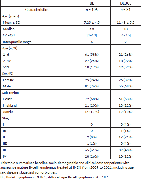
The cohort of mature aggressive B-cell lymphomas in children from the INEN diagnosed between 2009 and 2021 (N = 187) included 106 BL (57%) and 81 (43%) DLBCL NOS cases. Patients diagnosed with BL were generally younger (7.25 years; SD, 4.5) than DLBCL NOS patients (11.4 years; SD, 5.2), with a greater proportion of males and a higher representation from the coastal region (Table 1). BL cases tended to be in a more advanced stage, with a majority in stage III, compared to DLBCL NOS cases, where there is a more even distribution across different stages.
Table 1. Baseline socio-demographic and clinical characteristics of aggressive mature B-cell lymphoma cases at INEN (2009–2021).
In our review of 34 cases of high-grade B-cell lymphoma, 23 were confirmed as BL. The 11 excluded cases comprised eight MYC-FISH-negative cases, one case with 11q aberration, one case with unconfirmed MYC status and one MYC-FISH-positive case without an available clinical record (Figure 1). The median age of the patients included in the review was 5 years, and the mean was 6.7 ± 3.6 SD (range between 1 and 15 years); most of them were males (65%), and 48% were from the Peruvian coastal region (Figure 2). The positivity for infection by EBV was 61%, predominantly among males and in the 4–6 years group (Table 2). Similarly, most patients with EBV-negative status were male and aged between 7 and 10 years (n = 4). The mean age of infected patients was significantly younger than that of non-infected patients (5.2 years +/? 2.8 SD versus 9 years +/? 3.6 SD, respectively, p = 0.01); most patients (48%) came from the Peruvian coastal region, and 65% were male. The abdominal visceral region primary compromise accounted for 78% of instances, followed by the head and neck with 17% of cases, and the thorax with a single case accounting for the remaining 4% of patients. At diagnosis, all our cases were in advanced stages (III–IV) (Table 3).
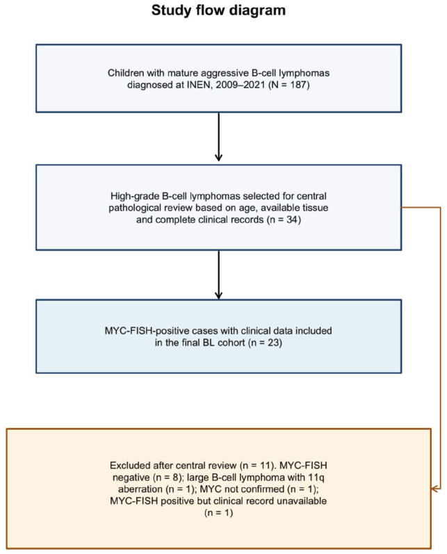
Figure 1. Study flow diagram showing the derivation of the final molecularly confirmed Burkitt lymphoma cohort.
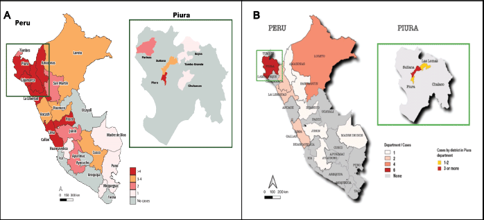
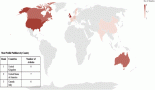
Figure 2. Geographic distribution of Burkitt lymphoma cases. A. The geographic distribution of Burkitt lymphoma cases. The magnified section in the upper right highlights an unusual increase in cases reported across several districts within the department of Piura, indicating a potential regional concentration that warrants further investigation. B. The geographic distribution of 23 Burkitt lymphoma cases with confirmed MYC translocation, based on place of birth. An unusual concentration of cases is observed in the district of Sullana, Piura.
Table 2. General socio-demographic and clinical characteristics of 23 cases with molecular diagnosis of MYC translocation and EBV status.
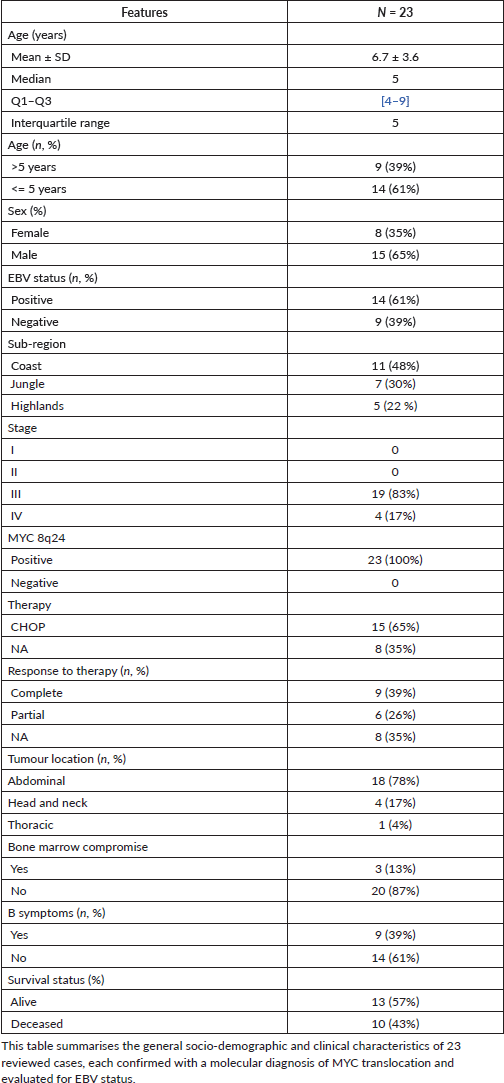
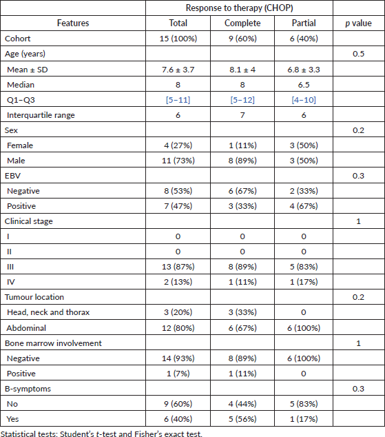
Cyclophosphamide, doxorubicin, vincristine and prednisolone (CHOP) was documented as first-line therapy in 15 patients (65%), and none received rituximab. Treatment information was not recorded for eight patients (35%); this does not necessarily indicate that they were untreated. Among the 15 patients with documented CHOP, 9 (60%) had a complete response and 6 (40%) had a partial response (Table 4). The total clinical-socio-demographic characteristics are presented in Table 3.
Table 3. Clinical and socio-demographic characteristics of patients with Epstein–Barr virus infection.
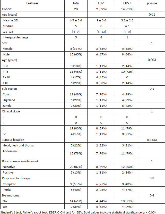
Table 4. Clinical and socio-demographic features versus response to therapy (CHOP).
Pathological features
Pathological analysis of the 23 BL cases revealed characteristic immunohistochemical profiles, demonstrating positive staining for CD20, BCL6 and CD10, while TDT, BCL2 and MUM1 were consistently negative. Ki67 was available for 20 cases, with a median value of 98.5% (range, 70%–100%); 19 of 20 cases had Ki67 values of at least 95%. Fluorescence in situ hybridisation (FISH) analysis confirmed the presence of MYC translocation (8q24-positive) in all BL cases (Figure 3). Among these, EBER was positive in 14 cases and negative in nine.
In the MYC-negative group of high-grade B-cell lymphomas, FISH analysis detected an 11q aberration in one case. The study also identified 10 cases of DLBCL NOS.
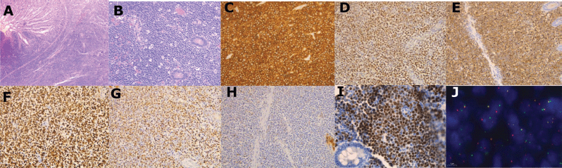
Figure 3. Ileocecal mucosal involvement in Burkitt lymphoma. (A) Hematoxylin and eosin (H&E) staining, panoramic view (5x), showing infiltration of the ileocecal mucosa by Burkitt lymphoma. (B) Higher magnification (10x) demonstrating the classic ‘starry sky’ pattern characteristic of Burkitt lymphoma. (C) CD20 immunostaining at 20x, highlighting predominantly membranous positivity. (D) BCL6 immunostaining with nuclear expression at 20x magnification. (E) CD10 immunostaining showing positive cytoplasmic expression at 20x. (F) Ki67 immunostaining indicating a proliferative index approaching 100% with nuclear expression at 20x magnification. (G) MYC immunostaining positive for nuclear expression at 20x. (H) MUM1 immunostaining negative at 20x magnification. (I) Chromogenic in situ hybridization (CISH) for Epstein–Barr virus (EBV) with positive nuclear brown staining (40x). (J) Fluorescence in situ hybridization (FISH) test MYC (100x) for 8q24 translocation detection.
Survival-related factors
Of the 23 patients included in this study, 10 (43%) died. With follow-up administratively truncated at 36 months, the median follow-up estimated by reverse Kaplan–Meier was 36 months (observed range, 0.3–36 months). Kaplan-Meier overall survival was 67.2% (95% CI, 43.1–82.8) at 6 months and 52.3% (95% CI, 29.5–71.0) at 12 months (Figure 4). Survival differed according to age (log-rank p = 0.009) and EBV status (log-rank p = 0.012). In univariable Cox analysis, EBV-positive status was associated with inferior survival (HR, 9.20; 95% CI, 1.15–73.58; p = 0.036), as was age ≤5 years (HR, 9.88; 95% CI, 1.22–79.93; p = 0.032). Documentation of CHOP was also associated with survival (HR, 0.19; 95% CI, 0.05–0.68; p = 0.011), but this comparison should not be interpreted as a treatment effect because missing treatment records do not indicate absence of treatment (Supplementary Figure 1). In the exploratory model including EBV status, age and treatment documentation, EBV was no longer statistically significant (adjusted HR, 3.69; 95% CI, 0.35–38.98; p = 0.278). The total survival estimates are presented in Table 5.
Bivariate analysis revealed that, in this series of patients, age (p = 0.01) was a factor associated with EBV infection. When stratified by age group, the presence of the virus was more frequent in the 4–6-year-old group (Table 3). In particular, there was a significant association with patients under 5 years of age. Most of the patients with the infection came from the jungle region of Peru. The complete results are presented in Table 3.
Discussion
In our review of 34 cases of high-grade B-cell lymphoma, 23 MYC-FISH-positive cases with clinical data were included as BL, one additional MYC-FISH-positive case lacked an available clinical record, one case was identified as large B-cell lymphoma with 11q aberration and nine cases were not molecularly confirmed as BL. This highlights the importance of molecular testing to confirm specific alterations within the diverse group of aggressive high-grade lymphomas.
Of the BL cases, 61% (14 of 23) were EBV-positive, consistent with rates reported across Latin America (29%–87%) [22]. With follow-up administratively truncated at 36 months, the observed mortality rate of 43% in our cohort is substantially higher than that reported in high-income countries, where paediatric treatment success rates are typically over 90%.
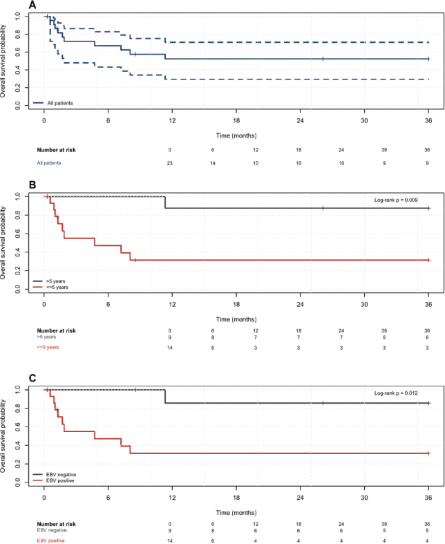
Figure 4. Kaplan–Meier curves for overall survival (A), age groups (B) and EBV status (C), with numbers at risk. Follow-up was administratively truncated at 36 months.
Table 5. Kaplan–Meier overall survival estimates according to clinical factors.
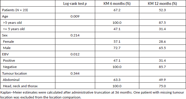
Survival outcomes were poorer among younger patients (log-rank p = 0.009) and in those with EBV-positive status (log-rank p = 0.012), with bivariate analysis further indicating a correlation between younger age and EBV positivity (p = 0.01). However, the EBV association was attenuated and was no longer statistically significant after exploratory adjustment for age and treatment documentation. In contrast to the findings of Richter et al [23], who associated EBV positivity in sporadic BL with older age [23], our study did not reveal significant associations between clinical or socio-demographic factors and treatment response. The small sample size, wide confidence intervals and incomplete treatment information limit the generalisability of these findings and do not allow EBV to be considered an independent prognostic factor.
Recent studies by Thomas et al [24] suggest that EBV status may be a stronger molecular predictor of BL prognosis than age alone [24]. EBV-positive cases have been linked to unique genetic features, including somatic hypermutation-mediated breakpoints and mutations in genes such as FOXO1 and BCR, which could contribute to distinct clinical behaviours warranting further investigation [24–26].
Phylogenetic analyses reveal geographical variations in EBV strains, with distinct sequence patterns observed in African, Asian and European lineages [27]. In Peru, where over 80% of the ancestry of the population has Indigenous traces, further research is needed to explore the co-evolution of EBV with host factors, particularly in high-prevalence areas like the jungle. In our cohort, an apparent concentration of sporadic BL cases was observed in the Piura region, specifically in Sullana. Environmental factors such as pesticide exposure, immunosuppression and malaria were not measured in this study, and their possible relationship with this concentration may warrant future investigation [28,29].
While the role of EBV infection in oncogenesis is established, the interaction between viral and host factors in BL development remains complex and not fully understood [30]. Clarifying the prevalence and impact of EBV in paediatric BL, particularly within Latin American populations, is essential for the development of targeted therapies that address viral oncogenic mechanisms and modulate immune responses to EBV-infected cells. This research represents a promising direction for improving outcomes in paediatric BL patients in EBV-endemic regions.
This study has several limitations. It was retrospective and conducted at a single referral centre, and the final cohort was small, with 23 patients and 10 deaths. Selection for central review depended on the availability of tissue and clinical records, which may have introduced selection bias. The low number of events, the association between EBV status and age, and incomplete treatment information limited the adjusted analysis. Treatment was not standardised, rituximab was not administered and information on treatment abandonment, treatment-related mortality and salvage therapy was incomplete. The technical details of EBER testing, the MYC-IHC threshold and the classification framework were not available in the records reviewed. Therefore, the survival findings should be considered exploratory.
Conclusion
In this Peruvian paediatric cohort of sporadic BL with molecular confirmation (N = 23), EBV-positive status was present in 61% of cases and was associated with younger age (p = 0.01) and inferior overall survival in univariate analysis (log-rank p = 0.012). Given the correlation between age and EBV status, incomplete treatment information and the limited sample size, the independent prognostic contribution of EBV could not be established, and these findings should be interpreted as exploratory. Our results support the need to strengthen access to standardised molecular diagnostics (EBER and MYC testing) and to conduct larger multicentre studies in Latin America to clarify the contribution of EBV and related factors to outcome.
Acknowledgments
The authors are grateful to all the patients whose participation was essential to the achievement of this study. The authors also thank the staff members of the National Tumour Bank at INEN for their leadership in aggregating medical information.
Funding
This work was supported by funds from the University of Huánuco’s Faculty of Health Sciences project ’Geolocation of lymphoma cases and possible associated epidemiological risk factors in the national institute of neoplastic diseases,’ International Joint Laboratory for Molecular Anthropology of Cancer and Oncogenic Viruses (LMI-LOAM), ITMO Cancer of the French National Alliance for Life Sciences and Health (Aviesan) and the French National Cancer Institute (INCa) on funds administered by the French National Institute of Health and Medical Research (Inserm), grant agreement 21CD025-00. The funders had no role in study design, data collection and analysis, decision to publish or preparation of the manuscript.
Conflicts of interest
The authors declare no conflicts of interest.
Author contributions
Conceptualisation: SCZ, JM, RDC, SB; Methodology: JH, RLP, JCG; Validation: JPC, CB, TV; Formal Analysis CL, JH, RLP, JBM; Data Curation JH, JCM; Writing – Origin al Draft Preparation: SCZ, JM, RDC, RLP; Writing – Review & Editing: CB, TV, JCM, SB; Visualisation: JH, JBM, JCM; Supervision: SCZ, SB; Project Administration: SCZ, SB; Funding Acquisition: SCZ, SB.
References
1. WHOG and Overview AN (2020) WHO Global Initiative for childhood cancer – India responds Pediatric Hematology Oncol J 5 145–150 https://doi.org/10.1016/j.phoj.2020.06.005
2. Steliarova-Foucher E, Colombet M, and Ries LAG, et al (2017) International incidence of childhood cancer, 2001–10: a population-based registry study Lancet Oncol 18 719–731 https://doi.org/10.1016/S1470-2045(17)30186-9 PMID: 28410997 PMCID: 5461370
3. Joko‐Fru WY, Parkin DM, and Borok M, et al (2018) Survival from childhood cancers in Eastern Africa: a population-based registry study Int J Cancer 143 2409–2415 https://doi.org/10.1002/ijc.31723
4. Ramos Muñoz WC, Guerrero Ramírez NN, and Medina Osis JL, et al (2020) Análisis de la situación del Cáncer en el Perú, 2018 1 ed Centro Nacional de Epidemiología, Prevención y Control de Enfermedades, Ministerio de Salud p 161 [https://pesquisa.bvsalud.org/portal/resource/pt/biblio-1146926]
5. Payet E, Perez P, and Poquioma E, et al (2021) Registro de Cáncer de Lima Metropolitana Incidencia y Mortalidad 2013–2015 (Surquillo: Instituto Nacional de Enfermedades Neoplasicas) Vol 6. [https://www.gob.pe/institucion/minsa/informes-publicaciones/3311511-registro-de-cancer-de-lima-metropolitana-incidencia-y-mortalidad-2013-2015]
6. De Martel C, Georges D, and Bray F, et al (2020) Global burden of cancer attributable to infections in 2018: a worldwide incidence analysis Lancet Global Health 8 e180–e190 https://doi.org/10.1016/S2214-109X(19)30488-7
7. Spector LG, Pankratz N, and Marcotte EL (2015) Genetic and nongenetic risk factors for childhood cancer Pediatric Clinics North Amer 62 11–25 https://doi.org/10.1016/j.pcl.2014.09.013
8. Pannone G, Zamparese R, and Pace M, et al (2014) The role of EBV in the pathogenesis of Burkitt’s lymphoma: an Italian hospital-based survey Infect Agents Cancer 9 34 https://doi.org/10.1186/1750-9378-9-34
9. Dunmire SK, Verghese PS, and Balfour HHJ (2018) Primary Epstein-Barr virus infection J Clin Virol 102 84–92 https://doi.org/10.1016/j.jcv.2018.03.001 PMID: 29525635
10. Piccaluga PP, Navari M, and De Falco G, et al (2016) Virus-encoded microRNA contributes to the molecular profile of EBV-positive Burkitt lymphomas Oncotarget 7 224–240 https://doi.org/10.18632/oncotarget.4399 PMCID: 4807994
11. Kennedy G, Komano J, and Sugden B (2003) Epstein–Barr virus provides a survival factor to Burkitt’s lymphomas Proc Natl Acad Sci USA 100 14269–74 https://doi.org/10.1073/pnas.2336099100
12. Molyneux EM, Rochford R, and Griffin B, et al (2012) Burkitt’s Lymphoma Lancet 379 1234–1244 https://doi.org/10.1016/S0140-6736(11)61177-X PMID: 22333947
13. Mbulaiteye SM, Biggar RJ, and Bhatia K, et al (2009) Sporadic Childhood Burkitt Lymphoma Incidence in the United States during 1992–2005 Pediatric Blood & Cancer 53 366–370 https://doi.org/10.1002/pbc.22047
14. Muhealdeen DN, Shwan A, and Yaqo RT, et al (2022) Epstein–Barr Virus and Burkitt’s Lymphoma: associations in Iraqi Kurdistan and twenty-two countries assessed in the international incidence of childhood cancer Infect Agents Cancer 17 46 https://doi.org/10.1186/s13027-022-00452-0
15. Broen K, Dickens J, and Trangucci R, et al (2023) burkitt lymphoma risk shows geographic and temporal associations with Plasmodium falciparum infections in Uganda, Tanzania, and Kenya Proc Nat Acad Sci United States Amer 120 e2211055120 https://doi.org/10.1073/pnas.2211055120
16. Redmond LS, Ogwang MD, and Kerchan P, et al (2020) Endemic Burkitt Lymphoma: a Complication of Asymptomatic Malaria in Sub-Saharan Africa Based on Published Literature and Primary Data from Uganda, Tanzania, and Kenya Malaria J 19 239 https://doi.org/10.1186/s12936-020-03312-7
17. Burkhardt B, Michgehl U, and Rohde J, et al (2022) Clinical Relevance of Molecular Characteristics in Burkitt Lymphoma Differs According to Age Nature Commun 13 3881 https://doi.org/10.1038/s41467-022-31355-8
18. Möker P, Zur Stadt U, and Zimmermann M, et al (2022) Characterization of IG-MYC Breakpoints and Their Application for Quantitative Minimal Disease Monitoring in High-Risk Pediatric Burkitt Lymphoma and Leukemia Leukemia 36 2343–3236 https://doi.org/10.1038/s41375-022-01626-w
19. Gru AA, Haverkos BH, and Freud AG, et al (2015) The Epstein–Barr Virus (EBV) in T Cell and NK Cell Lymphomas: time for a Reassessment Curr Hematologic Malignancy Rep 10 456–467 https://doi.org/10.1007/s11899-015-0292-z
20. Rodríguez-Pinilla SM, Barrionuevo C, and García J, et al (2011) Epstein-Barr virus-positive systemic NK/T-cell lymphomas in children: report of six cases Histopathology 59 1183–1193 https://doi.org/10.1111/j.1365-2559.2011.04047.x PMID: 22175898
21. Barrionuevo C, Zaharia M, and Martinez MT, et al (2007) Extranodal NK/T-Cell Lymphoma, Nasal Type: study of Clinicopathologic and Prognosis Factors in a Series of 78 Cases from Peru Appl Immunohistochemistry & Mol Morphol 15 38–44 https://doi.org/10.1097/01.pai.0000205062.27174.56
22. Wong Y, Meehan MT, and Burrows SR, et al (2022) Estimating the Global Burden of Epstein–Barr Virus-Related Cancers J Cancer Res Clin Oncol 148 31–46 https://doi.org/10.1007/s00432-021-03824-y
23. Richter J, John K, and Staiger AM, et al (2022) Epstein–Barr Virus Status of Sporadic Burkitt Lymphoma Is Associated with Patient Age and Mutational Features Br J Haematology 196 681–689 https://doi.org/10.1111/bjh.17874
24. Thomas N, Dreval K, and Gerhard DS, et al (2023) Genetic subgroups inform on pathobiology in adult and pediatric Burkitt lymphoma Blood 141 904–916 https://doi.org/10.1182/blood.2022016534 PMCID: 10023728
25. Panea RI, Love CL, and Shingleton JR, et al (2019) The whole-genome landscape of Burkitt lymphoma subtypes Blood 134 1598–1607 https://doi.org/10.1182/blood.2019001880 PMID: 31558468 PMCID: 6871305
26. Grande BM, Gerhard DS, and Jiang A, et al (2019) Genome-wide discovery of somatic coding and noncoding mutations in pediatric endemic and sporadic Burkitt lymphoma Blood 133(12) 1313–24 [https://doi.org/10.1182/blood-2018-09-871418]
27. Liao HM, Liu H, and Chin PJ, et al (2022) Epstein–Barr virus in Burkitt lymphoma in Africa reveals a limited set of whole genome and LMP-1 Sequence Patterns: analysis of Archival datasets and field samples from Uganda, Tanzania, and Kenya Front Oncol 12 812224 https://doi.org/10.3389/fonc.2022.812224
28. Harris DN, Song W, and Shetty AC, et al (2018) Evolutionary genomic dynamics of Peruvians before, during, and after the Inca Empire Proc Natl Acad Sci USA 115(28) E6526–E35 [https://doi.org/10.1073/pnas.1720798115]
29. Borda V, Alvim I, and Mendes M, et al (2020) The genetic structure and adaptation of Andean highlanders and Amazonians are influenced by the interplay between geography and culture Proc Natl Acad Sci USA 117 32557–32565 https://doi.org/10.1073/pnas.2013773117
30. Jox A, Rohen C, and Belge G, et al (1997) Integration of Epstein–Barr virus in Burkitt’s lymphoma cells leads to a region of enhanced chromosome instability Ann Oncol 8 S131–S135 https://doi.org/10.1093/annonc/8.suppl_2.S131
Supplementary figure
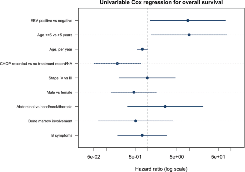
Supplementary Figure 1. Univariable Cox regression for overall survival. Hazard ratios are presented with 95% confidence intervals; estimates are exploratory because of the small cohort and low number of events. Epstein–Barr virus in paediatric sporadic Burkitt lymphoma: real-world experience from a Peruvian national referral centre