Quality indicators of pathology and laboratory medicine in cancer: a narrative synthesis of published literature
Chase Peng Yun Ng1, Asmita Raja2, Alicia Hunter2, Sai Kalpitha Eragamreddy2, Grace Tan3, Shahin Sayed4, Looi Lai-Meng5, Leke Folaranmi6, Beatriz Hornburg7, Meritxell Mallafre-Larrosa7, Julie Torode1, Ajay Aggarwal1, Kenneth Fleming8* and Richard Sullivan1*
1Institute of Cancer Policy, Kings College London, WC2R 2LS London, UK
2Imperial College Healthcare NHS Trust, W2 1NY London, UK
3University College London Hospitals NHS Foundation Trust, NW1 2PG London, UK
4Department of Pathology, The Aga Khan University, Nairobi 00100, Kenya
5Department of Pathology, Faculty of Medicine, University of Malaya, Kuala Lumpur 50603, Malaysia
6Department of Pathology, University of Ilorin Teaching Hospital, Ilorin 241102, Nigeria
7City Cancer Challenge, 1204 Geneva, Switzerland
8Green Templeton College, University of Oxford, OX2 6HG Oxford, UK
Abstract
Purpose: With the rise in global cancer incidence, inadequate access to pathology and laboratory medicine (PALM) is one of the largest gaps in the cascade of cancer care, especially in low- and lower-middle-income countries. Quality indicators (QIs) are essential to evaluate the accuracy, completeness, timeliness and cost-effectiveness of PALM services to ensure access and quality. To identify strengths and gaps, this review systematically identifies and synthesises published PALM-specific cancer QIs across cancer types and geography.
Methods: Following Preferred Reporting Items for Systematic reviews and Meta-analyses guidelines, a search of PubMed inclusive of Medline between 1990 and 2024 was conducted for studies that developed, applied or evaluated QIs for anatomical and molecular cancer pathology. Studies limited to chemopathology, immunopathology, haematopathology, haemato-oncology and paediatric oncology were excluded. Risk of bias was assessed using the Risk Of Bias In Non-randomised Studies - of Interventions tool.
Results: Of 8,685 records screened, 40 (0.5%) studies were included; 24 (61.5%) had moderate risk of bias. Nearly all the studies (97.5%) originated from high- or upper-middle-income countries, predominantly Europe and North America. Gynaecological and breast cancers were most frequently studied (n = 12 and 10), while prostate and lung cancers were underrepresented (n = 4 and 1, respectively). Most QIs focussed on diagnostic accuracy in the analytic phase (n = 26). Very few QIs assessed timeliness or cost-effectiveness (n = 2 and 3), particularly in pre-analytic and post-analytic phases.
Conclusion: Evidence for PALM-specific cancer QIs for timeliness and cost-effectiveness, in pre- and post-analytic phases, and across resource-limited settings is deficient. To facilitate global harmonisation, stakeholders from all resource settings should review international guidance to standardise QIs that are feasible, applicable and stratified to resource level. At the national level, Delphi processes are recommended to develop context-specific QIs, which inform national diagnostic strategy and cancer control planning.
Keywords : quality indicator, global oncology, pathology, laboratory medicine, cancer
Correspondence to: Chase Peng Yun Ng
Email: peng.ng3@nhs.net
Published: 06/10/2026
Received: 09/05/2026
Publication costs for this article were supported by ecancer (UK Charity number 1176307).
Copyright: © the authors; licensee ecancermedicalscience. This is an Open Access article distributed under the terms of the Creative Commons Attribution License (http://creativecommons.org/licenses/by/4.0), which permits unrestricted use, distribution, and reproduction in any medium, provided the original work is properly cited.
Introduction
Pathology and laboratory medicine (PALM) is fundamental to cancer care, underpinning diagnosis, staging, treatment selection and prognostication. Three intertwined aspects are necessary: access to PALM services, the quality of those services and the ability to measure and monitor that quality.
First, access to PALM across the cascade of cancer care remains highly inequitable. As the global cancer incidence rises – projected to reach 30.2 million new cases and 16.3 million cancer-related deaths by 2040 – health systems are increasingly strained [1, 2]. In low-income countries (LICs), up to 62% lack access to essential cancer diagnostics [3]. Despite long-standing recognition of this gap, progress has been slow. Limited visibility and low prioritisation of diagnostics from research to advocacy, alongside constrained infrastructure and workforce capacity, continue to limit access.[4] Only one in eight pathologists reported to have routine access to immunohistochemical (IHC) tests and none to molecular testing for female genitourinary cancers in LICs, despite their inclusion in the World Health Organisation (WHO) essential diagnostic list [5]. This nosologic shortcoming calls into question the feasibility and applicability of the diagnostic essential criteria.
Access to PALM needs to be accompanied by quality. The quality of PALM services can be defined by diagnostic accuracy, completeness, timeliness and cost-effectiveness of the diagnostics. The lack of quality can lead to misdiagnosis, delayed treatment, inappropriate therapy selection and increased healthcare costs, ultimately undermining patient outcomes even where services are available. Therefore, it is critical that the quality of PALM is being measured credibly, yet this remains an underdeveloped area. Quality indicators (QIs) are defined, measurable metrics with the purpose of evaluating or benchmarking the outcome, process, or structure of care for a particular type of patient or clinical condition [6]. QIs should capture variability across the entire testing process – from pre-analytic (specimen collection and handling), through analytic (testing and interpretation), to post-analytic (result reporting) phases. As such, QIs are essential instruments for governance, benchmarking and health system strengthening.
Importantly, cancer-specific PALM QIs present unique challenges that are not fully addressed by general laboratory QIs. Cancer PALM is particularly complex, integrating morphologic evaluation, tumour staging, and increasingly molecular and genomic profiling, all of which are required urgently and all of which directly influence clinical decisions and prognosis. Despite their importance, there is a paucity of evidence on the applicability of cancer-specific PALM QIs in different resource settings. Now is an apposite time to identify and characterise existing cancer-specific PALM QIs across geography and cancer type, as innovations, such as point-of-care diagnostics which democratise diagnostics; digital pathology, which promotes telepathology and facilitates training; and artificial intelligence, especially natural language processing which helps automate and structure pathology reporting, are emerging, collectively transforming the global cancer diagnostic landscape [7–9]. This narrative synthesis aims to inform the development of a more integrated, context-sensitive approach to QI design and application in future global cancer diagnostics.
Methods
The systematic review was conducted following the Preferred Reporting Items for Systematic reviews and Meta-analyses (PRISMA) guidelines. Studies published between 01/01/1990 and 31/12/2024 were searched on PubMed, inclusive of Medline. The core search terms used to identify relevant publications were: pathology, cancer and QI, with their respective expanded search strings included in the full search strategy (Appendix A).
Inclusion criteria
Studies focusing on anatomic pathology (histopathology, cytology and immunohistochemistry) and molecular pathology, written in English and focused on human adults, were considered. Publications were included if they reported measurable QIs used for evaluating or benchmarking outcomes, process or structure of cancer-related PALM services.
Exclusion criteria
Studies were excluded if they focused on chemical pathology, immunopathology or haematopathology. Although anatomical pathology includes autopsy and forensic investigation, they are excluded in this review given non-relevance to cancer. Non-primary research publications including review articles,
letters, conference proceedings, expert commentaries, non-clinical laboratory studies, protocols, case reports, editorials and pre-clinical studies were excluded. Research focused on paediatric oncology and haemato-oncology was also excluded.
Data selection
The titles and abstracts were assessed and screened by three reviewers (AR, AH and ER) to minimise reviewer errors and bias after records were identified. Full-text articles were then assessed for eligibility, and data were extracted by the three independent reviewers (AR, AH, ER). Inter-reviewer agreement was high (88%–95% across screening stages), although not formally quantified using statistical measures. Disagreements were resolved qualitatively through consensus with arbitration by senior authors (PN and RS).
Data extraction
The following variables were collected: author; reference year; country; study characteristics (including funding, research design and sample size); cancer type; domain of pathology (histopathology, cytopathology or molecular pathology); and the phase of pathology testing (pre-analytic, analytic or post-analytic). In addition, we extracted the aims and reported outcomes of the study, the purpose of the QIs (timeliness, completeness, accuracy or cost-effectiveness) and the specific QIs assessed.
Synthesis method
Given the heterogeneity of study designs, cancer types and QIs, a narrative synthesis was undertaken. Extracted data were organised thematically according to the domain of pathology, the phase of pathology testing and the stated purpose of QIs (timeliness, completeness, accuracy, cost-effectiveness). Findings were then synthesised to identify common patterns and gaps across studies.
Risk of bias assessment
Given the heterogeneity in the design of the included studies, no single validated risk-of-bias tool can be applied consistently across all the studies without losing validity or comparability [10]. The Risk Of Bias In Non-randomised Studies - of Interventions (ROBINS-I) tool was selected to assess methodological quality as it adopts a domain-based structure to enable consistent assessment of bias of all studies. The assessment focused on seven domains of bias: confounding, selection of participants, classification of intervention, deviations from intended interventions, missing data, measurement of outcomes and selection of reported results. Each domain was rated as low, moderate or not applicable (NA), with an overall judgment of risk of bias provided for each study. For studies without a defined intervention, domains relating to intervention and deviations from intended interventions (Domains 3 and 4) were NA and were therefore not assessed.
Results
Search strategy
Eight thousand six hundred and eighty-five studies were initially identified using PubMed, inclusive of Medline. Four hundred and one studies were deemed eligible after titles and abstracts were screened using the inclusion criteria. Three hundred and sixty-one studies were excluded according to the exclusion criteria, leaving 40 publications selected for inclusion, as illustrated in the PRISMA flow chart in Figure 1. The summary of all included studies is attached as Supplementary Table S1 (Appendix B).
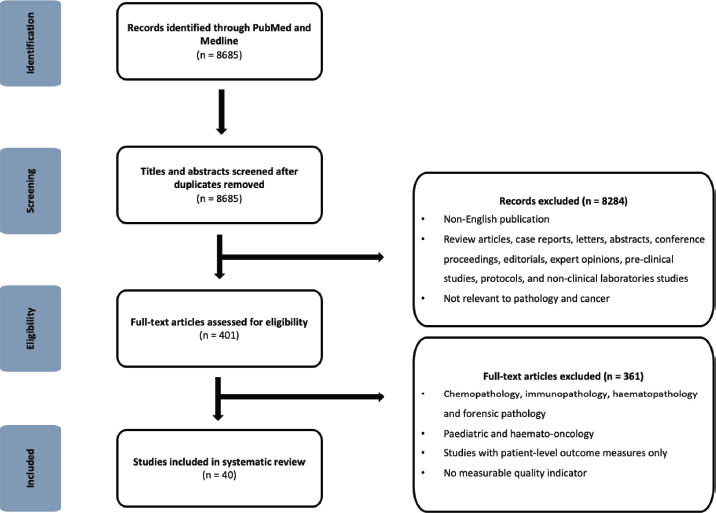
Figure 1. PRISMA flowchart of study selection.
Quality assessment of studies
Thirty-nine non-randomised studies were evaluated using the ROBINS-I tool. The breakdown and outcome of risk of bias assessment of each study was included as Supplementary Table S2 (Appendix C) and a summary of each domain of bias is displayed in a bar chart, as Supplementary Figure 1 (Appendix D). One study was not included in this assessment as it was a health economics study [11].
Twenty four (61.5%) studies were rated as having moderate overall risk of bias [13, 15, 18, 19, 21–23, 25–29, 34, 38–43, 45–49], primarily falling short due to confounders (n = 15, 38.5%) and/or missing data (n = 11, 28.2%). The remaining 15 (38.5%) studies were deemed to have low overall risk of bias, meaning that their risk is comparable to that of a well-conducted randomised trial [12, 14, 16, 17, 20, 24, 30–33, 35–37, 44, 50].
Location of studies
Thirty-nine of 40 (97.5%) of the studies were published in upper-middle-income countries (UMIC) or high-income countries (HIC) [11–26, 28–50]. The one study published in LMIC was from India [27]. Europe led as a continent overall, publishing 23 out of 40 (57.5%) of the studies [11, 13, 14, 16, 18, 19, 21–25, 28–34, 37, 40, 49, 50]; followed by North America, 13 (32.5%) [12, 17, 20, 34–36, 38, 39, 42, 44–47]. There were only three studies originated from Asia [15, 27, 41]; and none from Latin America or Africa. The United Kingdom was the most prolific country with eight studies (20%), while the United States of America was second with seven studies (17.5%) published. Canada and Italy both published six studies (15%), respectively (Figure 2).
Type of studies
Thirty-four of 40 studies (85%) were retrospective observational studies [13–18, 20–33, 35, 36, 38–49]; five (n = 12.5%), prospective observational studies [12, 19, 34, 37, 50]; and one (n = 2.5%), health economics study [11].
Cancer type
The number of studies varied significantly by cancer type, with gynaecological cancers representing the largest proportion (n = 12, 30%), as illustrated in Figure 3 [11–13, 15, 16, 19, 21, 22, 25, 27, 28, 37]. This was closely followed by breast cancer (n = 10, 25%) [17, 20, 23, 31, 33, 41–43, 48, 51]. Four (10%) of the studies focused on prostate cancer [14, 18, 34, 47]; three (7.5%), on colorectal cancer [29, 30, 47]; and three on skin cancers [24, 26, 45]. Two (5%) studies were on upper gastrointestinal cancer [35, 38], while only one (2.5%) study examines thyroid cancer [39], kidney cancer [34], adrenal cancer [40], and lung cancer [50]. Three (7.5%) of the studies addressed broader pathology quality metrics without focusing on a particular cancer type [36, 46, 49].
Purpose of QIs
The heatmap in Figure 4 reveals the distribution of QIs across the pre-analytic, analytic and post-analytic phases of pathology testing for cancer. The purpose of the QIs was predominantly to assess the accuracy of the pathology test, particularly in the analytic phase (n = 26, 65%), followed by its completeness. There are notable gaps in QIs focussing on the timeliness of pathology tests, with only two studies (5%) in the analytic phase and none reported for pre- and post-analytic phases. QIs focussing on cost-effectiveness of pathology tests were uniformly sparse, with one study (2.5%) per phase. The Sankey diagram, as illustrated in Figure 5, further delineates the flow of QIs from their pathology phases to respective sub-phases.
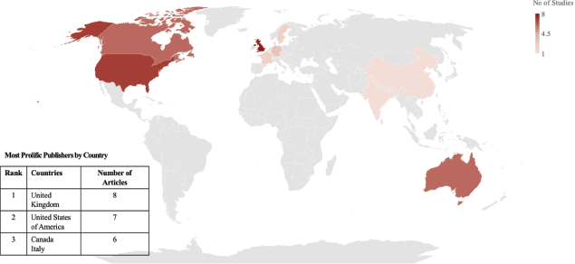
Figure 2. Global heatmap of studies related to pathology QIs in cancer.
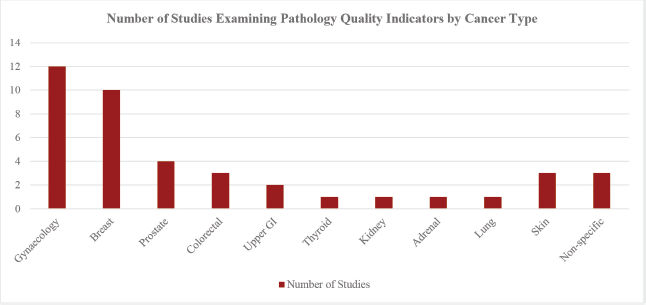
Figure 3. Barchart of studies examining pathology QIs by cancer type.
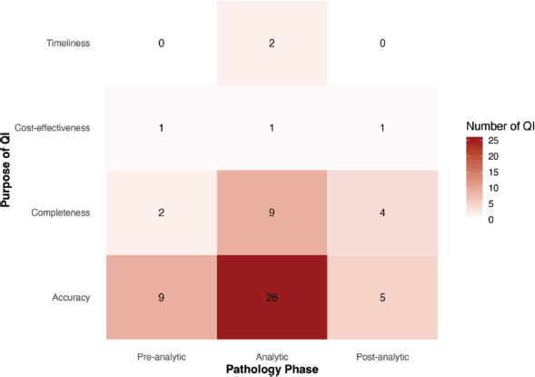
Figure 4. Heatmap of pathology QIs by purpose across phases of pathology testing.
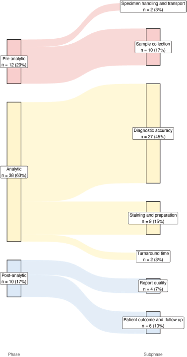
Figure 5. Sankey diagram depicting flow of QI from pathology phases to sub-phases. Line width represents QI density.
QIs by phase of testing
The array of pathology QIs was categorised based on their phase of pathology testing, as shown in Figure 6.
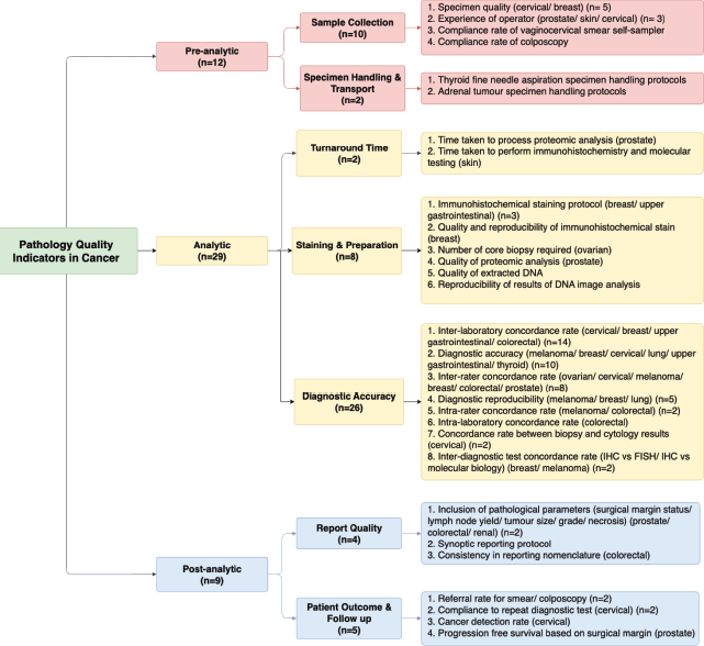
Figure 6. QIs in cancer pathology by phase of testing.
Pre-analytic
Five of the studies included QIs that ascertain the quality of specimen collected (vaginal, cervical and breast) [16, 23, 25, 28, 37]. Three focused instead on the experience of the operator who collected the specimens (prostatic, skin and cervical) [18, 26, 27]. One study measured the compliance rate of self-samplers for cervical smear [21]. Two studies employed QIs in assessing the adherence of protocol surrounding specimen handling for thyroid and adrenal lesions [39, 40].
Analytic
Only two studies assessed the turnaround time of pathology testing- one specifically on time taken to process proteomic analysis in prostatic specimens [14], while the other on time taken to perform IHC and molecular testing for skin biopsy with suspected melanoma [24]. Three of the studies looked at adherence to IHC staining protocol for breast and upper gastrointestinal specimens [32, 33, 35]. Quality and reproducibility of IHC, proteomic analysis, DNA image analysis, as well as the number of core biopsies required for tumour representation were also identified as QIs relevant to specimen preparation [12, 14, 41, 43, 46, 49].
The diagnostic accuracy of various pathology tests was the most studied QIs (n = 26, 65%). Inter-laboratory concordance rate of pathology results was the most commonly used QI, applied across cervical, breast, upper gastrointestinal and colorectal cancer [15–17, 20, 29, 31, 33, 35, 36, 42, 43, 48]. This is followed by the inter-rater concordance rate [12, 17, 30, 33, 34, 37, 45, 51] and the reproducibility of cancer diagnosis based on pathology tests [45, 48–50]. Concordance rates were also measured between different types of specimen (i.e., biopsy versus cytology) [15, 28], different diagnostic tests (i.e., IHC versus molecular testing versus fluorescence in situ hybridisation) [17, 20, 24] and within a laboratory but across different timepoints [16, 29].
Post-analytic
Four studies zeroed in on the quality of pathology reports [29, 38, 44, 47]; two specifically on the important pathological parameters including surgical margin status, lymph node yield, tumour size, tumour grade and other prognostic factors [44, 47]. The other two studies assessed the adherence to the synoptic reporting protocol and consistency in nomenclature, respectively [29, 38]. Two studies included referral rates for a repeat diagnostic test and/or another diagnostic test, while two included the compliance rate to a repeat diagnostic test [13, 22]. Long-term cancer detection rate, number of cancers averted and progression free survival based on surgical margin of prostate specimen were used as QIs in one study each [11, 19, 34]..
Discussion
This narrative synthesis identified a limited body of published evidence on QIs in cancer pathology, with only 40 studies (less than 0.5% of screened literature) directly evaluating or applying QIs. More than half (61.5%) of these studies were assessed as having low to moderate risk of bias (61.5%), reflecting variability in study design and reporting quality. The limited volume of QI-focused research reflects the historically low visibility and prioritisation of PALM within health systems [4]. PALM only accounts 3%–6% of overall healthcare expenditure and their professionals make up around 4% of the workforce in most healthcare systems [52]. This issue is critical as it may have implications for regulation for both laboratory accreditation and workforce professional standards and competencies in PALM.
In addition, there was a marked geographic disparity in research activity, with 97.5% of studies originating from high- and UMIC. This imbalance likely reflects the broader patterns of the trickle-down model in PALM, where innovations and quality frameworks are often developed in HICs before trickling down to the LMICs. The mismatch between available QIs and the needs of LMICs makes the countries vulnerable to suboptimal implementation and evaluation of PALM services. To overcome this, collaborative comparative studies on QIs between HIC and LMIC laboratories would provide insight into gaps within the country-specific context and should be encouraged.
Another observation is that QIs predominantly focused on diagnostic accuracy and completeness, with limited attention to timeliness and cost-effectiveness. While accuracy and completeness are essential, delays in diagnosis and inefficiencies in resource utilisation may significantly affect patient outcomes. The turnaround time and the cost-effectiveness of PALM are vital in assessing if the system caters to the needs of the poor, rural and marginalised communities in resource-limited settings. From a broader health systems perspective, expanding QIs to measure timeliness and cost-effectiveness of PALM would inform policymakers of bottlenecks in cancer diagnostics so resources could be allocated more transparently and efficiently between infrastructure and workforce capacity building. This might in turn strengthen accountability mechanisms and governance, the lack of which are recognised to impair delivery of PALM [53, 54].
In addition, QIs were unevenly distributed across cancer types, with greater representation in breast and gynaecological cancers compared to other high-burden cancers such as lung and prostate cancer [55]. This may be attributable to longstanding global investments in screening and early detection programmes, given that at least 15% of such investment was directly related to cervical and/or breast cancer screening programmes. This illustrates how coordinated international initiatives could drive positive downstream research and development of cancer diagnostics, fostering a cycle of innovation.
Recommendations for development of context-specific QIs in cancer pathology
A credible QI of cancer PALM should directly assess four aspects: the validity, reliability, timeliness and/or cost-effectiveness of a diagnostic service, across the three phases described above [56]. The validity of a diagnostic service should be based on clinical relevance (i.e., diagnostic accuracy, treatment selection and prognostication) and content validity, meaning measuring what it purports to measure. Its reliability should reflect operational clarity, from standardisation of procedures to synoptic reporting, to ensure completeness and to minimise subjective interpretation and discrepancy across observers, systems and time. The timeliness of a diagnostic service should be measurable and actionable, enabling prompt quality improvement action through near-real-time monitoring. Its cost-effectiveness should be modelled based on the benefit-cost ratio of the diagnostics.
On a health system level, the QIs to assess the above should be feasible, benchmarkable and sensitive so that the data collected can be utilised for public health epidemiological purposes, informing policy, regulation, financing, infrastructure and workforce planning. Last and most importantly, the benchmark set should be professionally endorsed by key stakeholders across the hierarchy of pathology services, from lab technicians and pathologists to surgeons, oncologists, industry, regulators and policymakers.
Based on the findings, we recommend a two-step approach to support the development and implementation of context-specific QIs in cancer-specific PALM. First, a multi-stakeholder international partnership is required to marshal the effort to establish a core set of global QIs for cancer pathology. The partnership should include international agencies such as the WHO, International Agency for Research on Cancer, International Organisation for Standards, professional pathology societies and Ministries of Health. To achieve global synoptic reporting and standardised cancer datasets, greater inclusion of LMIC stakeholders is essential to ensure feasibility and applicability of QIs across diverse health systems, thus facilitating regional harmonisation of cancer pathology. Stakeholders should review the existing international standards, such as the WHO Tumour Board for Cancer Staging and Classification, and the International Collaboration on Cancer Reporting [5, 57, 58].
Second, the QIs adopted should be adapted to local contexts based on country-specific health systems, including disease burden, resource availability and service delivery model. In this regard, lessons can be learnt from the recent implementation of the first national essential diagnostics list by India, which emulated the WHO diagnostic list but was adapted to meet Indian health needs through inclusion of diagnostic imaging [59]. The authors encouraged a non-hierarchical, consensus-based, participatory decision-making process involving key stakeholders across the diagnostic pathway in the co-development of the QIs. The QIs should be evidence-based, integrable and tiered to specific local, regional and national cancer networks, catering to the delivery of services grouped in specialised units or provided through a hub-and-spoke model. The final consensus could subsequently inform the External Quality Assurance Programme and be integrated into Managed Equipment Service Partnerships to on-board industry. The data would also be valuable in informing and monitoring national cancer control strategies.
Limitations
This review has several limitations. Firstly, this review is restricted to QIs reported in the literature and may under-represent QIs embedded within institutional audit systems or routine quality assurance processes. As discussed, this reflects a broader disconnect between operational quality evaluation and academic reporting. This gap limits the ability to translate operational practices into evidence, constraining the development, standardisation and scale-up of QIs across health systems. Secondly, this review did not focus on the contextual determinants that influence pathology quality, such as infrastructure, geographical access to diagnostic services, health system financing, laboratory information systems, as well as workforce capacity, which have been examined in-depth in existing literature [52, 60–62]. Thirdly, the use of a single PubMed/Medline database may have excluded relevant studies indexed in other databases, particularly those representing regional or LMIC-focused literature. Fourthly, the restriction to English-language publications may contribute to geographic bias and underrepresentation of evidence from non-English-speaking settings. Lastly, flow cytometry was not included, as its primary application lies in haematological malignancies. While it has some applications in non-haematological cancers such as DNA ploidy, cell cycle analysis and circulating tumour cells, these are not routine components of anatomic pathology practice for cancer diagnosis.
Conclusion
QIs in cancer PALM are deficient in the published literature, particularly in resource-limited settings. QIs assessing timeliness and cost-effectiveness in pre- and post-analytic phases of PALM are under-represented.
To facilitate global harmonisation of cancer pathology, international collaboration is required to standardise QIs that are feasible, applicable and stratified to resource level. Additionally, we recommend national efforts to undertake Delphi processes to develop context-specific QIs that are integrable into local workflow, address local health needs and inform national diagnostic strategy and cancer control planning.
Acknowledgment
RS and PN are funded by the Medical Research Council Global Alliance of Chronic Disease Grant ACCI No GACD-025. RS and PN are also funded by the BASO/Rosetrees Research Grant in Cancer Surgery. The funders have no role in study design; in the collection, analysis and interpretation of data; in the writing of the manuscript; and in the decision to submit the manuscript for publication.
Conflicts of interest
Not applicable.
Author contributions
PN, RS, KF conceived, designed and supervised the study. AR, AH and SKE searched, screened and assessed the publications. GT performed risk of bias assessment. PN analysed and visualised the data. RS and KF accessed and verified the data. PN drafted the manuscript. RS, KF, LF, SS, MM, BH and LL reviewed and edited the manuscript. All authors read and approved the final version.
References
1. National Cancer Institute (2025) Cancer statistics [Internet] [https://www.cancer.gov/about-cancer/understanding/statistics#:~:text=The%20rate%20of%20new%20cases,on%202013%E2%80%932017%20deaths
)] Date accessed: 01/03/2026
2. World Health Organisation, International Agency for Research on Cancer (2026) Estimated number of new cases from 2022 to 2045, both sexes, age [0-85+] [Internet] [https://gco.iarc.fr/tomorrow/en/dataviz/isotype?types=0&single_unit=500000] Date accessed: 01/03/2026
3. Cotache-Condor C, Rice HE, and Schroeder K, et al (2023) Delays in cancer care for children in low-income and middle-income countries: development of a composite vulnerability index Lancet Global Health 11(4) e505–e515 https://doi.org/10.1016/S2214-109X(23)00053-0 PMID: 36925171 PMCID: 10938288
4. Fleming KA, Horton S, and Wilson ML, et al (2021) The Lancet Commission on diagnostics: transforming access to diagnostics Lancet 398(10315) 1997–2050 https://doi.org/10.1016/S0140-6736(21)00673-5 PMID: 34626542 PMCID: 8494468
5. Rabban JT, Wadee R, and Plotkin A, et al (2025) International feasibility of applying diagnostic essential criteria of the fifth edition World Health Organization classification of female genital tumors: evidence for a global gap in health care equity Modern Pathol 38(9) 100798 https://doi.org/10.1016/j.modpat.2025.100798
6. Schuster MA, MC Glynn EA, and Brook RH (2005) How good is he quality of health care in the United States? Milbank Quart 83(4) 843–895 https://doi.org/10.1111/j.1468-0009.2005.00403.x
7. Bose R and Saxon LA (2019) The democratization of diagnosis: bringing the power of medical diagnosis to the masses EClinicalMedicine 8 6–7 https://doi.org/10.1016/j.eclinm.2019.03.004 PMID: 31193676 PMCID: 6537544
8. Razzano D, Puranam K, and Tomoka T, et al (2022) The role of telepathology in improving cancer diagnostic and research capacity in sub-Saharan Africa Front Med 9 978245 https://doi.org/10.3389/fmed.2022.978245
9. Park P, Choi Y, and Han N, et al (2025) Leveraging natural language processing for efficient information extraction from breast cancer pathology reports: single-institution study PLoS One 20(2) 318726 [https://doi.org/10.1371/journal.pone.0318726]
10. Sterne JA, Hernán MA, and Reeves BC, et al (2016) ROBINS-I: a tool for assessing risk of bias in non-randomised studies of interventions BMJ 4919 https://doi.org/10.1136/bmj.i4919
11. Legood R, Smith M, and Lew JB, et al (2012) Cost effectiveness of human papillomavirus test of cure after treatment for cervical intraepithelial neoplasia in England: economic analysis from NHS Sentinel Sites Study BMJ 345 7086 https://doi.org/10.1136/bmj.e7086
12. Hecht JL, Kotsopoulos J, and Gates MA, et al (2008) Validation of tissue microarray technology in ovarian cancer: results from the Nurses’ Health Study Cancer Epidemiol Biomark Prev 17(11) 3043–3050 https://doi.org/10.1158/1055-9965.EPI-08-0645
13. Ronco G, Ballegooijen MV, and Becker N, et al (2009) Process performance of cervical screening programmes in Europe Eur J Cancer 45(15) 2659–2670 https://doi.org/10.1016/j.ejca.2009.07.022 PMID: 19713100
14. Lexander H, Hellman U, and Palmberg C, et al (2006) Evaluation of two sample preparation methods for prostate proteome analysis Proteomics 6(13) 3918–3925 https://doi.org/10.1002/pmic.200600104 PMID: 16767792
15. Korach J, Menczer J, and Lusky A (1995) A comparison between referral and hospital cervical cytology laboratory reports Isr J Med Sci 31(6) 352–355 PMID: 7607854
16. Stanczuk G, Baxter G, and Currie H, et al (2016) Clinical validation of hrHPV testing on vaginal and urine self-samples in primary cervical screening (cross-sectional results from the Papillomavirus Dumfries and Galloway—PaVDaG study) BMJ Open 6(4) 10660 https://doi.org/10.1136/bmjopen-2015-010660
17. Diaz LK, Gupta R, and Kidwai N, et al (2004) The use of TMA for interlaboratory validation of FISH testing for detection of HER2 gene amplification in breast cancer J Histochem Cytochem 52(4) 501–507 https://doi.org/10.1177/002215540405200408 PMID: 15034001
18. Stabile A, Dell’Oglio P, and Gandaglia G, et al (2018) Not all multiparametric magnetic resonance imaging–targeted biopsies are equal: the impact of the type of approach and operator expertise on the detection of clinically significant prostate cancer Eur Urol Oncol 1(2) 120–128 https://doi.org/10.1016/j.euo.2018.02.002
19. Dillner J, Rebolj M, and Birembaut P, et al (2008) Long term predictive values of cytology and human papillomavirus testing in cervical cancer screening: joint European cohort study BMJ 337 1754 https://doi.org/10.1136/bmj.a1754
20. Fitzgibbons PL, Murphy DA, and Dorfman DM, et al (2006) Interlaboratory comparison of immunohistochemical testing for HER2: results of the 2004 and 2005 College of American Pathologists HER2 immunohistochemistry tissue microarray survey Arch Pathol Lab Med 130(10) 1440–2145 https://doi.org/10.5858/2006-130-1440-ICOITF PMID: 17090184
21. Gok M, Heideman DAM, and Van Kemenade FJ, et al (2010) HPV testing on self collected cervicovaginal lavage specimens as screening method for women who do not attend cervical screening: cohort study BMJ 340 c1040 https://doi.org/10.1136/bmj.c1040 PMID: 20223872 PMCID: 2837143
22. Ronco G, Giubilato P, and Naldoni C, et al (2006) Activity level and process indicators of organised programmes for cervical cancer screening in Italy Epidemiol Prev 30(1 Suppl 3) 27–40 PMID: 16937844
23. Anania G, Bazzocchi M, and Di Loreto C, et al (1997) Percutaneous large core needle biopsy versus surgical biopsy in the diagnosis of breast lesions Int Surg 82(1) 52–55 PMID: 9189803
24. Long E, Ilie M, and Lassalle S, et al (2015) Why and how immunohistochemistry should now be used to screen for the BRAFV 600E status in metastatic melanoma? The experience of a single institution (LCEP, Nice, France) Acad Dermatol Venereol 29(12) 2436–2443 https://doi.org/10.1111/jdv.13332
25. Harrison WN, Mohammed MA, and Wall MK, et al (2004) Analysis of inadequate cervical smears using Shewhart control charts BMC Public Health 29(1) 25 https://doi.org/10.1186/1471-2458-4-25
26. Keir J (2007) Diagnosing skin cancer in primary care: how do mainstream general practitioners compare with primary care skin cancer clinic doctors? Med J Aust 187(4) 215–220 https://doi.org/10.5694/j.1326-5377.2007.tb01202.x
27. Kottke TE and Trapp MA (1998) The quality of Pap test specimens collected by nurses in a breast and cervical cancer screening clinic Am J Prev Med 14(3) 196–200 https://doi.org/10.1016/S0749-3797(97)00062-7 PMID: 9569220
28. Orumaa M, Innos K, and Suurna M, et al (2022) quality assessment of cervical cytology practices in Estonia from 2007 to 2018 Cancer Control 29 10732748221141794 https://doi.org/10.1177/10732748221141794
29. Deans ZC, Tull J, and Beighton G, et al (2011) Molecular genetics external quality assessment pilot scheme for KRAS analysis in metastatic colorectal cancer Genet Test Mol Biomarkers 15(11) 777–783 https://doi.org/10.1089/gtmb.2010.0239 PMID: 21851273
30. Risio M, Bussolati G, and Senore C, et al (2010) Virtual microscopy for histology quality assurance of screen-detected polyps J Clin Pathol 63(10) 916–920 https://doi.org/10.1136/jcp.2010.080580 PMID: 20876325
31. Ellison G, Wallace A, and Kohlmann A, et al (2017) A comparative study of germline BRCA1 and BRCA2 mutation screening methods in use in 20 European clinical diagnostic laboratories Br J Cancer 117(5) 710–716 https://doi.org/10.1038/bjc.2017.223 PMID: 28751759 PMCID: 5572178
32. Wells JC, Sharma S, and Del Paggio JC, et al (2021) An analysis of contemporary oncology randomized clinical trials from low/middle-income vs high-income countries JAMA Oncol 7(3) 379 https://doi.org/10.1001/jamaoncol.2020.7478 PMID: 33507236 PMCID: 7844695
33. Von Wasielewski R, Mengel M, and Wiese B, et al (2002) Tissue array technology for testing interlaboratory and interobserver reproducibility of immunohistochemical estrogen receptor analysis in a large multicenter trial Am J Clin Pathol 118(5) 675–682 https://doi.org/10.1309/URLK-6AVK-331U-0V5P PMID: 12428786
34. Van Der Kwast TH, Collette L, and Van Poppel H, et al (2006) Impact of pathology review of stage and margin status of radical prostatectomy specimens (EORTC trial 22911) Virchows Arch 449(4) 428–434 https://doi.org/10.1007/s00428-006-0254-x PMID: 16941153
35. Sheffield BS, Garratt J, and Kalloger SE, et al (2014) HER2/neu testing in gastric cancer by immunohistochemistry: assessment of interlaboratory variation Arch Pathol Lab Med 138(11) 1495–1502 https://doi.org/10.5858/arpa.2013-0604-OA PMID: 25357111
36. Hsu FD, Nielsen TO, and Alkushi A, et al (2002) Tissue microarrays are an effective quality assurance tool for diagnostic immunohistochemistry Mod Pathol 15(12) 1374–1380 https://doi.org/10.1097/01.MP.0000039571.02827.CE PMID: 12481020
37. Ciatto S, Cariaggi MP, and Minuti AP, et al (1996) Interlaboratory reproducibility in reporting inadequate cervical smears—a multicentre multinational study Cytopathology 7(6) 386–390 https://doi.org/10.1111/j.1365-2303.1996.tb00543.x PMID: 8958471
38. Decoste R, Mcdonell LM, and Uzans D, et al (2020) Comparing pathology report quality indicators in 2 distinct Whipple resection specimen protocols Pancreas 49(6) 788–792 https://doi.org/10.1097/MPA.0000000000001574 PMID: 32541634
39. Scurry JP and Duggan MA (2000) Thin layer compared to direct smear in thyroid fine needle aspiration Cytopathology 11(2) 104–115 https://doi.org/10.1046/j.1365-2303.2000.00228.x PMID: 10772010
40. Johnsen IK, Hahner S, and Brière JJ, et al (2010) Evaluation of a standardized protocol for processing adrenal tumor samples: preparation for a European adrenal tumor bank Horm Metab Res 42(02) 93–101 https://doi.org/10.1055/s-0029-1241858
41. Wang YH, Lai CR, and Lien HC, et al (2020) Good staining quality ensuring the reproducibility of Ki67 assessment J Clin Pathol 73(7) 413–417 https://doi.org/10.1136/jclinpath-2019-206205
42. Parker RL, Huntsman DG, and Lesack DW, et al (2002) Assessment of interlaboratory variation in the immunohistochemical determination of estrogen receptor status using a breast cancer tissue microarray Am J Clin Pathol 117(5) 723–728 https://doi.org/10.1309/PEF8-GL6F-YWMC-AG56 PMID: 12090420
43. Regitnig P, Reiner A, and Dinges HP, et al (2002) Quality assurance for detection of estrogen and progesterone receptors by immunohistochemistry in Austrian pathology laboratories Virchows Archiv 441(4) 328–334 https://doi.org/10.1007/s00428-002-0646-5 PMID: 12404057
44. Shuch B, Pantuck AJ, and Pouliot F, et al (2011) Quality of pathological reporting for renal cell cancer: implications for systemic therapy, prognostication and surveillance BJU Int 108(3) 343–348 https://doi.org/10.1111/j.1464-410X.2010.09871.x
45. Elmore JG, Barnhill RL, and Elder DE, et al (2017) Pathologists’ diagnosis of invasive melanoma and melanocytic proliferations: observer accuracy and reproducibility study BMJ 2813 https://doi.org/10.1136/bmj.j2813
46. Hwang DH, Garcia EP, and Ducar MD, et al (2017) Next‐generation sequencing of cytologic preparations: an analysis of quality metrics Cancer Cytopathol 125(10) 786–794 https://doi.org/10.1002/cncy.21897 PMID: 28727266
47. McFadyen C, Lankshear S, and Divaris D, et al (2015) Physician level reporting of surgical and pathology performance indicators: a regional study to assess feasibility and impact on quality CJS 58(1) 31–40 https://doi.org/10.1503/cjs.004314
48. Wang M, Mclaren S, and Jeyathevan R, et al (2019) Laboratory validation studies in Ki-67 digital image analysis of breast carcinoma: a pathway to routine quality assurance Pathology 51(3) 246–252 https://doi.org/10.1016/j.pathol.2018.12.416 PMID: 30850279
49. Thunnissen FBJM, Ellis IO, and Jütting U (1997) Quality assurance in DNA image analysis on diploid cells Cytometry 27(1) 21–25 https://doi.org/10.1002/(SICI)1097-0320(19970101)27:1<21::AID-CYTO3>3.0.CO;2-O PMID: 9000581
50. Deans ZC, Bilbe N, and O’Sullivan B, et al (2013) Improvement in the quality of molecular analysis of EGFR in non-small-cell lung cancer detected by three rounds of external quality assessment J Clin Pathol 66(4) 319–325 https://doi.org/10.1136/jclinpath-2012-201227 PMID: 23378269
51. Wells CA, Sloane JP, and Coleman D, et al (2004) Consistency of staining and reporting of oestrogen receptor immunocytochemistry within the European Union—an inter-laboratory study Virchows Arch 445(2) 119–128 https://doi.org/10.1007/s00428-004-1063-8 PMID: 15221370
52. Horton S, Sullivan R, and Flanigan J, et al (2018) Delivering modern, high-quality, affordable pathology and laboratory medicine to low-income and middle-income countries: a call to action Lancet 391(10133) 1953–1964 https://doi.org/10.1016/S0140-6736(18)30460-4 PMID: 29550030
53. Gadre A, Shukla A (2016) Dissenting Diagnosis: Voices of Conscience from the Medical Profession (Penguin Books India).
54. Rose-Ackerman S and Tan Y (2014) Corruption in the procurement of pharmaceuticals and medical equipment in China: the incentives facing multinationals, domestic firms and hospital officials UCLA Pac Basin Law J 32(1) [https://doi.org/10.5070/P8321028003]
55. International Agency for Research on Cancer (2022), World Health Organisation Age-standardized rate (World) per 100 000, incidence and mortality, both sexes, in 2022 [Internet]. [https://gco.iarc.fr/today/en/dataviz/bars?types=0_1&mode=cancer&group_populations=1&sort_by=value1] Date accessed: 12/09/25
56. Donabedian A (2005) Evaluating the quality of medical care Milbank Quart 83(4) 691–729 [10.1111/j.1468-0009.2005.00397.x] https://doi.org/10.1111/j.1468-0009.2005.00397.x
57. World Health Organisation (2026) WHO classification of tumours [Internet] [https://tumourclassification.iarc.who.int/welcome/] Date accessed: 01/03/2026
58. International Collaboration on Cancer Reporting (2026) Improving outcomes of cancer patients by standardising pathology reporting [Internet] [https://www.iccr-cancer.org/] Date accessed: 01/03/2026
59. Vijay S, Gangakhedkar RR, and Shekhar C, et al (2021) Introducing a national essential diagnostics list in India Bull World Health Organ 99(3) 236–238 https://doi.org/10.2471/BLT.20.268037 PMID: 33716346 PMCID: 7941112
60. Ntiamoah P, Monu NR, and Abdulkareem FB, et al (2019) Pathology services in Nigeria: cross-sectional survey results from three cancer consortia JGO (5) 1–9 https://doi.org/10.1200/JGO.19.00138 PMID: 31479341 PMCID: 6733183
61. Tucci J, Jacobs JW, and Ibrahim ZU, et al (2025) Survey of anatomic and clinical pathology services and infrastructure in Nigeria Am J Clin Pathol 163(4) 586–600 https://doi.org/10.1093/ajcp/aqae156 PMCID: 12009665
62. Fleming KA, Naidoo M, and Wilson M, et al (2016) An essential pathology package for low- and middle-income countries Am J Clin Pathol 143 https://doi.org/10.1093/ajcp/aqw143
Supplementary material
Supplementary material for this article can be viewed at: https://docs.google.com/document/d/168hnPEFKM9DT4FHTxsX0V9bbF1885YLV/edit?usp=sharing&ouid=104381241870033180956&rtpof=true&sd=true