External validation of the International Prognostic Score (IPS-7) for classical Hodgkin lymphoma: a retrospective study from an Indonesian cohort
Agus Jati Sunggoro1a, Felicia Renata2b, Garwita Anindya Restisa2c and Devina Ravelia Tiffany Subroto2d
1Division of Hematology and Medical Oncology, Department of Internal Medicine, Dr. Moewardi General Hospital/Sebelas Maret University Faculty of Medicine, Surakarta, Central Java 57126, Indonesia
2Sebelas Maret University Faculty of Medicine, Surakarta, Central Java 57126, Indonesia
ahttps://orcid.org/0000-0002-4410-6291
bhttps://orcid.org/0000-0001-8695-7660
chttps://orcid.org/0009-0008-2506-4680
dhttps://orcid.org/0000-0002-8963-3626
Abstract
Background: The International Prognostic Score (IPS-7) is widely used for risk stratification in classical Hodgkin lymphoma (cHL), but its applicability in low- and middle-income countries remains unclear. This study aimed to externally validate IPS-7 and assess its prognostic performance for overall survival (OS) in an Indonesian cohort.
Methods: This retrospective cohort study included patients aged ≥15 years with histologically confirmed cHL across Ann Arbor stages I–IV, treated at a tertiary cancer referral centre between January 2019 and June 2022. IPS-7 was determined at diagnosis. OS at 1–3 years was estimated using Kaplan–Meier and compared across risk groups using the log-rank test. Univariate Cox regression evaluated each IPS-7 component, and model discrimination was assessed using Harrell’s C-index.
Results: Ninety-eight patients were analysed, with 34 events (34.7%). Median OS for low-, intermediate-, and high-risk groups was 42, 30, and 16 months, respectively. OS was 88%, 80%, and 74% at 1–3 years in the low-risk group; 70.5%, 67.4%, and 54.5% in the intermediate group; and 35.1% at 1 year in the high-risk group. Survival differed significantly among risk groups (log-rank p = 0.015). No single IPS-7 variable was significant on Cox analysis. Harrell’s C-index for IPS-7 in the overall cohort was 0.547 (95% confidence interval (CI) 0.394–0.701), indicating poor to borderline discrimination. In an exploratory analysis, IPS-3 showed comparable performance, with a C-index of 0.590 (95% CI 0.326–0.855). In the advanced-stage subgroup (Ann Arbor stage III–IV), both IPS-7 and IPS-3 demonstrated moderate discriminative ability, with C-index of 0.605 (95% CI 0.445–0.765) and 0.603 (95% CI 0.423–0.783), respectively. Conclusion: IPS-7 significantly stratified OS, with limited discrimination in the overall mixed-stage cohort but modest performance in the advanced-stage subgroup, supporting its cautious use for baseline risk stratification in resource-limited settings.
Keywords: Hodgkin lymphoma, prognosis, risk assessment, reproducibility of results, cohort studies
Correspondence to: Devina Ravelia Tiffany Subroto
Email: devinaravelia@gmail.com
Published: 15/07/2026
Received: 10/03/2026
Publication costs for this article were supported by ecancer (UK Charity number 1176307).
Copyright: © the authors; licensee ecancermedicalscience. This is an Open Access article distributed under the terms of the Creative Commons Attribution License (http://creativecommons.org/licenses/by/4.0), which permits unrestricted use, distribution, and reproduction in any medium, provided the original work is properly cited.
Background
Hodgkin lymphoma (HL) is a B-cell malignancy, predominantly derived from germinal centre cells [1]. HL is characterised by the presence of Hodgkin and Reed–Sternberg cells within an inflammatory background, exhibiting a bimodal age distribution with peaks at 20–40 years and above 55 years, and generally associated with a favourable prognosis [2]. The malignant cells represent only a minor portion of the tumour and are encompassed by a reactive inflammatory background consisting of lymphocytes, plasma cells, neutrophils, eosinophils, and histiocytes [3].
Based on data from the Global Cancer Observatory 2022, HL ranks 26th in global cancer incidence with 82,469 new cases and 28th in cancer-related mortality with 22,733 deaths worldwide, with the highest incidence reported in Asia [4].
The International Prognostic Score (IPS-7) is the standard risk stratification tool for advanced HL, IPS-7 was determined based on the number of adverse factors present at diagnosis, including serum albumin <4 g/dL, haemoglobin <10.5 g/dL, male sex, age ≥45 years, Ann Arbor stage IV disease, white blood cell count ≥15,000/mm³, and lymphocyte count <600/mm³ or <8% of total white cells [5]. The IPS-7, introduced in 1998 and simplified to IPS-3 in 2015, consists of adverse factors that collectively predict overall survival (OS) and freedom from progression, but its prognostic accuracy has progressively declined with evolving treatment strategies and improved outcomes in classical Hodgkin lymphoma (cHL) [5, 6].
The IPS-7 has shown lower prognostic discrimination in the modern therapeutic era, prompting the development of the simplified IPS-3, which incorporates age, stage, and haemoglobin to improve survival prediction [6]. An external validation study in 2012 confirmed that the IPS remained a useful prognostic tool for advanced HL, but its predictive range has narrowed due to improved outcomes with modern therapies such as ABVD [7]. Although newer prognostic models have been proposed, IPS-7 continues to be used in Indonesia and other resource-limited settings, maintaining its practical relevance in contemporary clinical practice.
Due to variations in demographics, healthcare systems, and treatment access, validating the IPS-7 in Indonesian patients with cHL is essential. Therefore, the study was conducted to externally validate the IPS-7 in an Indonesian cohort with cHL. We hypothesised that IPS-7 would maintain significant prognostic value for risk stratification in an Indonesian cohort. To our knowledge, this is among the first studies to externally validate IPS-7 and explore IPS-3 performance in an Indonesian cHL cohort, including both all-stage and advanced-stage populations.
Methods
Study population
This was a single-centre, retrospective cohort study conducted at Dr. Moewardi General Hospital, a tertiary cancer referral centre in Surakarta, Indonesia. Ethical approval was obtained from the institutional review board prior to data collection (1.512/VII/HREC/2025), and patient confidentiality was maintained throughout the study.
Data were collected from medical records of eligible patients diagnosed between January 2019 and June 2022, with follow-up through June 2025. A formal sample size calculation was not undertaken, as the study employed a total sampling approach. Eligible patients were aged ≥15 years, had a histologically confirmed diagnosis of cHL across Ann Arbor stages I–IV, and received at least one cycle of chemotherapy. Patients were excluded if they had incomplete IPS-7 variable data or an indeterminate survival status during the 3-year follow-up period. To minimise selection bias, all consecutive patients meeting the eligibility criteria within the study period were included. Data extraction was performed using a predefined standardised data collection form to reduce information bias. A subgroup analysis was performed including only patients with advanced-stage disease (Ann Arbor stage III–IV).
Predictors
The predictor variables were the seven components of IPS-7, all measured at diagnosis: age ≥45 years, male sex, Ann Arbor stage IV, haemoglobin <10.5 g/dL, leukocyte ≥15,000/mm³, lymphocyte count <600/mm³ or <8% of total leukocytes, and albumin <4.0 g/dL. The total IPS-7 score ranged from 0 to 7 and was categorised into three risk groups: low (0–2), intermediate (3–4), and high (5–7). In addition, IPS-3 was calculated as an exploratory comparator using three adverse prognostic factors: age ≥45 years, Ann Arbor stage IV disease, and haemoglobin <10.5 g/dL. The IPS-3 score ranged from 0 to 3.
Outcomes
The primary outcome was OS at 1–3 years, defined as the time from the date of first chemotherapy to death from any cause or censoring at 3 years. OS was defined from the start of first chemotherapy to maintain consistency with the original methodology of Hasenclever and Diehl [5].
Statistical analysis
Descriptive statistics were used to summarise baseline characteristics. Categorical variables were presented as frequencies and percentages, and continuous variables as mean ± standard deviation or median with interquartile range, depending on normality assessed by the Shapiro–Wilk test. Survival distributions were estimated using the Kaplan–Meier method and compared across IPS-7 risk groups using the log-rank test. Median OS and survival probabilities at 1–3 years were reported with 95% confidence intervals (CIs). A predefined subgroup analysis was performed in patients with advanced-stage disease, defined as Ann Arbor stage III–IV, to evaluate the prognostic performance of IPS-based models in the population for which these scores were originally developed and primarily validated.
Univariate Cox proportional hazards regression was performed to assess the association of each IPS-7 component and the total IPS-7 score with OS. Hazard ratios (HRs) with 95% CIs were reported. The proportional hazards assumption was assessed using Schoenfeld residuals and graphical inspection of log-minus-log survival plots. Variables with p < 0.10 in univariate analysis were included in the multivariable model.
Model discrimination was assessed using Harrell’s C-index with corresponding 95% CIs. The discriminative performance of IPS-7 and IPS-3 was evaluated in both the overall cohort and the advanced-stage subgroup. A C-index value of less than 0.60 was classified as poor, a value between 0.60 and 0.69 as moderate, and a value of 0.70 or higher as good discriminatory performance. Comparisons between IPS-7 and IPS-3 were descriptive and exploratory, and no formal statistical comparison between C-indices was performed. All Harrell’s C-index values were calculated using R version 4.5.1 (The R Foundation for Statistical Computing, Vienna, Austria), and 95% CIs were estimated for each model.
The alpha level was set at 0.05. All statistical tests were two-sided, and a p-value <0.05 was considered statistically significant. Given the retrospective design and total sampling of all eligible patients during the study period, a formal a priori power analysis was not conducted. However, the number of observed events (n = 34) allowed exploratory assessment of survival differences and model discrimination in this external validation study. Descriptive, survival, and regression analyses were performed using IBM® SPSS Statistics for Windows version 26.0 (IBM Corp., Armonk, NY, USA).
Results
Patients
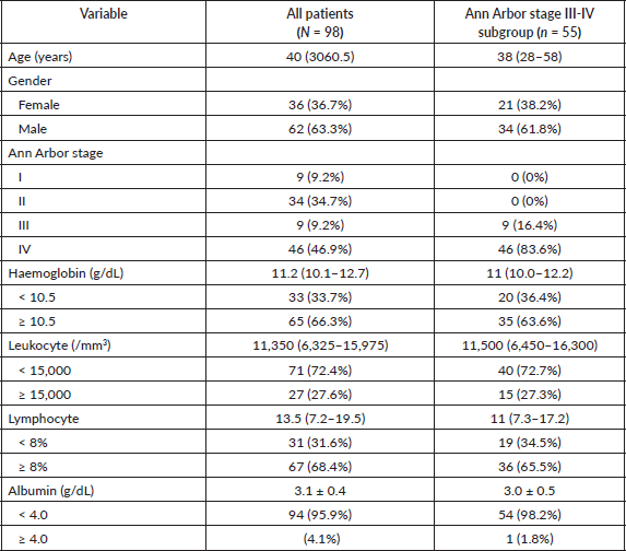
A total of 98 patients with cHL were included in the analysis, with 34 death events (34.7%) and 64 patients (65.3%) censored. For the final analysed cohort, there were no missing data for any of the clinical or laboratory variables of interest, as the presence of complete medical record data for all seven IPS-7 components was a predefined inclusion criterion. The distribution of IPS-7 risk categories showed that 26 patients (26.5%) were classified as low risk, 52 (53.0%) as intermediate risk, and 20 (20.5%) as high risk. More than half of the cohort (approximately 56%) had advanced-stage disease (Ann Arbor stage III–IV). Advanced disease stage (Ann Arbor stage IV) and hypoalbuminemia were the most frequent adverse prognostic factors. Baseline patient characteristics are summarised in Table 1.
Table 1. Baseline characteristics of patients.
Overall survival
Kaplan–Meier analysis showed a significant difference in OS between IPS-7 risk groups (log-rank p = 0.015) (Figure 1). The median OS was approximately 42 months for the low-risk group, 30 months for the intermediate-risk group, and 16 months for the high-risk group. Observed OS based on Kaplan–Meier estimates were approximately 88%, 80%, and 74% at 1–3 respectively, for the low-risk group; 70.5%, 67.4%, and 54.5% at 1–3 years for the intermediate-risk group; and 35.1% at 1 year for the high-risk group.
In the advanced-stage subgroup (Ann Arbor stage III–IV), Kaplan–Meier analysis demonstrated a trend towards worse OS with increasing IPS-7 risk category. Mean survival times decreased progressively from 19.7 months in the low-risk group to 15.6 months in the intermediate-risk group and 5.4 months in the high-risk group. However, differences in survival distributions across IPS-7 groups did not reach statistical significance (log-rank p = 0.056) (Figure 2).
Univariate analysis of IPS-7 components
On univariate Cox proportional hazards analysis (Table 2), none of the IPS-7 components reached statistical significance for OS (p < 0.05). Stage IV disease had the highest (HR 1.757; p = 0.127), followed by male sex (HR 1.629; p = 0.196), leukocytosis ≥15,000/mm³ (HR 1.627; p = 0.191), and lymphocyte count <8% (HR 1.585; p = 0.213). Other variables, including age ≥45 years, haemoglobin <10.5 g/dL, and albumin <4 g/dL, also did not show significant associations with mortality. As no variable met the predefined significance threshold (p < 0.10), multivariate analysis was not performed.
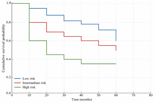
Figure 1. Kaplan–Meier curves of OS by IPS-7 risk group.
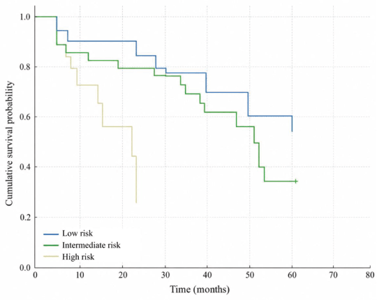
Figure 2. Kaplan–Meier curves of OS by IPS-7 risk group in the advanced-stage subgroup.
Table 2. Univariate Cox proportional hazards analysis of IPS-7 components.
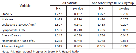
Table 3. Multivariable Cox proportional hazards analysis in the advanced-stage subgroup (Ann Arbor stage III–IV).

In the advanced-stage subgroup analysis (Ann Arbor stage III–IV), univariate Cox proportional hazards analysis demonstrated that age ≥45 years and lymphocyte count <8% were significantly associated with worse OS. Patients aged ≥45 years had a 1.786-fold higher risk of mortality compared with younger patients (HR 1.786, p = 0.045), while lymphocyte count <8% was associated with a 1.935-fold increased mortality risk (HR 1.935, p = 0.032) (Table 3). Other IPS-7 components were not significantly associated with OS in this subgroup. As variables with p <0.10 in univariate analysis were prespecified for multivariable modelling, age ≥45 years and lymphocyte count <8% were subsequently included in a multivariable Cox regression model.
Multivariate analysis of IPS-7 components
In multivariable Cox regression analysis of the advanced-stage subgroup, neither age ≥45 years nor lymphocyte count <8% remained independently associated with OS. Lymphocyte count <8% showed a borderline association with worse survival (adjusted HR 1.821, 95% CI 0.990–3.352; p = 0.054), as did age ≥45 years (adjusted HR 1.681, 95% CI 0.946–2.984; p = 0.076). However, the overall multivariable model was statistically significant (omnibus test p = 0.019).
Model discrimination
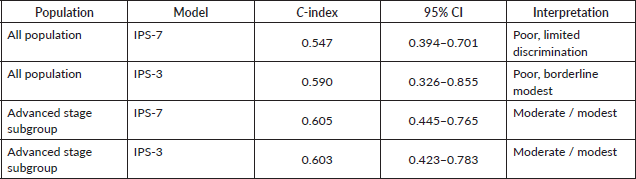
In the overall cohort, Harrell’s C-index for IPS-7 was 0.547 (95% CI 0.394–0.701), indicating poor to borderline discriminative performance. In an exploratory analysis, IPS-3 yielded a C-index of 0.590 (95% CI 0.326–0.855) in the same population, which was similarly classified as poor to borderline discrimination. The wide CIs for both scores reflect the limited number of events (n = 34) and should be interpreted with caution.
In the advanced-stage subgroup (Ann Arbor stage III–IV, n = 55), discriminative performance improved for both models. IPS-7 demonstrated a C-index of 0.605 (95% CI 0.445–0.765), and IPS-3 showed a comparable C-index of 0.603 (95% CI 0.423–0.783), both consistent with moderate discrimination. The improvement in IPS-3 performance from the overall cohort (0.590) to the advanced-stage subgroup (0.603) (Table 4), alongside the narrowing of CIs, suggests that both models may perform more consistently when applied to the population for which IPS-7 was originally developed.
Table 4. Harrell’s C-index values for IPS-7 and IPS-3 in the overall cohort and advanced-stage subgroup.
Discussion
This cohort of 98 patients with cHL was characterised by a median age at diagnosis of 40 years, with advanced-stage disease and hypoalbuminemia as the most frequent adverse prognostic features. A large single-institution study from the United States involving 727 patients with cHL reported a median age of 35 years, while an Indian cohort of 200 patients showed a median age of 28 years [8, 9]. An analysis of the Surveillance, Epidemiology, and End Results database in the United States showed that most cHL cases were diagnosed at stage II [10]. The high proportion of advanced-stage cases in this study suggests possible delayed diagnosis, a pattern frequently reported in low and middle income countries, where Obtel et al [11] found that limited access to healthcare services, lack of health insurance coverage, and low socioeconomic status act as significant barriers to timely diagnosis and treatment. At diagnosis, most patients with cHL present with supradiaphragmatic lymphadenopathy involving cervical, supraclavicular, mediastinal, or axillary nodes, often accompanied by systemic B symptoms with occasional extranodal involvement of the spleen, lungs, liver, or bone marrow [12]. The baseline profile of this study highlights epidemiological and clinical disparities that may influence the performance and validation of the IPS-7 in diverse populations. Serum albumin levels below 4.0 g/dL indicate poorer prognosis in cHL and other hematologic malignancies, reflecting decreased hepatic protein synthesis caused by IL-6–mediated acute-phase responses, elevated proinflammatory cytokines (IL-6, TNF-α, IL-1RA), or impaired nutritional status that limits amino acid availability [13].
In the overall cohort, univariate Cox proportional hazards analysis showed that none of the IPS-7 components reached statistical significance for OS. These findings suggest that individual IPS-7 components may have limited prognostic value when assessed separately, possibly due to differences in population characteristics, treatment heterogeneity, and the modest sample size. Diefenbach et al [6] evaluated 854 patients with advanced-stage cHL treated in the modern era and found that only age, stage, and haemoglobin remained significant predictors of OS in multivariable analysis, while other IPS-7 components lost prognostic relevance. Cellini et al [14] proposed an improved prognostic scoring system for cHL patients treated with PET/CT-guided ABVD with four variables (stage IV disease, leukocytosis, anaemia, low lymphocyte-to-monocyte ratio (LMR)) for progression-free survival (PFS) and three variables (male sex, stage IV disease, lymphopenia) for OS, improves risk prediction compared to IPS, particularly in identifying patients at high risk of recurrence after therapy, highlighting the need to update prognostic models to reflect modern treatment outcomes and incorporating easily measurable factors like lymphocyte-to-monocyte ratio and neutrophil-to-lymphocyte ratios for broader clinical use [14]. In a Chinese cohort of patients with advanced HL, the IPS still predicted outcomes but with reduced discrimination, as only stage IV disease and low haemoglobin remained significant, and a simplified two-factor score offered limited improvement [15]. In the advanced-stage subgroup of the present study, age ≥45 years and lymphocyte count <8% were associated with inferior OS in univariate analysis, but neither remained statistically significant after multivariable adjustment. Although the borderline adjusted associations suggest potential prognostic relevance, the limited subgroup sample size and number of events may have reduced statistical power. These findings should therefore be interpreted as exploratory and hypothesis-generating rather than definitive evidence of independent prognostic effects.
Kaplan–Meier analysis demonstrated a significant difference in OS between IPS-7 risk groups. In the advanced-stage subgroup, Kaplan–Meier analysis showed a trend towards progressively inferior OS with increasing IPS-7 risk category, although the difference did not reach statistical significance. This borderline finding may reflect reduced statistical power after subgroup stratification, given the smaller sample size and limited number of events. Nevertheless, the observed survival gradient suggests that IPS-7 may still capture some clinically relevant prognostic separation, although this finding did not reach conventional statistical significance in the advanced-stage subgroup. In a retrospective series of 314 patients with advanced‐stage cHL treated with ABVD, patients with IPS ≥3 had significantly worse 5-year OS (75.8%) than those with lower scores [16]. Rose et al [8] reported that the median OS of patients with early-stage and advanced-stage cHL was 19 and 12.9 years, respectively, with older age, advanced disease stage, presence of B symptoms, and a higher IPS identified as adverse prognostic factors for OS. In a study of elderly patients (age ≥60 years) with cHL, the median OS was 11.3 years, with significantly lower 5-year OS compared to younger patients (70.5% versus 99.4%), and worse outcomes linked to high comorbidity, lymphopenia, and non-anthracycline therapy [17]. The lower median OS observed in our cohort compared with Western data may reflect a higher proportion of advanced-stage disease and limited access to optimal treatment.
In this study, the IPS-7 showed limited discriminative performance in the overall cohort, with a Harrell’s C-index of 0.547 (95% CI, 0.394–0.701). IPS-3 showed a slightly higher C-index of 0.590 (95% CI, 0.326–0.855); however, its performance remained limited, and the wide, overlapping CIs preclude any definitive conclusion regarding superiority over IPS-7. Hasenclever and Diehl first introduced the IPS in 1998, showing strong prognostic separation among patients treated with ABVD-based regimens [5]. Diefenbach et al [6] re-evaluated the score in modern cohorts and found only age, stage, and haemoglobin remained independently prognostic, forming a simplified IPS-3 model. Several studies evaluating IPS-3 have reported C-index values higher than those of IPS-7 [18, 19]. The limited discrimination observed for both scores may reflect the modest sample size, limited number of events, cohort heterogeneity, and population-specific differences, particularly because the overall cohort included patients across all disease stages, whereas previous IPS-3 studies were largely restricted to advanced-stage cHL.
The exploratory advanced-stage subgroup analysis provides additional context for interpreting the stage-dependent performance of the IPS-3 score and its comparability with IPS-7 in this cohort. When the analysis was restricted to patients with advanced-stage disease, IPS-7 and IPS-3 demonstrated nearly identical modest discriminative performance, with C-index values of 0.605 (95% CI, 0.445–0.765) and 0.603 (95% CI, 0.423–0.783), respectively. This finding suggests that the prognostic utility of IPS-based scores may be more evident when applied to the clinical population for which these models were originally developed and evaluated, namely patients with advanced-stage cHL. Nevertheless, given the modest sample size, limited number of events, and wide CIs, these subgroup findings should be interpreted as exploratory and hypothesis-generating rather than definitive evidence of equivalent performance between IPS-3 and IPS-7.
These findings are consistent with the broader clinical heterogeneity of the present all-stage cHL cohort, which included patients with Ann Arbor stages I–IV and reflects the real-world case mix encountered in Indonesian tertiary referral hospitals within an low to moderate income country (LMIC) setting. In this setting, patients may present with variable disease stages, limited diagnostic and treatment resources, variability in guideline implementation, and restricted access to novel therapies [20, 21]; consequently, traditional IPS-based scores may remain useful for baseline risk stratification, but they have limited ability to provide precise individualised prognostication in an all-stage population. These findings emphasise the continued relevance of externally validating established and clinically familiar prognostic tools such as IPS-7 in local clinical contexts, while also highlighting the need for contemporary prognostic models adapted to diverse real-world populations.
Recent advances in prognostic modelling for cHL have introduced more contemporary and data-driven approaches aimed at improving risk stratification. The machine learning-based prognostic model for advanced-stage HL showed higher C-index and time-varying area under curve values than the IPS models, indicating better predictive accuracy within the first 5 years after diagnosis, although the improvement over the A-HIPI model was modest [18, 22]. Cellini et al [14] demonstrated that their new prognostic scoring system had better predictive accuracy than the IPS, with higher C-index values for PFS (0.71 versus 0.68) and comparable results for OS (0.79 versus 0.77) [14]. These results suggest that modern, data-driven models may provide better discrimination of survival outcomes compared with traditional IPS-based scores. However, their applicability in LMIC settings requires further validation.
Several limitations should be considered when interpreting the findings of this study. First, the retrospective single-centre design and reliance on medical record data may have introduced information bias and limited generalisability. Second, because the original IPS-7 was derived in advanced-stage HL, inclusion of patients across all disease stages in our cohort may have introduced heterogeneity and reduced comparability with the derivation population. Although an advanced-stage subgroup analysis was performed, the limited sample size and the number of events reduced statistical precision and resulted in wide CIs. Third, OS was defined from the start of first chemotherapy rather than from the time of diagnosis, which may not fully capture the prognostic impact of diagnosis-to-treatment delay in LMIC settings. Fourth, formal calibration analysis could not be performed, in accordance with TRIPOD recommendations, because IPS-7 was designed as a points-based score without published regression coefficients or baseline hazard functions, which precludes estimation of absolute survival probabilities. Fifth, detailed treatment-related variables, including chemotherapy regimen, treatment completion, dose delays, radiotherapy use, interim PET response, and salvage therapy, were not fully incorporated into the analysis. Therefore, these findings should be interpreted as supportive rather than definitive evidence of prognostic utility. Future prospective multicentre studies with larger sample sizes are needed to allow for comprehensive validation and recalibration of prognostic models in the contemporary treatment era.
Conclusion
In this retrospective cohort study of Indonesian patients with cHL, the IPS-7 significantly stratified OS across risk groups but demonstrated limited discriminative performance in the overall all-stage cohort. In the advanced-stage subgroup, IPS-7 showed modest discrimination, consistent with its intended clinical application in advanced-stage disease. Although individual IPS-7 components were not independently associated with mortality, the composite score maintained modest discriminative ability, suggesting that it remains a clinically useful baseline risk stratification tool, particularly in resource-limited settings. The relatively high proportion of advanced-stage disease observed in this cohort highlights potential diagnostic delays and disparities in healthcare access in low- and middle-income settings. These findings suggest that while IPS-7 continues to provide practical prognostic information, its predictive performance may be limited in the contemporary treatment era. Larger prospective multicentre studies incorporating contemporary biomarkers are needed to improve prognostic prediction in cHL.
Acknowledgments
The authors would like to express their sincere appreciation to all healthcare professionals and administrative staff at Dr. Moewardi General Hospital, Surakarta, Indonesia, for their support in the retrieval and management of clinical data used in this study, whose cooperation was essential to the completion of this research. This study received no external funding and was conducted using independent resources provided by the authors. The funding source had no role in the study design, data collection, analysis, or interpretation, manuscript preparation, or the decision to submit the manuscript for publication. Author contributions were as follows: Agus Jati Sunggoro (AJS): Conceptualisation, Methodology, Writing – Original Draft, Review & Editing; Felicia Renata (FR): Methodology, Formal Analysis, Data Curation, Writing – Original Draft, Review & Editing; Garwita Anindya Restisa (GAR): Methodology, Formal Analysis, Data Curation, Writing – Original Draft, Review & Editing; Devina Ravelia Tiffany Subroto (DRTS): Review & Editing, Project Administration. All authors have read and approved the final version of the manuscript. The authors declare that artificial intelligence–assisted technologies were not used to generate the scientific content of this manuscript. AI tools were used only for language editing and grammar improvement, and all content was reviewed and approved by the authors.
Conflicts of interest
The authors declare that there are no conflicts of interest related to this study. No author has any financial or personal relationships that could inappropriately influence or bias the design, conduct, interpretation, or publication of the research.
Funding
The authors declare that no financial support or funding was received for this study. The authors have no financial relationships, including research support, employment, stocks, consultancies, honoraria, or patents/patent applications, that could be perceived as influencing the work reported in this manuscript.
Disclosure of results at a meeting
Parts of this study were previously presented at the Role of Internist in Cancer Management (ROICAM) 12, 2025 (Jakarta, Indonesia) Scientific Meeting in the form of an oral presentation. The presentation was limited to a scientific conference format and did not constitute prior publication. Therefore, it does not conflict with the submission of this manuscript to ecancermedicalscience.
Institutional review
All authors confirm that they have read and approved the final version of the manuscript. This study was conducted in accordance with the ethical principles outlined in the Declaration of Helsinki. Ethical approval was obtained from the Institutional Review Board of Dr. Moewardi General Hospital, Surakarta, Indonesia (Approval No. 1.512/VII/HREC/2025) prior to data collection. As this was a retrospective cohort study using anonymised medical record data, the requirement for informed consent was waived by the ethics committee. Patient confidentiality and data privacy were strictly maintained throughout the study.
Author contributions
AJS: Conceptualisation, Methodology, Writing, Review & Editing.
FR: Methodology, Formal Analysis, Data Curation, Writing, Review & Editing.
GAR: Methodology, Formal Analysis, Data Curation, Writing, Review & Editing.
DRTS: Review & Editing, Project Administration.
References
1. Wang HW, Balakrishna JP, and Pittaluga S, et al (2019) Diagnosis of Hodgkin lymphoma in the modern era Br J Haematol 184(1) 45–59 https://doi.org/10.1111/bjh.15614
2. Kaseb H and Babiker HM (2023) Hodgkin Lymphoma in StatPearls [Internet] (Treasure Island: StatPearls Publishing)
3. Shanbhag S and Ambinder RF (2018) Hodgkin lymphoma: a review and update on recent progress CA Cancer J Clin 68(2) 116–132
4. Ferlay J, Ervik M, and Lam F, et al (2024) Global Cancer Observatory: Cancer Today [Internet] (Lyon: International Agency for Research on Cancer) [https://gco.iarc.who.int/today]
5. Hasenclever D, Diehl V, and Armitage JO, et al (1998) A prognostic score for advanced Hodgkin’s disease N Engl J Med 339(21) 1506–1514 https://doi.org/10.1056/NEJM199811193392104 PMID: 9819449
6. Diefenbach CS, Li H, and Hong F, et al (2015) Evaluation of the international prognostic score (IPS-7) and a simpler prognostic score (IPS-3) for advanced Hodgkin lymphoma in the modern era Br J Haematol 171(4) 530–538 https://doi.org/10.1111/bjh.13634 PMID: 26343802 PMCID: 4881845
7. Moccia AA, Donaldson J, and Chhanabhai M, et al (2012) International prognostic score in advanced-stage Hodgkin’s lymphoma: altered utility in the modern era J Clin Oncol 30(27) 3383–3389 https://doi.org/10.1200/JCO.2011.41.0910 PMID: 22869887
8. Rose A, Grajales-Cruz A, and Lim A, et al (2021) Classical Hodgkin lymphoma: clinicopathologic features, prognostic factors, and outcomes from a 28-year single institutional experience Clin Lymphoma Myeloma Leuk 21(2) 132–138 https://doi.org/10.1016/j.clml.2020.08.018
9. Jacob LA, Begum T, and Halder A, et al (2024) Clinical profile and outcome of adult classical Hodgkin’s lymphoma: real world single centre experience Indian J Hematol Blood Transfus 40(3) 392–399 https://doi.org/10.1007/s12288-024-01735-9 PMID: 39011262 PMCID: 11246344
10. Toro-Vélez E, Velez-Mejia C, and Rosas D, et al (2023) Classical Hodgkin lymphoma: a surveillance, epidemiology, and end results (SEER) database analysis comparing racial and ethnic disparities with emphasis on Hispanics J Clin Oncol 41 e19514 https://doi.org/10.1200/JCO.2023.41.16_suppl.e19514
11. Obtel M, Berraho M, and Abda N, et al (2017) Factors associated with delayed diagnosis of lymphomas: experience with patients from Hematology Centers in Morocco Asian Pac J Cancer Prev 18(6) 1603–1610 PMID: 28669176 PMCID: 6373802
12. Ansell SM (2022) Hodgkin lymphoma: 2023 update on diagnosis, risk-stratification, and management Am J Hematol 97(11) 1478–1488 https://doi.org/10.1002/ajh.26717 PMID: 36215668
13. Cuccaro A, Bartolomei F, and Cupelli E, et al (2014) Prognostic factors in Hodgkin lymphoma Mediterr J Hematol Infect Dis 6(1) e2014053 https://doi.org/10.4084/mjhid.2014.053 PMID: 25045461 PMCID: 4103502
14. Cellini A, Cavarretta CA, and Angotzi F, et al (2024) Baseline prognostic predictors in classical Hodgkin lymphoma: a retrospective, single-center analysis on patients treated with PET/CT-guided ABVD Front Oncol 14 1419118 https://doi.org/10.3389/fonc.2024.1419118 PMID: 39301543 PMCID: 11410762
15. Wang Q, Qin Y, and Kang SY, et al (2016) Decreased prognostic value of International Prognostic Score in Chinese advanced Hodgkin lymphoma patients treated in the contemporary era Chin Med J (Engl) 129(23) 2780–2785 https://doi.org/10.4103/0366-6999.194661 PMID: 27900988 PMCID: 5146782
16. Andjelic BM, Mihaljevic BS, and Jakovic LR (2012) ABVD as the treatment option in advanced Hodgkin’s lymphoma patients older than 45 years Pathol Oncol Res 18(3) 675–680 https://doi.org/10.1007/s12253-011-9494-4 PMID: 22234624
17. Sykorova A, Mocikova H, and Lukasova M, et al (2020) Outcome of elderly patients with classical Hodgkin’s lymphoma Leuk Res 90 106311 https://doi.org/10.1016/j.leukres.2020.106311
18. Koca O, Ozyurt B, and Umar A, et al (2025) The validation of advanced-stage Hodgkin lymphoma international prognostic index (A-HIPI) in Turkish patients with classical Hodgkin lymphoma Ann Hematol 104(3) 1765–1775 https://doi.org/10.1007/s00277-025-06292-3 PMID: 40069436 PMCID: 12031855
19. Almeida C, Sousa RCE, and Roque A, et al (2022) IPS-7 or IPS-3 to identify very-high risk patients in advanced classical Hodgkin’s lymphoma: which score to choose Hemasphere 6 5–6 https://doi.org/10.1097/01.HS9.0000890612.51878.ae
20. Chan JY, Fujimoto A, and Gan GG, et al (2025) Advanced classical Hodgkin lymphoma management in East and Southeast Asia: real-world challenges and aspirations of the Asian Lymphoma Study Group JCO Glob Oncol 11 GO-25-00288 PMID: 41160782
21. Relecom A, Federico M, and Connors JM, et al (2020) Resources-stratified guidelines for classical Hodgkin lymphana Int J Environ Res Public Health 17(5) 1783 https://doi.org/10.3390/ijerph17051783 PMID: 32182952 PMCID: 7084688
22. Rask Kragh Jørgensen R, Bergström F, and Eloranta S, et al (2024) Machine learning-based survival prediction models for progression-free and overall survival in advanced-stage Hodgkin lymphoma JCO Clin Cancer Inf 8 e2300255