Lymphoepithelioma epidermoid carcinoma of the uterine cervix: surgical management of an isolated case and review of the literature
Tenazoa -Villalobos José Richard1,2,a , Yan-Quiroz Edgar Fermín3,2,b , Ordoñez-Chinguel Augusto4,5,c , Prado-Cucho Sofia Leonor4,5,d and Villoslada-Terrones Vladimir4,5
1Oncological Surgery Area of the Víctor Lazarte Echegaray Hospital – EsSalud, Trujillo 13013, Peru
2Faculty of Medicine, Antenor Orrego Private University, Trujillo 13008, Peru
3Oncological Surgery Area of the Virgen de la Puerta High Complexity Hospital - EsSalud, La Esperanza 13013, Peru
4National Institute of Neoplastic Diseases, Lima 15036, Peru
5Faculty of Medicine, Cayetano Heredia Peruvian University, Lima 150135, Peru
ahttps://orcid.org/0000-0003-3622-9408
bhttps://orcid.org/0000-0002-9128-4760
chttps://orcid.org/0000-0001-8682-4948
dhttps://orcid.org/0000-0003-3042-0340
ehttps://orcid.org/0000-0001-6877-5441
Abstract
Cervical cancer is the gynecological malignancy that ranks third worldwide. It consists histologically of multiple subtypes, such as squamous cell carcinoma, which is the most common (65%), then adenocarcinoma (15%) and other types such as neuroendocrine, adenosquamous and carcinosarcoma tumours, which are less common. According to the World Health Organisation, lymphoepithelioma-type carcinoma has been described as an uncommon subtype and a variant of squamous cell carcinoma of the cervix. Its pathogenesis is related to the presence of the human Epstein-Barr virus and human papillomavirus. We present the case of a woman diagnosed with squamous cell lymphoepithelioma-like carcinoma of the cervix that was comprehensively managed with radical hysterectomy alone, presenting a good response and without recurrence.
Keywords: uterine cervical neoplasms, squamous cell carcinoma, hysterectomy
Correspondence to: Tenazoa -Villalobos José Richard
Email: josertenov@gmail.com
Published: 22/08/2025
Received: 14/01/2025
Publication costs for this article were supported by ecancer (UK Charity number 1176307).
Copyright: © the authors; licensee ecancermedicalscience. This is an Open Access article distributed under the terms of the Creative Commons Attribution License (
Introduction
It is estimated that cervical cancer reached an incidence of 660,000 new cases and more than 300,000 deaths worldwide in 2022. Eighty-five percent of these cases arose in low-resource countries, where cervical cancer is the second most frequent cancer and the third in mortality [1, 2].
There are two main histological types, adenocarcinoma and squamous cell carcinoma, in almost all cases associated with persistent infection with human papillomavirus (HPV). Lymphoepithelioma-like carcinoma, according to the WHO tumour classification, is a rare subtype that is considered a variant of epidermoid carcinoma. The incidence of this malignant neoplasm is 3.3% in women with FIGO 1 cervical cáncer. The pathogenesis is associated with high-risk HPV infection; some studies suggest that Epstein-Barr virus (EBV) is probably also involved, as well as in gastric and nasopharyngeal lymphoepithelioma epidermoid carcinoma (LEC), but this is still controversial and cases have been seen in Asian women [3–8].
We present the case of a 38-year-old woman with a 2 cm cervical tumour that was treated with radical hysterectomy alone and the anatomopathological study reveals the presence of lymphoepithelioma carcinoma of the cérvix. She did not receive chemotherapy or radiotherapy and currently attends periodic check-ups without showing signs of recurrence.
Case presentation
A 38-year-old woman from Lambayeque, Tumbes, Peru, with no medical or surgical history of importance, with a gynecological history of menarche at age 14, G5P4014, first sexual intercourse at age 17 with only one partner, which she has had until now. She manifests 5 months of illness characterised by abnormal vaginal bleeding and post coital bleeding associated with pelvic pain, lancinating type of moderate intensity not irradiated, which is why she goes to the health center of origin, where she is evaluated by a physician and the gynecological examination reveals a 2 cm tumour in the cervix, no parametrial involvement. The biopsy at the health center reveals a small cell infiltrating squamous cell carcinoma.
She is subsequently referred to the National Institute of Neoplastic Diseases (22.08.2016) and is seen in the gynecologic oncology service, where she is evaluated. Patient ECOG 1, no peripheral adenopathies. External genitalia preserved, at speculoscopy 2 cm tumour is observed dependent on the posterior lip of the cervix. Digitorectal examination: Parametrium free. Complementary studies with tomography of the thorax, abdomen and pelvis (30.08.2016): Uterus with regular borders and contours. Prominent cervix of 5.3 cm of greater diameter, in the posterior lip, there is a hypocaptive area of contrast in an extension of 2 cm, which could correspond to a lesion. No adenopathies are observed in the iliac chains. No free liquid is observed in the cavity (Figure 1). MRI of the pelvis with contrast (2.9.2016): thickening at the level of the cervix of 2 × 2 cm that shows restriction to diffusion and enhances with the contrast substance. No parametrial involvement (Figure 2).
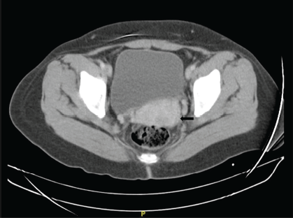
Figure 1. Chest, abdomen and pelvis tomography with contrast. Transverse section. Prominent uterine cervix, in the posterior lip there is a 2 × 2 cm area of contrast uptake (→).
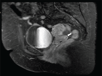
Figure 2. Contrast MRI of the pelvis. Sagittal view. Thickening of 2 × 2 cm at the level of the uterine cervix, predominance of the posterior lip that shows diffusion restriction and enhances with the contrast substance (→).
A repeat biopsy was performed, identifying a malignant neoplasm with a squamous cell component associated with severe lymphoplasmacytic inflammatory infiltrate. The squamous cell component presented nests of indistinct cell borders, scarce cytoplasm, finely granular, round or oval nuclei and prominent nucleoli, marginalised chromatin; while the inflammatory infiltrate presented predominance of lymphocytes (Figure 3A). Immunohistochemistry was performed, showing positive staining for P63 in the squamous cell component and negative staining in the inflammatory component. Also, sinpatophysin and chromogranin studies were performed, being negative for both stains (Figure 3B–D).
Patient is admitted to the operating room (14.09.2016) where Radical Hysterectomy type C1 plus oophoropexy plus bilateral pelvic lymphadenectomy is performed, the operative findings reveal a 2 × 2 cm tumour dependent on the posterior lip of the cervix, no adenopathies are observed. The patient tolerated the operative procedure, presenting a favourable evolution and was discharged on her second operative day.
Pathological anatomy results are obtained:
- Microscopy (Figure 4).
– Histologic type: Nonkeratinising infiltrating epidermoid carcinoma – Lymphoepithelioma-like carcinoma: Stromata of the uterine cervix occupied by clustered epithelioid cells bordered by a dense inflammatory infiltrate. In addition, single cells and nests consisting of abundant inflammatory infiltrate such as histiocytes, eosinophils, lymphocytes and plasmacytes, epithelioid cells in the center of these clusters are arranged syncytically. Atypical squamous epithelial cells infiltrate the stroma, which would correspond to a lymphoepithelioma-like carcinoma variant.
– Histologic grade: Poorly differentiated.
– Infiltration: Invasion 18 mm. Extension 13 mm.
– Absent lymphovascular invasion.
– Free surgical margins.
– Uterine body: Endometrium in secretory phase, myometrium without alterations.
– Parametrium: Right and left free, 3 left intraparametrial lymph nodes are free of malignant neoplasm are identified.
– Free right and left trunk.
– Right (8) and left (9) pelvic lymph nodes are free of malignancy.
After these findings, a diagnosis of lymphoepithelioma of the uterine cervix carcinoma type FIGO I B1 was confirmed. Due to the clinical stage of the disease, a medical meeting was held where it was decided that, due to the early stage of the disease, the patient would remain under observation, with strict controls and would not require adjuvant chemotherapy or radiotherapy.
The patient attends periodic check-ups until the present, showing favourable evolution with no alteration in lifestyle, preserved urinary function and no signs of recurrence, metastasis or suspicious lesions.
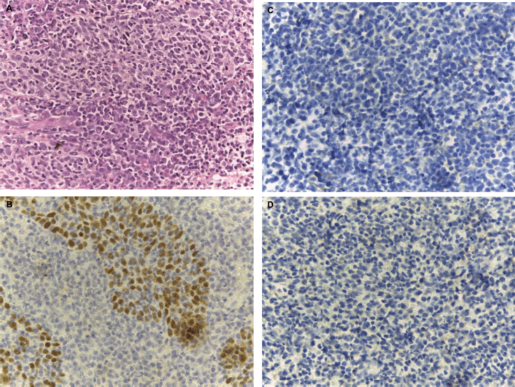
Figure 3. Biopsy microscopy. (a): Hematoxylin-eosin image at 40X: Malignant neoplasm composed of carcinoma component and abundant chronic inflammatory component, interspersed with nests of squamous cells. Immunohistochemistry 40X. (b): P63: Positive expression is observed in the epithelial component and negative staining in the inflammatory component. Biopsy microscopy. Immunohistochemistry 40X. (c): Synpatophysin and (d): Chromogranin, negative staining for both.
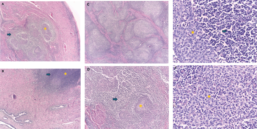
Figure 4. Microscopic image. Hematoxylin-Eosin-4X: In image (a): in the stroma of the uterine cervix there is proliferation of epithelioid cells (*) arranged in groups and surrounded by a dense inflammatory infiltrate (→). The exocervical epithelium does not present significant alterations. Image (b): shows few clusters of epithelioid cells with a marked inflammatory component and adjacent to these there are endocervical glands without atypia. Microscopic image. Hematoxylin-Eosin-4X: Image (c): shows nests formed by abundant inflammatory infiltrate and in the center of these groups of epithelioid cells arranged as syncytia. In image (d): at a magnification of 10X the nests are formed by epithelial cells that can form small groups (*) or be arranged in isolated cells (→). These cells present ample eosinophilic cytoplasm and the inflammatory infiltrate is predominantly formed by lymphocytes and plasmacytes. Microscopic image. Hematoxylin-Eosin-40X: Image (e): shows a nest of atypical squamous epithelial cells (*), with eosinophilic cytoplasm and indistinguishable borders, large, uniform, vesicular nuclei and prominent nucleoli, which are surrounded by a dense inflammatory infiltrate formed by lymphocytes, plasmacytes, also few eosinophils and histiocytes can be observed (→). In image (f): atypical squamous epithelial cells (*) are observed infiltrating the stroma, which would correspond to poorly differentiated infiltrating squamous epidermoid carcinoma variant Carcinoma type lymphoepithelioma.
Discussion
Lymphoepithelioma carcinoma is an extremely rare subtype of epidermoid carcinoma of the cérvix. Microscopically, it presents similar characteristics to its counterpart in the nasopharynx and other locations. The squamous cells are undifferentiated and arranged in syncytial growth and present indistinguishable borders, the nuclei are large, monotonous and nucleolus-like with frequent mitoses, asyloid cells with poorly differentiated nuclei that are infiltrated by lymphocytes can also be found, even previously it has been designated as an inflammatory cervical neoplasm. On the other hand, the inflammatory infiltrate is dense and is composed of lymphocytes, monocytes, histiocytes and eosinophils; these inflammatory cells can present high values of CD3+ and CD8+ [6, 9].
The pathogenesis of LEC is uncertain, this neoplasm can be associated to the presence of the EBV gene, a study in Asian women has registered up to 73.3% of cases of cervical LEC had positivity for antibodies directed against EBV, in comparison with 26.7% registered in typical epidermoid carcinomas; however, there are no definitive findings that point to EBV as the main etiological factor [9–11]. Likewise, it is known that there is an association with the human papillomavirus (HPV) [16–18]. When the gene has been sequenced, an association of around 80% has been found in typical epidermoid carcinomas versus 20% found in LEC [12].
This type of neoplasm manifests in patients younger than 40 years, which is an earlier age than the other types of malignant tumours of cervical carcinoma, a 10-year study in one institution evaluated 630 patients, 17 of which were cervical LEC (3.3% of all cases in FIGO stage I), all patients were directed to surgery and none had neoadjuvant therapy, the mean age was 49.6 years (range 32–67 years). Also, in all women, radical hysterectomy was performed, plus complete pelvic lymph node dissection after that received adjuvant radiotherapy. 47.1% tested positive for HPV and EBV staining. The FIGO IB1 stage was found in 82.4%, followed by 17% as FIGO IB2, 76% had no lymph node metastasis and 17% had positive lymph nodes. It should also be noted that the incidence is much higher in Asia than in other countries such as Latin America, and the overall 5-year survival rate for this neoplasm is around 76.4%. Our patient was 38 years old at the time of diagnosis, which is younger than the mean age described, with a stage and management similar to that described, providing excellent survival [13–15].
Although the histologic features of cervical LEC presume aggressiveness, the prognosis is, on the contrary, better than any other type of squamous cell carcinoma of the uterine cervix. Compared to other cancers at the same FIGO stage, LEC is distinguished by a very low metastasis to regional lymph nodes. As in other organs, LEC has been associated with favourable patient outcomes. It is believed that the good prognosis is associated with both cellular and humoral immune reactions induced by the tumour, and it is even suggested that the immune reaction is associated with the response to antigens produced by the tumour. The lymphocytic infiltrate that lodges in the stroma reflects the humoral and cellular response against the tumours. This reaction reduces metastasis to regional lymph nodes and increases overall survival. In the case of our patient, there was no lymphovascular or perineural invasion, free surgical margins and, above all, no lymph node or distant metastasis [9–16].
The biopsy on two occasions revealed an epidermoid carcinoma of the cervix. It should be noted that when a sample is obtained from an inflammatory lesion, it is complex to make an accurate diagnosis; therefore, a complete analysis of an operative specimen from a hysterectomy is required for a definitive diagnosis. LEC is an important differential diagnosis and confusion with other types that carry a poor prognosis should be avoided. In our patient, the initial biopsies showed an epidermoid carcinoma and the diagnosis was confirmed after performing a radical hysterectomy, which is the surgical treatment of choice, and we also preserved the ovaries to avoid early menopause [17].
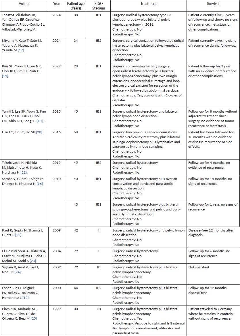
LEC of the uterine cervix is a rare entity; more knowledge is needed to determine the appropriate treatment for this type of neoplasm. The patient has been closely monitored since then, and no signs of recurrence have been observed. While radiotherapy has specific indications at any stage of the disease, surgery is the management of choice for early stages and LECs have better survival than other histological types of cervical cancer [6]. One study shows that cervical LECs have a lower frequency of recurrence and nodal metastasis [18]. Table 1 lists the cases of SCC managed surgically with good results.
Radical hysterectomy is the management used to treat cervical LEC. In our case, Radical Hysterectomy type C1 with oophoropexy and bilateral pelvic lymphadenectomy was performed. Surgery is planned based on imaging findings, preferably pelvic MRI. Lymph node freezing or sentinel lymph node biopsy is not performed in this case because indocyanine green is not available in our institution, and this procedure is still being validated, so it is not a standard treatment yet, and it is being performed in isolation. Oophoropexy is an acceptable management for the patient due to the age of presentation, thus avoiding early menopause and the wave of symptoms that this entails. Considering that cervical LEC is an infrequent neoplasm, many elements remain to be taken into account, so further research is needed to understand the optimal treatment for cervical LEC.
In our case, there is evidence of safe management by radical hysterectomy for LEC of the cervix, being notorious that the patient did not need chemotherapy or radiotherapy and to date shows no signs of recurrence, nodal or distant metastasis.
Table 1. Described cases of epidermoid cervical cancer of the lymphoepithelioma type reported in Pubmed managed with surgery and optimal survival.
Conclusion
LEC is an extremely rare entity that, despite the morphological characteristics that appear aggressive, has a favourable prognosis in relation to other types of squamous cervical cancer. It should be included in the differential and thus provide adequate management. Due to the rarity of this neoplasm, some aspects are unknown, so it is necessary to carry out extensive studies in this field.
Conflicts of interest
The authors declare that they have no conflicts of interest associated with the manuscript.
Funding
The case presented was entirely self-financed by the authors and did not receive any external funding.
Ethical aspects
The written informed consent was approved by the patient for the purpose of publication of the case presented, as well as the attached images. We have the approval of the Ethics Committee of the National Institute of Neoplastic Diseases.
Author contributions
JRTV Y EFYQ Preparation, creation and presentation of the paper, drafting of the manuscript and supervision. AOC & SPC Provision of anatomic pathology materials and drafting of manuscript. JRTV, EFYQ and VVT drafting of manuscript. JRTV & EFYQ Approval of manuscript submission.
References
1. Bray F, Laversanne M, and Sung H, et al (2024) Global cancer statistics 2022: GLOBOCAN estimates of incidence and mortality worldwide for 36 cancers in 185 countries CA Cancer J Clin 74(3) 229–263 https://doi.org/10.3322/caac.21834 PMID: 38572751
2. Torre LA, Bray F, and Siegel RL, et al (2015) Global cancer statistics, 2012 CA Cancer J Clin 65(2) 87–108 https://doi.org/10.3322/caac.21262 PMID: 25651787
3. International Collaboration of Epidemiological Studies of Cervical Cancer (2007) Comparison of risk factors for invasive squamous cell carcinoma and adenocarcinoma of the cervix: collaborative reanalysis of individual data on 8,097 women with squamous cell carcinoma and 1,374 women with adenocarcinoma from 12 epidemiological studies Int J Cancer 120(4) 885–891 https://doi.org/10.1002/ijc.22357
4. Noel J, Lespagnard L, and Fayt I, et al (2001) Evidence of human papilloma virus infection but lack of Epstein-Barr virus in lymphoepithelioma-like carcinoma of uterine cervix: report of two cases and review of the literature Hum Pathol 32(1) 135–138 https://doi.org/10.1053/hupa.2001.20901 PMID: 11172309
5. Kohrenhagen N, Eck M, and Höller S, et al (2008) Lymphoepithelioma-like carcinoma of the uterine cervix: absence of Epstein-Barr virus and high-risk human papilloma virus infection Arch Gynecol Obstet 277(2) 175–178 https://doi.org/10.1007/s00404-007-0427-0
6. Bais AG, Kooi S, and Teune TM, et al (2005) Lymphoepithelioma-like carcinoma of the uterine cervix: absence of Epstein-Barr virus, but presence of a multiple human papillomavirus infection Gynecol Oncol 97(2) 716–718 https://doi.org/10.1016/j.ygyno.2005.01.016 PMID: 15863191
7. Pinto A, Huang M, and Nadji M (2019) Lymphoepithelioma-like carcinoma of the uterine cervix: a pathologic study of eight cases with emphasis on the association with human papillomavirus Am J Clin Pathol 151(2) 231–239 https://doi.org/10.1093/ajcp/aqy130
8. Chao A, Tsai CN, and Hsueh S, et al (2009) Does Epstein-Barr virus play a role in lymphoepithelioma-like carcinoma of the uterine cervix? Int J Gynecol Pathol 28(3) 279–285 https://doi.org/10.1097/PGP.0b013e31818fb0a9 PMID: 19620947
9. Tseng CJ, Pao CC, and Tseng LH, et al (1997) Lymphoepithelioma-like carcinoma of the uterine cervix: association with Epstein-Barr virus and human papillomavirus Cancer 80(1) 91–97 https://doi.org/10.1002/(SICI)1097-0142(19970701)80:1<91::AID-CNCR12>3.0.CO;2-A PMID: 9210713
10. Yun HS, Lee SK, and Yoon G, et al (2017) Lymphoepithelioma-like carcinoma of the uterine cervix Obstet Gynecol Sci 60(1) 118–123 https://doi.org/10.5468/ogs.2017.60.1.118 PMID: 28217683 PMCID: 5313355
13. Hasumi K, Sugano H, and Sakamoto G, et al (1977) Circumscribed carcinoma of the uterine cervix, with marked lymphocytic infiltration Cancer 39(6) 2503–2507 https://doi.org/10.1002/1097-0142(197706)39:6<2503::AID-CNCR2820390629>3.0.CO;2-M PMID: 872050
14. Yordanov A, Karamanliev M, and Karcheva M, et al (2019) Single-center study of lymphoepithelioma-like carcinoma of uterine cervix over a 10-year period Medicine (Kaunas) 55(12) 780 https://doi.org/10.3390/medicine55120780
15. Martorell MA, Julian JM, and Calabuig C, et al (2002) Lymphoepithelioma-like carcinoma of the uterine cervix Arch Pathol Lab Med 126(12) 1501–1505 https://doi.org/10.5858/2002-126-1501-LLCOTU PMID: 12456211
16. Saroha V, Gupta P, and Singh M, et al (2010) Lymphoepithelioma-like carcinoma of the cervix J Obstet Gynaecol 30(7) 659–661 https://doi.org/10.3109/01443615.2010.500421 PMID: 20925604
17. Miyama Y, Kato T, and Sato M, et al (2024) Cervical lymphoepithelioma-like carcinoma with deficient mismatch repair and loss of SMARCA4/BRG1: a case report and five related cases Diagn Pathol 19(1) 6 https://doi.org/10.1186/s13000-023-01429-2 PMID: 38178127 PMCID: 10765828
18. van Nagell JR Jr, Donaldson ES, and Wood EG, et al (1978) The significance of vascular invasion and lymphocytic infiltration in invasive cervical cancer Cancer 41(1) 228–234 https://doi.org/10.1002/1097-0142(197801)41:1<228::AID-CNCR2820410131>3.0.CO;2-6 PMID: 626931
19. Kim SH, Yoon HJ, and Lee NK, et al (2022) A fertility-sparing surgery in lymphoepithelioma-like carcinomas of the uterine cervix: a case report Medicine (Baltimore) 101(45) e31579 https://doi.org/10.1097/MD.0000000000031579 PMID: 36397341 PMCID: 9666136
20. Hsu LC, Lin JC, and Ho SP (2016) A case of lymphoepithelioma-like carcinoma of the uterine cervix in a patient with a history of high-grade squamous intraepithelial lesion Taiwan J Obstet Gynecol 55(3) 453–454 https://doi.org/10.1016/j.tjog.2016.04.029 PMID: 27343338
21. Takebayashi K, Nishida M, and Matsumoto H, et al (2015) A case of lymphoepithelioma-like carcinoma in the uterine cervix Rare Tumors 7(1) 5688 https://doi.org/10.4081/rt.2015.5688 PMID: 25918614 PMCID: 4387360
22. Kaul R, Gupta N, and Sharma J, et al (2009) Lymphoepithelioma-like carcinoma of the uterine cervix J Cancer Res Ther 5(4) 300–301 https://doi.org/10.4103/0973-1482.59916
23. El Hossini Soua A, Trabelsi A, and Laarif M, et al (2004) Carcinoma de type lymphoepithelioma-like carcinoma of the uterine cervix. A propos d'un cas [Lymphoepithelioma-like carcinoma of the uterine cervix: case report] J Gynecol Obstet Biol Reprod (Paris) 33(1 Pt 1) 47–50 https://doi.org/10.1016/S0368-2315(04)96312-0 PMID: 14968055
24. Saylam K, Anaf V, and Fayt I, et al (2002) Lymphoepithelioma-like carcinoma of the cervix with prominent eosinophilic infiltrate: an HPV-18 associated case Acta Obstet Gynecol Scand 81(6) 564–566 https://doi.org/10.1034/j.1600-0412.2002.810616.x PMID: 12047313


