
Primary neuroendocrine neoplasms of the breast: a clinicopathological appraisal from a tertiary cancer centre of North India
Neha Singh1, Sujata Agrawal1, Zachariah Chowdhury2, Shashikant Patne2, Ipsita Dhal2, Paramita Paul2, Sadaf Haiyat2, Akhil Kapoor3, Anuj Gupta3, Bal Krishna Mishra3, Mayank Tripathi4 and Bipinesh Sansar3
1Department of Pathology , Institute of Medical Sciences, Banaras Hindu University, Varanasi 221005, India
2Departments of Oncopathology, Mahamana Pandit Madan Mohan Malaviya Cancer Centre and Homi Bhabha Cancer Hospital, Homi Bhabha National Institute, Varanasi 221005, India
3Medical Oncology, Mahamana Pandit Madan Mohan Malaviya Cancer Centre and Homi Bhabha Cancer Hospital, Homi Bhabha National Institute, Varanasi 221005, India
4Surgical Oncology, Mahamana Pandit Madan Mohan Malaviya Cancer Centre and Homi Bhabha Cancer Hospital, Homi Bhabha National Institute, Varanasi 221005, India
Abstract
Background: Neuroendocrine neoplasms (NENs) of the breast are rare, heterogeneous malignancies varying from well-differentiated neuroendocrine tumours (NETs) to poorly differentiated neuroendocrine carcinomas (NECs). Diagnosis is challenging due to histopathologic overlap with invasive breast carcinoma of no special type and the absence of consensus-based treatment guidelines. The objective of this study was to characterise the clinicopathologic spectrum, immunophenotype, management strategies and outcomes of primary breast NENs treated at a tertiary cancer centre in North India.
Methods: We retrospectively reviewed 12 histologically confirmed cases of breast NENs diagnosed between 2019 and 2024. Clinical, radiologic, histopathologic, immunohistochemical, staging, treatment and survival data were tabulated and analysed to identify any distinctive patterns, specifically clinical presentation, tumour grade, neuroendocrine markers and overall survival.
Results: Median age was 47.5 years (interquartile range: 43–61.5); right breast involvement predominated (75%). Histologic subtypes included small-cell NEC (n = 7), NET-G2 (n = 3) and large-cell NEC (n = 2). All tumours expressed ≥1 neuroendocrine marker, with INSM1 and synaptophysin most consistently positive. Hormone receptor expression was heterogeneous, while all tumours were HER2-negative. Axillary lymph node involvement occurred in 83% of patients, and distant metastases occurred in 33%, with clinical staging (American Joint Committee on Cancer Tumor,Nodes,Metastasis TNM) ranging from cT1cN0M0 to cT4bN2M1. Treatment included neoadjuvant chemotherapy (42%) and surgery (58%), followed by adjuvant systemic therapy or radiotherapy as indicated. At a median follow-up of 24 months, 33% of patients were alive with disease, 25% were alive without disease and 25% had died of progressive disease or treatment-related complications.
Conclusion: Breast NENs remain diagnostically challenging and clinically aggressive in a subset of patients, as evidenced by the death of a quarter of patients at 2-year follow-up. Accurate classification requires meticulous histopathologic evaluation supported by immunohistochemistry. Treatment mirrors standard breast cancer regimens, but outcomes remain variable, underscoring the need for collaborative research and standardised management algorithms.
Keywords: neuroendocrine neoplasm, breast cancer, small-cell neuroendocrine carcinoma, INSM1, synaptophysin, case series
Correspondence to: Neha Singh
Email: nsunra77@gmail.com
Published: 13/10/2026
Received: 27/01/2026
Publication costs for this article were supported by ecancer (UK Charity number 1176307).
Copyright: © the authors; licensee ecancermedicalscience. This is an Open Access article distributed under the terms of the Creative Commons Attribution License (http://creativecommons.org/licenses/by/4.0), which permits unrestricted use, distribution, and reproduction in any medium, provided the original work is properly cited.
Background
Primary neuroendocrine neoplasms (NENs) of the breast are a rare and underrecognised entity characterised by distinctive neuroendocrine morphology and confirmed through immunohistochemical (IHC) expression of neuroendocrine markers such as chromogranin A and synaptophysin [1]. The reported incidence rates range from <0.1% to as high as 20% [2–4], a variation largely stemming from historical inconsistencies in diagnostic standards. A mini-review by Gallo et al [4] highlighted that <200 cases have been recorded in the literature[4]. The presence of neuroendocrine differentiation in breast carcinomas was first noted by Feyrter and Hartmann in 1963, particularly in mucinous carcinomas. Since then, several classifications have been proposed, culminating in the 2019 WHO fifth edition classification, which formally recognises NENs as a distinct entity. Tumours with over 90% neuroendocrine histological features and/or neuroendocrine marker expression – excluding solid papillary and hypercellular mucinous carcinomas – are classified as NENs. NENs are further categorised into neuroendocrine tumours (NETs) and neuroendocrine carcinomas (NECs), including small-cell (SCNEC) and large-cell (LCNEC) subtypes [1].
Owing to their low incidence, current knowledge of these tumours is largely derived from isolated case reports and small retrospective series, resulting in limited consensus regarding their definition, true prevalence, biological behaviour and prognostic implications. Although acknowledged in the 2019 World Health Organization (WHO) classification, the diagnostic criteria for neuroendocrine breast carcinoma remain somewhat ambiguous, and no standardised treatment algorithms have been specifically validated for this subset of breast malignancies. Most breast NENs are mixed neoplasms containing both neuroendocrine and conventional mammary carcinoma components. Tumours with 10%–90% SCNEC are classified as mixed invasive carcinomas (NST or other special types) and SCNEC, with the percentage of neuroendocrine differentiation noted in the pathology report. Tumours with >90% neuroendocrine differentiation are classified solely as NET or NEC [1,5].
Histologically and immunohistochemically, breast NENs are almost indistinguishable from those originating in the lung. NETs of the breast are characterised by solid nests, trabeculae and dense cellularity separated by delicate fibrovascular stroma. Tumour cells often display spindle, plasmacytoid, or polygonal morphology with moderate to abundant eosinophilic and granular cytoplasm [6]. These tumours are graded using the Nottingham grading system, with the majority classified as Grade 2 (G2). Unlike NETs from other organs, breast NETs are not categorised into G1, G2, or G3 based on mitotic count, Ki-67 index, or necrosis, indicating distinct grading criteria [7]. Although the grading system is not utilised for breast NETs, the mitotic count continues to serve as the key factor influencing tumour grade.
The diagnosis of NENs within the breast is particularly challenging on core needle biopsies, where architectural features may be subtle and IHC profiles can overlap with other subtypes, such as invasive breast carcinoma of no special type. Furthermore, the prognostic relevance of neuroendocrine differentiation in breast cancer continues to be a subject of debate, with studies reporting variable outcomes depending on the tumour grade and receptor status [7].
In this retrospective case series, we present 12 histologically confirmed cases of primary breast NENs encountered at a tertiary cancer centre. The objective was to highlight the diagnostic complexities, clinicopathological spectrum and therapeutic considerations associated with these rare tumours, while contributing to the growing yet still limited literature on this uncommon neoplastic entity.
Materials and methods
This is a retrospective study conducted at a tertiary cancer care centre in North India after due approval from the Institutional Ethics Committee vide letter no OIEC/11000971/2025/00001. Data pertaining to patients diagnosed over a 5-year period from January 2019 to January 2024 were collected. All patients diagnosed with primary NENs of the breast, including well-differentiated NETs and poorly differentiated NECs, were included in the study. Case identification was performed by retrieving file numbers from the archives of the Department of Oncopathology.
Demographic and clinical details, including age, gender, presenting symptoms, radiological findings (including ultrasound, mammography and positron emission tomography-computed tomography where available), tumour laterality, nodal status, metastatic involvement, treatment received and follow-up outcomes, were obtained from the institutional electronic medical records system. Histopathologic diagnoses were confirmed by review of haematoxylin and eosin-stained sections and IHC analysis. IHC markers evaluated included synaptophysin, chromogranin A, insulinoma-associated protein 1 (INSM1), Ki-67, estrogen receptor (ER), progesterone receptor (PR) and HER2/neu, among others. Tumours were classified as NETs or NECs in accordance with the 5th edition of the WHO Classification of Breast Tumours, 2019.
Clinical staging was assigned based on the American Joint Committee on Cancer (AJCC) 8th edition Tumor, Nodes, Metastasis staging system for breast carcinoma. Treatment details, including neoadjuvant chemotherapy, surgical intervention, adjuvant therapy and palliative care, were recorded. Response to therapy was assessed clinically and radiologically. Follow-up data were reviewed to document recurrence, metastasis, survival status and duration of follow-up. In cases of loss to follow-up, telephonic contact was attempted where possible.
Statistical analyses were performed using International Business Machines Corporation Statistical Package for the Social Sciences Statistics, version 30. Survival estimates were calculated by the Kaplan–Meier method. Median follow-up duration was calculated by the reverse Kaplan–Meier method.
Progression-free survival (PFS) was calculated from the date of histological diagnosis to the date of disease progression or death. Recurrence was defined by any radiological evidence of local or systemic disease. Overall survival (OS) was calculated from the date of diagnosis to death due to any cause. Patients who defaulted after diagnosis or during treatment were censored at the date of last documented follow-up.
Results
During the 5-year study period, 9,247 breast specimens were examined, among which 12 cases were diagnosed as NENs, accounting for ~0.13% of all breast cases. All patients were female, with a median age of 47.5 years (interquartile range: 43–61.5). The most frequent clinical presentation was a palpable breast lump, occasionally associated with pain, systemic symptoms, or evidence of metastasis. The right breast was involved in 9/12 cases (75%) and the left in 3/12 (25%), with no bilateral disease. Tumours predominantly affected the upper outer quadrant (10/12, 83%), while two patients (2/12, 17%) presented with diffuse lesions and overlying skin involvement. Tumour size ranged from 2.0 to 11.5 cm (Table 1).
Histologically, 7/12 cases (58%) were classified as SCNEC, 2/12 (17%) as LCNEC and 3/12 (25%) as (NET, grade 2). Two NET cases were initially misdiagnosed as poorly differentiated invasive breast carcinoma of no special type (IBC-NST) on core needle biopsy but were reclassified after resection, based on detailed histopathology and immunohistochemistry (Figures 1 and 2).
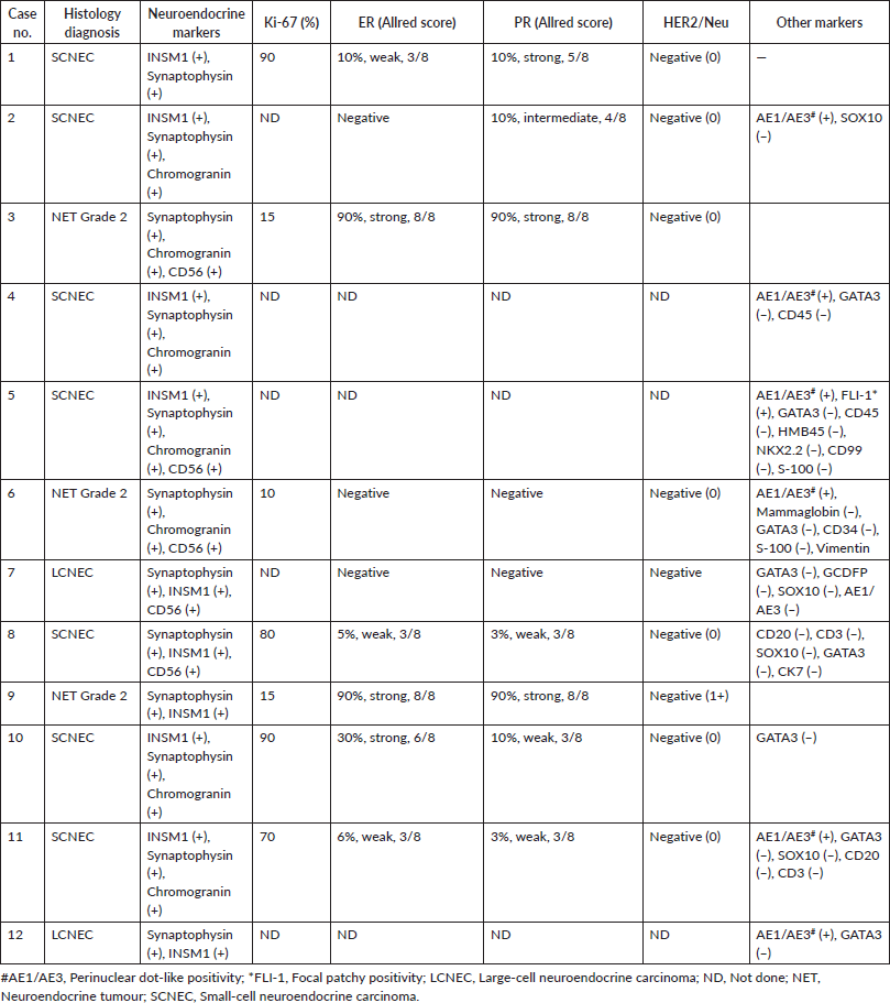
All tumours expressed at least two neuroendocrine markers; the choice and use of markers were varied, and hence, not all markers were used in all patients. Synaptophysin was positive in 9/9 (100%), chromogranin in 7/7 (100%), CD56 in 4/4 (100%) and INSM1 in 11/11 (100%). AE1/AE3 was positive in 7/8 (88%), typically with a perinuclear dot-like pattern. One SCNEC showed focal nuclear FLI-1 positivity, initially raising suspicion of a haemato-lymphoid neoplasm. Negative immunostaining for cytokeratin 7 (CK7), GATA binding protein 3 (GATA3), gross cystic disease fluid protein-15 (GCDFP-15), mammaglobin, CD45, CD3, CD20, CD34, vimentin, S-100, SRY-box transcription factor 10 (SOX10) and NK2 homeobox 2 (NKX2.2) helped exclude differentials such as IBC-NST, lymphoma, sarcoma and Ewing family tumours (Table 2).
Hormone receptor analysis (n = 9) revealed strong ER and PR positivity (Allred score 7/8–8/8) in 2/9 cases (22%), while the remaining 7/9 (78%) showed variable or absent expression. HER2/neu was negative in all 9/9 (100%) tested cases. These markers were not done for three other patients. The Ki-67 index ranged from 10% to >80%, with higher values observed in SCNECs.
Axillary lymph node involvement was present in 10/12 cases (83%), most frequently in NECs. Only 2/12 (17%), were node-negative, both of which were NET. Beyond axillary nodes, 5/12 (42%) demonstrated supraclavicular, mediastinal, paratracheal, hilar and epiphrenic nodal metastases. Distant metastases occurred upfront in 4/12 patients (33%) – three SCNEC and one NET – involving lungs, pleura, pancreas, vertebra and skin. Clinical staging (AJCC TNM) ranged from cT1cN0M0 to cT4bN2M1, highlighting presentations from localised to advanced disease.
Treatment and outcomes
Surgery was performed in seven cases (7/12, 58%), including breast-conserving surgery, modified radical mastectomy and wide local excision. Five patients received neoadjuvant treatment. Of these, four received standard protocols for invasive ductal carcinoma, that is, four cycles of anthracycline cyclophosphamide followed by taxanes for four cycles (AC-T). Of these, one patient progressed to metastatic disease on treatment, while two others progressed within 3 months post-surgery. Only one patient is currently on observation, 11 months post-surgery.
Table 1. Summary of demographic, pathologic and treatment details of 12 cases of primary breast neuroendocrine neoplasms.
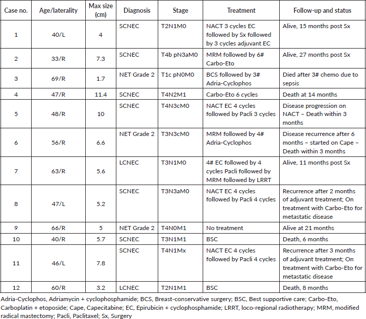
Only one patient received etoposide plus (EP) carboplatin as neoadjuvant therapy and is currently disease-free for 15 months post-surgery. Another patient received adjuvant EP and is currently on observation 27 months after surgery. Two other patients received adjuvant treatment with the AC-T regimen; one patient died from a complication of adjuvant treatment, while the other progressed after 6 months. These patients received adjuvant radiotherapy when indicated.
Four patients were upfront metastatic. Of these, one patient was histologically NET grade 2 and is currently alive 21 months post-diagnosis without any treatment. One patient was started on an EP regimen, responded and is currently on follow-up post 12 months since diagnosis. Two other patients who were not willing to undergo treatment survived for 6 and 8 months, respectively. Of the four patients who progressed or recurred post-NACT or adjuvant therapy, two were started on an EP regimen and are currently on treatment. The other two patients expired within 3 months of progression.
The median follow-up was 15 months (95% confidence interval [CI], 11.1–18.9). The median OS was 14 months (95% CI, 11.7–16.3 months). The median progression-free survival was 10 months (95% CI, 6.6–13.4 months).
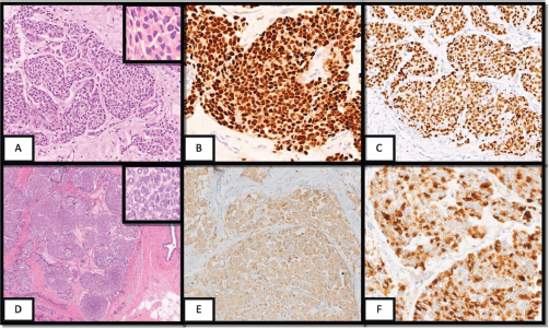
Figure 1. (A) Core biopsy showing tumour cells arranged in solid nests; inset displays cells with moderate nuclear pleomorphism and eosinophilic cytoplasm (H&E, ×10; inset ×40). (B, C) Tumour cells exhibit strong nuclear immunoreactivity for ER and PR (IHC, ×20). (D) MRM section of NET-2 demonstrating densely cellular, solid nests separated by delicate fibrovascular stroma (insular pattern); inset shows ovoid nuclei with characteristic salt-and-pepper chromatin (H&E, ×2; inset ×40). (E, F) Diffuse cytoplasmic positivity for synaptophysin and chromogranin (IHC, ×10 and ×20).
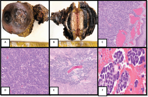
Figure 2. Gross and microscopic features of the breast NEC. (A) Mastectomy specimen showing an ulcerated, fungating mass involving the skin. (B) Cut surface revealing a solid, grey-white, lobulated tumour. (C, D) Low-power views showing an infiltrative tumour beneath ulcerated epidermis, composed of solid nests and trabeculae of uniform polygonal cells separated by delicate fibrovascular septa (H&E, 4× and 10×). (E) Peritumoral area with extensive necrosis. (F) Pleural fluid cell block showing tumour cells (H&E, 40×).
Table 2. Histological and immunohistochemical features of twelve patients with NENB.
Discussion
In this case series, the clinical presentation of breast NENs did not demonstrate any unique features that distinguished them from other breast carcinoma types, consistent with current literature. None of the patients displayed clinical syndromes indicative of hormone secretion. As previously reported, patients were predominantly in their fourth to sixth decade of life, reinforcing that NENs typically occur in a similar age range as other breast carcinomas.
In our series, two NET cases were initially misdiagnosed as poorly differentiated IBC-NST on core needle biopsy and were subsequently reclassified after evaluation of the resection specimens and immunohistochemistry. These findings highlight the diagnostic challenges associated with breast NENs and emphasise the importance of careful morphological assessment and immunohistochemistry in establishing an accurate diagnosis [8].
Immunohistochemistry played a pivotal role in confirming the diagnosis in our series. Neuroendocrine differentiation was consistently demonstrated by positivity for INSM1, synaptophysin, chromogranin and CD56 [9,10]. Cytokeratin showed perinuclear dot-like positivity in most cases. NENs of the breast typically exhibit characteristics consistent with the luminal A subtype, expressing hormone receptors (ER/PR) while being negative for HER2/neu (ERBB2) [11,12]. This observation aligns with our findings, with the intriguing distinction that NECs exhibit comparatively lower hormone receptor expression scores than NETs.
Differentiating breast NENs from solid papillary carcinoma (SPC) may occasionally be challenging because both can express neuroendocrine markers. However, SPC is generally characterised by fibrovascular cores, retained hormone receptor and GATA3 expression and an indolent clinical course. According to the 2019 WHO classification, SPC is excluded from the NEN category unless neuroendocrine differentiation exceeds 90% [13].
Hypercellular mucinous carcinoma (HMC) may mimic breast NENs because of overlapping morphological and IHC features [14]. However, it is excluded from the NEN category in the 2019 WHO classification unless neuroendocrine differentiation exceeds 90%. Clinically, HMC generally has a more favourable prognosis than NECs, making accurate diagnosis important for appropriate patient management [15,16].
Currently, there is no specific or tailored treatment regimen for breast NENs, and they are managed similarly to invasive breast carcinomas, using a combination of surgery, radiotherapy and systemic therapies based on tumour biology and stage [17]. Treatment recommendations are derived from established breast cancer protocols, with outcomes primarily influenced by tumour grade, hormone receptor status and HER2 expression [18–23]. Multidisciplinary management is essential for optimising patient outcomes. However, as seen in our limited database, the patients (2/8, 25%) who received neoadjuvant/adjuvant regimens for NECs, that is, EP regimen, are currently disease-free. However, in the patients (6/8, 75%) who received the standard regimen for breast cancer, that is, AC-T, only one patient is disease-free. Although these numbers are too small to reach any definitive conclusion, a definite trend of superiority of the EP regimen used for NENs can be observed. A few patients of upfront metastatic NEC did not take any treatment due to COVID-19-induced disruption, further diluting our data for this group [24]. For NECs, regimens similar to those used for small-cell lung carcinoma are currently advocated by many studies, and based on our data, a similar approach should be explored for breast NECs in both the adjuvant and metastatic settings. Furthermore, as with other GI NENs and triple-negative breast cancers, it is important to perform genetic testing in breast NENs because of their association with hereditary syndromes [25,26].
We compared our data to some other recently published case series. Most of them comprised a similar, if not fewer, number of patients, <15, highlighting rarity and possibly underdiagnosis and heterogeneity in diagnosis. Jeon et al reported median disease-free survival of 14.2 months and median OS of 23.6 months, respectively. This is slightly better than our data, as none of these patients were upfront metastatic. Batty et al reported data from a series of only metastatic breast NEN cases with a PFS of 5.8 months and OS of 31.8 months. The shorter median PFS than that observed in our study can be explained because all cases were metastatic. The longer OS could be explained by >50% of patients in that series having Ki-67 <50%, while in our series, only 25% patients had similar Ki-67 values. Thus, our study highlights the treatment and reporting heterogeneity in these patients, reflecting the need for a larger collaborative dataset [27–29].
The heterogeneity is further highlighted by one of our patients having metastatic breast NET upfront with only lung metastases and surviving for 21 months at the time of data compilation without any treatment or symptoms.
Conclusion
Our case series underscores the complexity of treating breast NENs. The variability in treatment responses and outcomes highlights the need for a more individualised therapeutic approach that integrates both standard and innovative strategies. Long-term follow-up and strict adherence to treatment protocols are critical for improving patient outcomes. Further research is necessary to refine treatment guidelines and enhance our understanding of these rare tumours, ultimately improving our ability to predict and manage their clinical behaviour effectively.
This series reinforces several key points:
-
Diagnostic challenge: Two cases were initially misdiagnosed as IBC-NST on core biopsies, underlining the importance of comprehensive IHC, including INSM1, which shows good sensitivity. Differentiation from SPC and HMC is crucial, given their more indolent behaviour. The use of artificial intelligence may be used in this regard [30].
-
Aggressive biology: High rates of nodal (83%) and distant metastasis (33%) were observed, particularly in NECs, aligning with previous studies. SCNECs behave aggressively, paralleling their pulmonary counterparts, and may warrant platinum-based chemotherapy as first-line treatment.
-
Therapeutic uncertainty: No site-specific guidelines exist; treatment is individualised. Hormone receptor-positive NETs may benefit from endocrine therapy, but optimal regimens for triple-negative NECs remain undefined.
-
Need for collaborative data: Pooling of cases through multi-institutional registries is necessary to clarify prognostic factors, define molecular profiles and explore targeted therapies (e.g. somatostatin analogues, PARP inhibitors in HRD tumours).
In summation, primary NENs of the breast are rare, diagnostically demanding and clinically heterogeneous. Multidisciplinary evaluation, extensive IHC work-up and individualised therapy are essential. Future studies should integrate molecular profiling to refine classification and guide targeted treatment strategies.
Conflicts of interest
The author(s) declare that they have no conflicts of interest.
Funding
The authors declare that no funds, grants or other support were received during the preparation of this manuscript.
Consent for publication
All authors who contributed to the manuscript have provided consent for its submission.
Consent to participate
As this was a retrospective analysis, a waiver of informed consent was obtained and approved by the Institutional Ethics Committee.
Ethics approval
This study was conducted in accordance with the principles of the Declaration of Helsinki. Ethical approval was obtained from the Institutional Ethics Committee (approval letter no. OIEC/11000971/2025/00001).
Author contributions
Neha Singh, Sujata Agrawal, Zachariah Chowdhury, Ipsita Dhal, Shashikant Patne: concept, design, draft writing; Paramita Paul, Sadaf Haiyat, Akhil Kapoor, Anuj Gupta, Bal Krishna Mishra, Bipinesh Sansar, Mayank Tripathi: data collection and analysis; Neha Singh, Sujata Agrawal, Zachariah Chowdhury, Bipinesh Sansar: analysis, interpretation, draft writing; Neha Singh, Sujata Agrawal, Bipinesh Sansar, Zachariah Chowdhury: critical revisions, final approval.
References
1. World Health Organization Classification of Tumours Editorial Board (2019) WHO classification of tumours of the breast. 5th ed. Vol 2. Lyon: international Agency for Research on Cancer 5th edn (Lyon: International Agency for Research on Cancer) 2 [https://www.iarc.who.int/news-events/who-classification-of-tumours-5th-edition-volume-2-breast-tumours]
2. Wang J, Wei B, and Albarracin CT, et al (2014) Invasive neuroendocrine carcinoma of the breast: a population-based study from the Surveillance, Epidemiology and End Results database BMC Cancer 14 147 https://doi.org/10.1186/1471-2407-14-147
3. Bogina G, Munari E, and Brunelli M, et al (2016) Neuroendocrine differentiation in breast carcinoma: clinicopathological features and outcome Histopathology 68(3) 422–432 https://doi.org/10.1111/his.12766
4. Gallo M, Campione S, and Vito VD, et al (2021) Primary neuroendocrine neoplasms of the breast: still open issues Front Endocrinol (Lausanne) 11 610230 https://doi.org/10.3389/fendo.2020.610230 PMID: 33584543 PMCID: 7874233
5. Zheng F, Chen AC, and Berger D, et al (2018) Metastatic adrenocorticotropic hormone–secreting breast cancer treated with bilateral adrenalectomy AACE Clin Case Rep 4(5) e402–e406
6. Kelten Talu C, Leblebici C, and Kilicaslan Ozturk T, et al (2018) Primary breast carcinomas with neuroendocrine features: clinicopathological features and analysis of tumor growth patterns in 36 cases Ann Diagn Pathol 34 122–130 https://doi.org/10.1016/j.anndiagpath.2018.03.010 PMID: 29661717
7. Rindi G, Klimstra DS, and Abedi-Ardekani B, et al (2018) A common classification framework for neuroendocrine neoplasms: an IARC and WHO expert consensus proposal Mod Pathol 31(12) 1770–1886 https://doi.org/10.1038/s41379-018-0110-y PMCID: 6265262
8. Singh N, Hayat S, and Kumar S, et al (2024) Breast lymphomas: clinical and pathological insights from a tertiary cancer care center in India Indian J Pathol Microbiol 67(4) 787–793 https://doi.org/10.4103/ijpm.ijpm_233_23 PMID: 38727411
9. Kawasaki T and Kaira K (2021) Insulinoma-associated protein 1 (INSM1) expression in breast carcinomas with neuroendocrine morphologies: application and future perspective Virchows Arch 479(1) 191–194 https://doi.org/10.1007/s00428-020-02935-0
10. Bellizzi AM (2019) Immunohistochemistry in the diagnosis and classification of neuroendocrine neoplasms: what can brown do for you? Hum Pathol 96 8–33 https://doi.org/10.1016/j.humpath.2019.12.002 PMID: 31857137 PMCID: 7177196
11. Shin SJ, DeLellis RA, and Ying L, et al (2000) Small cell carcinoma of the breast: a clinicopathologic and immunohistochemical study of nine patients Am J Surg Pathol 24(9) 1231–1238 https://doi.org/10.1097/00000478-200009000-00006 PMID: 10976697
12. Mccullar B, Pandey M, and Yaghmour G, et al (2016) Genomic landscape of small cell carcinoma of the breast contrasted to small cell carcinoma of the lung Breast Cancer Res Treatment 158(1) 195–202 https://doi.org/10.1007/s10549-016-3867-z
13. Guo S, Wang Y, and Rohr J, et al (2016) Solid papillary carcinoma of the breast: a special entity needs to be distinguished from conventional invasive carcinoma to avoid overtreatment Breast 26 67–72 https://doi.org/10.1016/j.breast.2015.12.015 PMID: 27017244
14. Capella C, Eusebi V, and Mann B, et al (1980) Endocrine differentiation in mucoid carcinoma of the breast Histopathology 4(6) 613–630 https://doi.org/10.1111/j.1365-2559.1980.tb02957.x PMID: 6254868
15. Di Saverio S, Gutierrez J, and Avisar E (2008) A retrospective review with long-term follow-up of 11,400 cases of pure mucinous breast carcinoma Breast Cancer Res Treatment 111(3) 541–547 https://doi.org/10.1007/s10549-007-9809-z
16. Barkley CR, Ligibel JA, and Wong JS, et al (2008) Mucinous breast carcinoma: a large contemporary series Am J Surg 196(4) 549–551 https://doi.org/10.1016/j.amjsurg.2008.06.013 PMID: 18809061
17. Inno A, Bogina G, and Turazza M, et al (2016) Neuroendocrine carcinoma of the breast: current evidence and future perspectives Oncologist 21(1) 28–32 https://doi.org/10.1634/theoncologist.2015-0309
18. Kalwar A, Sharma B, and Satyanarayan, et al (2013) Five-year retrospective survival analysis of triple-negative breast cancer in North-West India Indian J Cancer 50(4) 330–332 https://doi.org/10.4103/0019-509X.123616 PMID: 24369211
19. Gupta A, Sansar B, and Mishra BK, et al (2025) Real-world data on trastuzumab emtansine efficacy and safety: results of a single-centre retrospective study of HER2-positive metastatic breast cancer patients Sci Rep 15 18669 https://doi.org/10.1038/s41598-025-97923-2
20. Kapoor A, Gupta A, and Sansar B, et al (2024) Unravelling eribulin’s role in metastatic breast cancer: real-world outcomes in resource-constrained settings Ecancermedicalscience 18 1804 https://doi.org/10.3332/ecancer.2024.1804
21. Wadasadawala T, Joshi S, and Rath S, et al (2024) Tata Memorial Centre evidence-based management of breast cancer Indian J Cancer 61(Suppl 1) S52–S79 https://doi.org/10.4103/ijc.ijc_55_24
22. Das S, Al-Toubah T, and Strosberg J (2021) Chemotherapy in neuroendocrine tumors Cancers (Basel) 13(19) 4872 https://doi.org/10.3390/cancers13194872 PMID: 34638356 PMCID: 8507720
23. Ye S, Li J, and Xu J (2025) Treatment strategies for advanced neuroendocrine neoplasms: current status and future prospects Cancer Biol Med 22(1) 14–20 PMID: 39749725 PMCID: 11795266
24. Noronha V, Shah D, and Mokal S, et al (2022) Deviation from standard cancer treatment during the first wave of the COVID-19 pandemic in India Cancer Res Stat Treat 5(2) 212–219 https://doi.org/10.4103/crst.crst_115_22
25. Kapoor A, Kapoor AR, and Rungta A, et al (2025) Navigating the genetic frontier: establishing genetic clinics for comprehensive hereditary cancer management in resource-limited settings Indian J Med Paediatr Oncol 46(3)
26. O’Shea T and Druce M (2017) When should genetic testing be performed in patients with neuroendocrine tumours? Rev Endocr Metab Disord 18(4) 499–515 https://doi.org/10.1007/s11154-017-9430-3
27. Özdirik B, Kayser A, and Ullrich A, et al (2020) Primary neuroendocrine neoplasms of the breast: case series and literature review Cancers (Basel) 12(3) 733 [10.3390/cancers12030733] https://doi.org/10.3390/cancers12030733 PMID: 32244940 PMCID: 7140078
28. Jeon Y, Kim JY, and Ahn JS, et al (2024) Breast cancers with neuroendocrine differentiation: retrospective case studies series from a single institution based on the 2019 WHO classification Cancer Treat Res Commun 42 100857 [10.1016/j.ctarc.2024.100857] PMID: 39719737
29. Batty K, Taylor AM, and Bernard EJ, et al (2023) Metastatic primary breast neuroendocrine neoplasms: a case series Intern Med J 53(10) 1813–1818 [10.1111/imj.15961] https://doi.org/10.1111/imj.15961
30. Kapoor A, Kapoor AR, and Kumar A, et al (2023) Molecular oncology and the role of artificial intelligence in advancing cancer treatment Int J Mol Immuno Oncol 8(3) 89–92 https://doi.org/10.25259/IJMIO_17_2023


