Temporal trends and high mortality of tumour lysis syndrome in gastrointestinal cancers: a national inpatient study, 2017–2022
Edwin Saji¹, Jason Jacob¹, Udaya Kumar Damodaran¹, Sabah Kulsum², Sharanya Tripathi¹, Akshat Saxena¹, Panah Parab¹, Utkarsh Dayal¹, Priyal Dilip Mehta¹ and Aju Mathew³
¹Department of Internal Medicine, Saint Vincent Hospital, 123 Summer Street, Worcester, MA 01608, USA
²Department of Internal Medicine, New York Medical College, St. Mary’s General Hospital/Saint Clare’s Health, Passaic, NJ 07055, USA
³Department of Oncology, MOSC Kolenchery, Medical College Road, PO Kolenchery, Kochi, Kerala 682311, India
Abstract
Background: Tumour lysis syndrome (TLS) is an oncologic emergency classically associated with hematologic malignancies but increasingly recognised in solid tumours. Contemporary population-level data describing TLS among patients with gastrointestinal (GI) malignancies remain limited.
Methods: We performed a retrospective serial cross-sectional analysis of the National Inpatient Sample from 2017 to 2022. Adult hospitalisations with TLS were identified and classified according to the presence of oesophageal, gastric, small-intestinal, colorectal, liver/biliary or pancreatic malignancy. Survey-weighted analyses estimated national hospitalisation rates and temporal patterns. Multivariable models compared mortality, length of stay (LOS), total charges, clinical severity and disposition between TLS hospitalisations with and without GI malignancy and evaluated factors associated with mortality among GI-TLS hospitalisations.
Results: An estimated 90,680 adult TLS hospitalisations were identified, including 3,920 (4.3%) associated with GI malignancy. From 2017 to 2022, GI-TLS increased from 1.53 to 2.89 per 100,000 adult hospitalisations and from 8.72 to 14.37 per 10,000 GI-cancer hospitalisations; however, the linear temporal trend did not reach conventional statistical significance (OR per year, 1.04; 95% confidence intervals (CI), 1.00–1.09; p = 0.051). GI-TLS was associated with higher in-hospital mortality than TLS without GI malignancy (34.6% versus 23.9%; adjusted OR, 1.42; 95% CI, 1.21–1.67; p < .001), despite shorter LOS (adjusted difference, −4.62 days) and lower total charges (−$86,864; both p < 0.001). Among GI-TLS hospitalisations, metastatic malignancy was independently associated with mortality. After incorporation of acute severity measures, metastatic disease, sepsis, mechanical ventilation and renal replacement therapy remained associated with death, whereas age, sex, race/ethnicity and GI cancer site were not.
Conclusion: GI-associated TLS is uncommon but carries substantial excess inpatient mortality. Its national hospitalisation burden increased numerically over time, while mortality was more strongly associated with metastatic disease and acute physiologic deterioration than with age or anatomic cancer site.
Keywords: tumour lysis syndrome, gastrointestinal malignancies, national inpatient sample, inpatient mortality, cancer epidemiology
Correspondence to: Edwin Saji
Email: edwinsaji1996@gmail.com
Published: 08/10/2026
Received: 28/01/2026
Publication costs for this article were supported by ecancer (UK Charity number 1176307).
Copyright: © the authors; licensee ecancermedicalscience. This is an Open Access article distributed under the terms of the Creative Commons Attribution License (http://creativecommons.org/licenses/by/4.0), which permits unrestricted use, distribution, and reproduction in any medium, provided the original work is properly cited.
Introduction
Tumour lysis syndrome (TLS) is a life-threatening oncologic emergency characterised by rapid release of intracellular contents following tumour cell breakdown, which can result in profound metabolic abnormalities, acute kidney injury, cardiac arrhythmias, seizures and death [1, 2]. Although classically associated with hematologic malignancies, TLS is increasingly recognised in solid tumours, particularly in patients with high tumour burden, rapidly proliferating disease or marked treatment response [3–6].
The treatment landscape for gastrointestinal (GI) malignancies has expanded substantially with the use of multi-agent cytotoxic chemotherapy regimens, targeted therapies and immune checkpoint inhibitors [7–9]. TLS has been reported in association with several GI malignancies and treatment settings; however, much of the available literature consists of case reports and small observational series [10–14]. Consequently, the national burden, temporal patterns and inpatient outcomes of TLS occurring in the setting of GI malignancy remain incompletely characterised.
Characterising GI-associated TLS at the population level may help identify patient and disease features associated with adverse outcomes and clarify whether its inpatient burden has changed over time. Such information may also inform clinical recognition, risk stratification and resource planning for patients with GI malignancies who develop TLS.
Using the National Inpatient Sample (NIS), we aimed to characterise adult hospitalisations with GI-associated TLS in the United States from 2017 to 2022. Specifically, we evaluated temporal trends and site-specific hospitalisation rates, compared mortality, resource utilisation, clinical severity and disposition between TLS hospitalisations with and without GI malignancy and examined factors associated with in-hospital mortality among patients with GI-associated TLS.
Methods
Study design and data source
We conducted a retrospective serial cross-sectional study using 2017–2022 data from the NIS, Healthcare Cost and Utilization Project (HCUP), Agency for Healthcare Research and Quality [15]. The NIS is a nationally representative, stratified sample of hospital discharges from U.S. community hospitals. Discharge-level weights provided by the HCUP were applied to generate national estimates. The unit of analysis was the hospitalisation rather than the individual patient.
Study population
We included hospitalisations of adults aged ≥18 years with a diagnosis of TLS. GI-associated TLS was defined as a TLS hospitalisation with a concurrent qualifying GI malignancy in any diagnosis position. TLS hospitalisations without a qualifying GI malignancy comprised the comparison group.
GI malignancies included oesophageal, gastric, small-intestinal, colorectal, liver/biliary and pancreatic cancers. For site-specific analyses, hospitalisations were assigned to the first-listed qualifying GI malignancy diagnosis, creating mutually exclusive cancer-site categories.
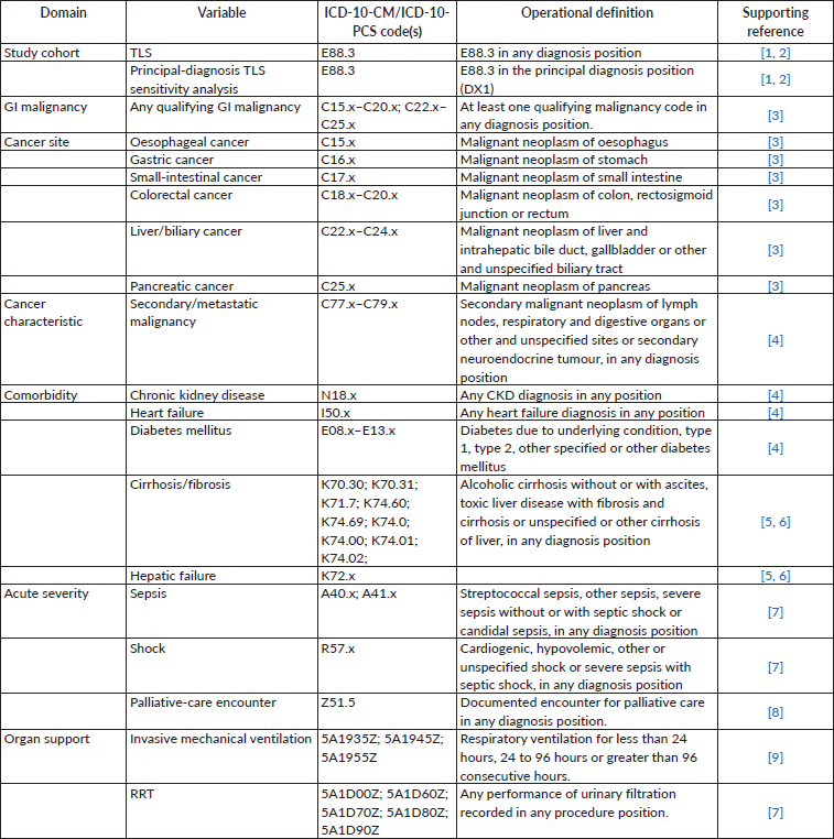
TLS, GI malignancies, comorbidities, acute clinical conditions and procedure-based outcomes were identified using International Classification of Diseases, Tenth Revision, Clinical Modification (ICD-10-CM) and Procedure Coding System (ICD-10-PCS) definitions. Published administrative definitions were used to support case definitions where available. All ICD-10-CM and ICD-10-PCS definitions used in the study, together with supporting references, are provided in Supplementary Table S1.
Because the NIS does not contain the laboratory values required to independently apply laboratory-based TLS criteria, TLS in this study represents clinician-coded TLS documented during the hospitalisation. As a sensitivity analysis, we restricted the cohort to hospitalisations in which TLS was recorded as the principal diagnosis.
Patient and hospital characteristics
Patient-level characteristics included age, sex, race/ethnicity, primary payer, median household income quartile for the patient’s ZIP code and Charlson comorbidity index category. Race/ethnicity was categorised as White, Black, Hispanic and Asian/Pacific Islander, Native American or Other. Additional clinical characteristics examined among GI-TLS hospitalisations included metastatic malignancy, chronic kidney disease, heart failure, diabetes mellitus, cirrhosis/hepatic fibrosis and hepatic failure. Definitions for diagnosis-based comorbidities are provided in Supplementary Table S1. Hospital-level characteristics included geographic region, rural location, teaching status, bed size and weekend admission.
Outcomes
The primary epidemiologic outcomes were the annual hospitalisation rate of GI-associated TLS per 100,000 adult hospitalisations, the rate per 10,000 hospitalisations involving a GI malignancy and the proportion of all TLS hospitalisations associated with GI malignancy. Site-specific rates were calculated per 10,000 hospitalisations involving the corresponding GI cancer site. Clinical outcomes included in-hospital mortality, length of stay (LOS) and total hospital charges. Additional outcomes included mechanical ventilation, renal replacement therapy (RRT), shock, sepsis, an Intensive Care Unit (ICU) -level-care proxy, palliative-care encounters and discharge disposition. Diagnosis- and procedure-based definitions for these outcomes are detailed in Supplementary Table S1.
The ICU-level-care proxy was defined by the presence of mechanical ventilation, RRT or shock during the hospitalisation and was used as a marker of high-acuity care rather than a direct measure of ICU admission.
Discharge disposition was categorised as home or home-health discharge, discharge to a facility or transfer to another short-term hospital. Because in-hospital death precludes discharge disposition, the primary disposition analysis was restricted to hospitalisations surviving to discharge. An analysis including all TLS hospitalisations was performed as a sensitivity analysis.
Factors associated with mortality among GI-TLS hospitalisations
To identify factors associated with in-hospital mortality among GI-TLS hospitalisations, we constructed sequential survey-weighted logistic regression models. The first model included age, sex, race/ethnicity, Charlson comorbidity category, metastatic malignancy, GI cancer site, hospital teaching status, rural location and calendar year.
A second model additionally incorporated markers of acute clinical severity and organ support, including sepsis, shock, mechanical ventilation and RRT. Because these events may develop during the hospitalisation, they were interpreted as factors associated with mortality rather than baseline causal risk factors.
Age-stratified analyses
To evaluate whether age was associated with differences in comorbidity burden, clinical severity or mortality, GI-TLS hospitalisations were stratified into patients aged <50 and ≥50 years. Comorbidities and clinical outcomes were compared between age groups, and age-by-year interaction terms were used to evaluate whether temporal patterns in GI-associated TLS or in-hospital mortality differed by age.
Sequential mortality models evaluated the association between age group and in-hospital death after adjustment for calendar year; patient, cancer and hospital characteristics; and subsequently acute severity variables. An additional sensitivity model replaced the Charlson comorbidity category with individual comorbid conditions to examine whether specific age-related comorbidities influenced the association between age and mortality.
Sensitivity analyses
Several sensitivity analyses were performed. First, the mortality analysis was repeated among hospitalisations in which TLS was recorded as the principal diagnosis. Second, discharge disposition was analysed both among survivors and among all TLS hospitalisations. Third, an individual-comorbidity mortality model was evaluated among GI-TLS hospitalisations in place of the aggregate Charlson comorbidity category.
Statistical analysis
All analyses accounted for the complex NIS survey design using discharge weights, hospital strata and hospital-level sampling units. Survey subpopulation methods were used for analyses restricted to TLS hospitalisations, GI-TLS hospitalisations, specific cancer sites or hospitalisations surviving to discharge.
National weighted counts and annual hospitalisation rates were calculated using NIS discharge weights. Annual and site-specific rates were independently verified using weighted numerators and denominators. Temporal trends in the proportion of TLS hospitalisations associated with GI malignancy were evaluated using survey-weighted logistic regression, with calendar year examined both categorically and as a continuous variable.
Categorical outcomes were evaluated using survey-weighted logistic regression and are reported as adjusted odds ratios (aORs) with 95% confidence intervals (CIs). LOS and total hospital charges were evaluated using survey-weighted linear regression and are reported as adjusted absolute differences with 95% CIs.
Primary comparative models adjusted for age, sex, race/ethnicity, primary payer, ZIP-code income quartile, Charlson comorbidity category, hospital region, rural location, teaching status, bed size, weekend admission and calendar year.
No imputation was performed; observations with missing values required for a given model were excluded from that analysis. In accordance with HCUP reporting requirements, estimates based on ≤10 unweighted hospitalisations were suppressed in published subgroup tables. All analyses were performed using Stata 19 (StataCorp, College Station, TX). Statistical tests were two-sided, and a p value <0.05 was considered statistically significant.
Results
Study population and baseline characteristics
From 2017 to 2022, the NIS identified an estimated 90,680 adult hospitalisations with TLS, of which 3,920 (4.3%) were associated with a GI malignancy and 86,760 (95.7%) were not. Cohort derivation is shown in Figure 1. The mean age was 61.8 years among GI-TLS hospitalisations compared with 63.5 years among TLS hospitalisations without GI malignancy.
GI-TLS hospitalisations differed substantially in underlying disease burden. Metastatic malignancy was present in 76.3% of GI-TLS hospitalisations compared with 17.2% of non-GI TLS hospitalisations, and 95.0% of GI-TLS hospitalisations had a Charlson comorbidity category ≥3 compared with 71.9% of non-GI TLS hospitalisations. GI-TLS hospitalisations also had higher prevalences of hepatic failure (23.2% versus 7.4%), cirrhosis/hepatic fibrosis (10.3% versus 2.4%) and diabetes mellitus (29.8% versus 24.0%). Women accounted for 35.6% and 37.5% of the two groups, respectively. Black patients represented a greater proportion of GI-TLS hospitalisations than non-GI TLS hospitalisations (24.5% versus 14.2%). Additional patient and hospital characteristics are summarised in Table 1.
Temporal trends in GI-associated TLS
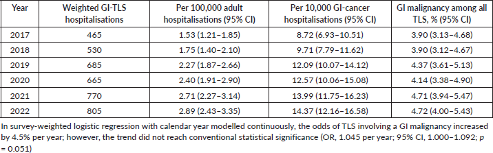
The weighted number of GI-TLS hospitalisations increased from approximately 465 in 2017 to 805 in 2022. Over the same period, the hospitalisation rate increased from 1.53 to 2.89 per 100,000 adult hospitalisations and from 8.72 to 14.37 per 10,000 GI-cancer hospitalisations. GI malignancies accounted for 3.90% of all TLS hospitalisations in 2017 and 4.72% in 2022 (Table 2). The corresponding annual estimates and 95% CIs are shown in Figure 2 and Table 2.
Although these measures increased numerically over the study period, the temporal association did not meet conventional statistical significance. In the continuous-year model, the odds of a TLS hospitalisation being associated with a GI malignancy increased by approximately 4.5% per year (OR, 1.045; 95% CI, 1.000–1.092; p = 0.051), while the overall categorical year effect was not significant (p = 0.462). Thus, the observed increase is best interpreted as a descriptive temporal pattern rather than a statistically confirmed upward trend.
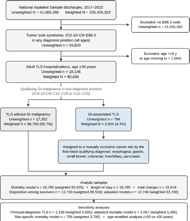
Figure 1. Cohort derivation and analytic flow. Legend: Derivation of the study cohort from the NIS, 2017 to 2022. Counts are unweighted discharge records and survey-weighted national estimates. TLS was identified by ICD-10-CM E88.3 in any diagnosis position, and GI-associated TLS by a concurrent qualifying malignancy code (C15 to C20 or C22 to C25) in any diagnosis position. For site-specific analyses, hospitalisations were assigned to the first-listed qualifying GI malignancy to create mutually exclusive categories. Analytic sample sizes differ across models because observations with missing values on a required covariate were excluded from that model without imputation. Incidence denominators were approximately 174.9 million weighted adult hospitalisations and 3,285,579 weighted GI-cancer hospitalisations across 2017 to 2022, each summed from the annual survey-weighted denominators.
Table 1. Baseline characteristics of adult TLS hospitalisations with and without GI malignancy, NIS 2017–2022.
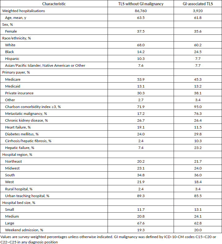
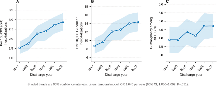
Figure 2. Annual burden of GI-associated TLS, 2017 to 2022. Legend: Survey-weighted annual estimates with 95% CIs. (a): GI-associated TLS per 100,000 adult hospitalisations. (b): GI-associated TLS per 10,000 hospitalisations involving a GI malignancy. (c): Proportion of all TLS hospitalisations associated with a GI malignancy. In a survey-weighted linear temporal model, the odds of a TLS hospitalisation involving a GI malignancy increased by 4.5% per year (OR 1.045; 95% CI, 1.000 to 1.092; p = 0.051), and the categorical year effect was not significant (p = 0.462). No trend line is fitted because the temporal association did not reach conventional statistical significance.
Table 2. Annual burden and hospitalisation rates of GI-associated TLS, 2017–2022.
Site-specific hospitalisation rates
TLS hospitalisation rates varied across GI cancer sites (Table 3). Among colorectal cancer hospitalisations, the rate increased numerically from 4.49 per 10,000 in 2017 to 13.44 per 10,000 in 2022. In contrast, liver/biliary cancer rates were relatively stable, from 15.89 in 2017 to 14.77 per 10,000 in 2022. Pancreatic cancer rates fluctuated during the study period, from 7.21 per 10,000 in 2017 to 11.93 per 10,000 in 2022. Oesophageal cancer rates were 15.54 and 18.73 per 10,000 in 2017 and 2022, respectively. Gastric cancer rates were 15.51, 28.91 and 19.66 per 10,000 in 2020, 2021 and 2022, respectively. Several oesophageal, gastric and small-bowel site-year estimates were suppressed because they were based on ≤10 unweighted hospitalisations; no TLS hospitalisations involving small-bowel cancer were observed in 2018 or 2019.
Differences in the numerical trajectories across cancer sites should be considered descriptive rather than evidence of differential temporal trends.
Clinical outcomes, resource utilisation and disposition
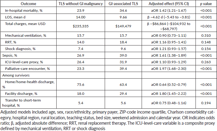
GI-TLS hospitalisations had higher crude in-hospital mortality than TLS hospitalisations without GI malignancy (34.6% versus 23.9%). After adjustment for patient demographics, comorbidity burden, hospital characteristics, weekend admission and calendar year, GI malignancy remained associated with greater odds of in-hospital death (aOR, 1.42; 95% CI, 1.21–1.67; p <0.001). Adjusted predicted mortality was 30.6% for GI-TLS and 23.8% for TLS without GI malignancy.
Despite higher mortality, GI-TLS hospitalisations had a shorter mean LOS (9.66 versus 14.00 days) and lower mean total hospital charges ($149,479 versus $235,335). After multivariable adjustment, GI-TLS was associated with a 4.62-day shorter LOS (β, −4.62; 95% CI, −5.43 to −3.81; p < 0.001) and $86,864 lower total charges (β, −$86,864; 95% CI, −$104,932 to −$68,797; p < 0.001).
Acute clinical characteristics also differed between groups. GI-TLS hospitalisations had higher unadjusted rates of RRT (18.4% versus 14.0%), shock (9.6% versus 7.4%), sepsis (39.0% versus 26.9%), the ICU-level-care proxy (31.9% versus 26.4%) and palliative-care encounters (39.4% versus 23.3%), whereas mechanical ventilation rates were nearly identical (15.7% in both groups). After multivariable adjustment, GI-TLS remained associated with greater odds of sepsis (aOR, 1.61; 95% CI, 1.38–1.89) and palliative-care encounters (aOR, 1.97; 95% CI, 1.68–2.30), whereas differences in mechanical ventilation, RRT, shock and the ICU-level-care proxy were not statistically significant.
Among hospitalisations surviving to discharge, GI-TLS was associated with lower rates of discharge home or with home health (63.4% versus 75.6%) and higher rates of facility discharge (29.4% versus 18.0%), whereas transfer to another short-term hospital was similar (5.6% versus 5.4%). After adjustment, GI-TLS was associated with lower odds of home/home-health discharge (aOR, 0.64; 95% CI, 0.52–0.79; p < 0.001) and higher odds of facility discharge (aOR, 1.80; 95% CI, 1.45–2.22; p < 0.001), without a significant difference in interhospital transfer (aOR, 0.75; 95% CI, 0.48–1.16; p = 0.194). These clinical, resource and disposition outcomes are summarised in Figure 3 and Table 4.
Table 3. Site-specific GI-TLS hospitalisation rates per 10,000 cancer hospitalisations, 2017–2022.
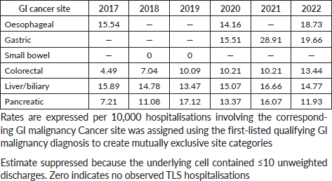
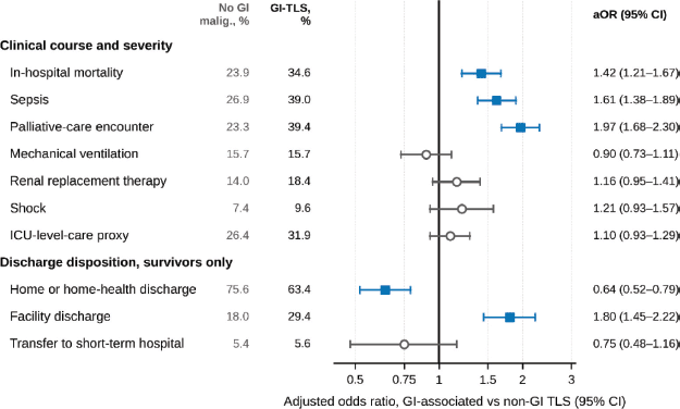
Figure 3. Clinical outcomes, resource use and disposition in TLS hospitalisations with versus without GI malignancy. Legend: aORs and 95% CIs comparing GI-associated TLS with TLS without GI malignancy, NIS 2017 to 2022. Unadjusted survey-weighted percentages for each group are shown to the left of the plot. Models adjusted for age, sex, race/ethnicity, primary payer, ZIP-code income quartile, Charlson comorbidity category, hospital region, rural location, teaching status, bed size, weekend admission and calendar year. Disposition outcomes were restricted to hospitalisations surviving to discharge. Blue markers indicate estimates whose 95% CI excludes 1; grey markers indicate estimates whose interval includes 1. The x-axis is on a logarithmic scale. The ICU-level-care proxy was defined by mechanical ventilation, RRT or shock. LOS and total charges are reported as adjusted absolute differences in Table 4 and are not shown here because they are on a different scale.
Factors associated with mortality among GI-TLS hospitalisations
Within GI-TLS hospitalisations, metastatic malignancy was independently associated with in-hospital mortality in the case-mix model (aOR, 2.00; 95% CI, 1.30–3.10; p = 0.002). In contrast, age, sex, race/ethnicity, Charlson comorbidity category, cancer site, hospital teaching status, rural location and calendar year were not independently associated with mortality. Race/ethnicity was not significant as a group (p = 0.321), nor was Charlson comorbidity category (p = 0.801) or GI cancer site (p = 0.297).
After markers of acute severity and organ support were added, metastatic malignancy remained associated with greater mortality (aOR, 2.54; 95% CI, 1.55–4.16; p < 0.001). Sepsis (aOR, 2.47; 95% CI, 1.73–3.54; p < 0.001), mechanical ventilation (aOR, 8.27; 95% CI, 4.82–14.17; p < 0.001) and RRT (aOR, 1.58; 95% CI, 1.01–2.46; p = 0.046) were also associated with mortality, whereas shock was not statistically significant (aOR, 1.60; 95% CI, 0.88–2.92; p = 0.125). The acute severity variables were jointly associated with mortality (p < 0.001). These analyses are summarised in Figure 4 and Table 5.
Age-stratified analyses
Among GI-TLS hospitalisations, patients aged ≥50 years had higher prevalences of chronic kidney disease (28.9% versus 13.7%) and diabetes mellitus (33.7% versus 10.7%) than patients aged <50 years, whereas hepatic failure (21.6% versus 31.3%) and metastatic malignancy (74.3% versus 86.3%) were more frequent among younger patients. In-hospital mortality was 33.8% among patients aged ≥50 years and 37.4% among those aged <50 years; mechanical ventilation, RRT, shock, sepsis and the ICU-level-care proxy were also broadly similar between the age groups (Supplementary Table S2).
Table 4. Clinical outcomes, resource use and disposition in TLS hospitalisations with and without GI malignancy, NIS 2017–2022.
Age <50 years was not independently associated with mortality after adjustment for demographics, comorbidity, metastatic disease, cancer site, hospital characteristics and calendar year (aOR, 1.01; 95% CI, 0.67–1.54; p = 0.948) and remained unassociated with mortality after acute severity variables were added (aOR, 0.94; 95% CI, 0.59–1.51; p = 0.806). There was no evidence that mortality trends differed by age group over time (age-by-year interaction p = 0.287). Similarly, the temporal pattern in the proportion of TLS hospitalisations associated with GI malignancy did not significantly differ between patients aged <50 and ≥50 years (year-by-age interaction p = .413).
Principal-diagnosis TLS sensitivity analysis
In the sensitivity analysis restricted to hospitalisations in which TLS was recorded as the principal diagnosis, there were an estimated 5,695 hospitalisations, of which 5.88% were associated with a GI malignancy (Supplementary Table S3). The direction of the mortality association was consistent with the primary analysis but was attenuated in precision and was no longer statistically significant (aOR, 1.72; 95% CI, 0.89–3.30; p = 0.105). Adjusted predicted mortality was 21.2% for GI-associated principal-diagnosis TLS and 13.9% for principal-diagnosis TLS without GI malignancy.
Disposition sensitivity analysis
When discharge disposition was analysed among all TLS hospitalisations rather than only those surviving to discharge, the findings were directionally consistent with the primary survivor analysis (Supplementary Table S4). GI-TLS hospitalisations were less likely to be discharged home or with home health and more likely to be discharged to a facility after multivariable adjustment, whereas interhospital transfer did not differ significantly. This consistency supports the primary survivor-restricted disposition findings.
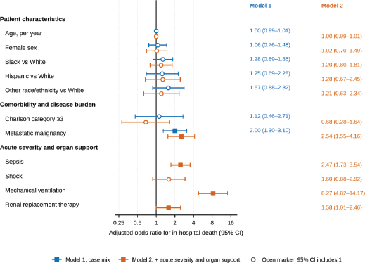
Figure 4. Factors associated with in-hospital mortality among GI-associated TLS hospitalisations. Legend: aORs and 95% CIs for in-hospital death among hospitalisations with GI-associated TLS, NIS 2017 to 2022. Model 1 adjusted for age, sex, race/ethnicity, Charlson comorbidity category, metastatic malignancy, GI cancer site, hospital teaching status, rural location and calendar year. Model 2 additionally included sepsis, shock, mechanical ventilation and RRT. Filled squares indicate estimates whose 95% CI excludes 1; open circles indicate estimates whose interval includes 1. The x-axis is on a logarithmic scale. GI cancer site was included in both models and was not independently associated with mortality (joint p = 0.297). Acute severity measures may develop during the hospitalisation and are interpreted as factors associated with mortality rather than causal risk factors.
Site-specific mortality
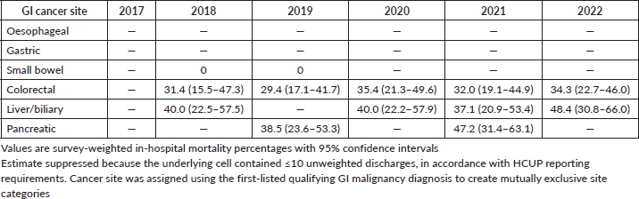
Overall weighted mortality among GI-TLS hospitalisations was high, but there was no significant difference in mortality across GI cancer sites in the design-based comparison (p = 0.548), and cancer site was not independently associated with mortality in the adjusted model (joint p = 0.297) (Figure 5).
Site- and year-specific mortality estimates are presented in Supplementary Table S5. Because many site-year mortality estimates were based on ≤10 unweighted deaths, those estimates were suppressed in accordance with HCUP reporting requirements. Among reportable cells, mortality remained substantial, but the estimates varied across years and had wide CIs; these descriptive fluctuations should therefore not be interpreted as evidence of cancer-site-specific temporal trends.
Discussion
In this contemporary, nationally representative analysis of adult hospitalisations with TLS, GI malignancies accounted for approximately 4% of TLS hospitalisations but were associated with a disproportionate burden of inpatient mortality. GI-associated TLS was associated with 42% greater adjusted odds of in-hospital death compared with TLS without GI malignancy, despite shorter hospital stays and lower total hospital charges. Within GI-TLS, metastatic disease was the principal baseline disease characteristic associated with mortality, while sepsis, mechanical ventilation and RRT identified a particularly high-risk clinical course. Conversely, age, sex, race/ethnicity, overall Charlson comorbidity burden and the anatomic site of the GI primary were not independently associated with mortality. Together, these findings suggest that once TLS develops in a patient with GI cancer, prognosis may be determined less by the organ of origin than by the extent of malignant disease and the severity of subsequent physiologic decompensation.
Table 5. Factors associated with in-hospital mortality among GI-TLS hospitalisations.
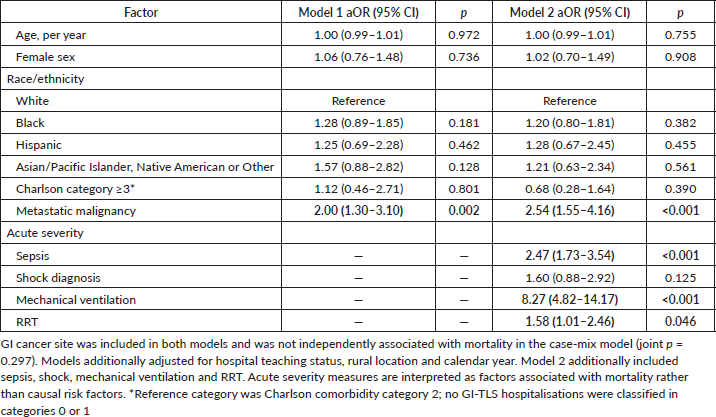
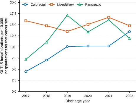
Figure 5. Site-specific rates of GI-associated TLS per 10,000 cancer hospitalisations. Legend: Survey-weighted annual rates of TLS per 10,000 hospitalisations for the corresponding GI cancer site. Only colorectal, liver/biliary and pancreatic cancer are plotted, because oesophageal, gastric and small-bowel estimates were suppressed in multiple years under HCUP reporting requirements for cells containing ten or fewer unweighted discharges; those sites are reported in Table 3. Cancer site was assigned using the first-listed qualifying GI malignancy diagnosis. Differences in trajectory across sites are descriptive and were not formally tested for differential temporal trends.
These findings extend a literature on solid-tumour TLS that has largely been derived from heterogeneous case reports and small pooled series. In an earlier NIS analysis spanning 2010–2013, Durani et al [10] reported 21% mortality among all TLS hospitalisations and 28% mortality among those with solid tumours. More recent systematic review data from 132 published cases of solid-tumour TLS reported mortality of 54%, with metastatic disease present in three quarters of cases [11]. A GI-specific pooled analysis similarly demonstrated high mortality across GI malignancies, but was necessarily limited by the small number and selective reporting of published cases [16]. Against this background, the approximately 35% mortality observed in the present national cohort remains striking while being lower than estimates derived from case-report literature. This difference may reflect the well-recognised tendency of case reports to capture unusually severe or clinically distinctive presentations and underscores the value of population-level data in defining the broader clinical spectrum of GI-TLS. Prior national data confirm the substantial morbidity and mortality associated with TLS overall, while published solid-tumour series disproportionately comprise metastatic and fatal presentations.
Metastatic disease emerged as a particularly important finding. More than three-quarters of GI-TLS hospitalisations in our cohort involved metastatic malignancy, and metastatic disease was associated with approximately twofold greater odds of death in the case-mix model; this association persisted after inclusion of acute severity measures. This is concordant with prior reports in solid tumours, in which metastatic disease is among the most frequently observed characteristics of TLS [11]. A recent scoping review of spontaneous TLS in solid tumours similarly found that metastatic disease was common and that metastatic involvement, particularly of the liver or lungs, was associated with worse outcomes [17]. These observations are biologically plausible: greater tumour burden provides a larger reservoir of intracellular metabolites that can be released abruptly with spontaneous or treatment-related cell lysis, while advanced malignancy may coexist with impaired physiologic reserve and organ dysfunction [1]. Importantly, the NIS does not provide direct measurements of tumour volume, cancer stage, hepatic metastatic burden or treatment response; metastatic disease should therefore be interpreted as a marker of advanced malignant burden rather than a mechanistic explanation for mortality. Contemporary reviews identify large tumour burden and effective tumour cell killing as central contributors to TLS risk, while the published solid-tumour literature has repeatedly identified metastatic disease in severe presentations.
The acute severity analysis provides a complementary perspective. After accounting for baseline patient and cancer characteristics, sepsis, mechanical ventilation and RRT were independently associated with mortality, with mechanical ventilation carrying the largest association. These variables should not be interpreted as pretreatment risk factors because they may represent consequences of TLS, the underlying malignancy, intercurrent illness or combinations thereof. Rather, they characterise the clinical trajectory of a patient with GI-TLS who has progressed to severe systemic illness. Collectively, these findings support a conceptual distinction between baseline disease risk, represented in our data by metastatic malignancy and evolving clinical severity, reflected by organ support and sepsis. This distinction may be more clinically informative than stratification according to cancer site alone.
Indeed, GI cancer site was not independently associated with mortality, and the overall site comparison was nonsignificant. This finding contrasts with the impression created by isolated reports in which pancreatic, hepatobiliary, gastric or other GI cancers have appeared to carry exceptionally high mortality [16]. Because published experience with many individual GI malignancies comprises only a handful of cases, apparent differences between sites may largely reflect small sample sizes, disease stage, treatment context and publication selection. Our findings instead suggest that once GI-TLS has occurred, advanced disease burden and physiologic deterioration may be more important determinants of outcome than the anatomic origin of the malignancy.
We observed a clear numerical increase in the hospitalisation burden of GI-associated TLS during the study period. Weighted GI-TLS hospitalisations increased from approximately 465 in 2017 to 805 in 2022, and the rate per 10,000 GI-cancer hospitalisations increased from 8.72 to 14.37. However, the continuous temporal model narrowly failed to meet conventional statistical significance, and the categorical year effect was also nonsignificant. Thus, these data support a descriptive increase in burden, but not a definitive conclusion that GI-TLS incidence is increasing over time. This distinction is particularly important because the NIS does not capture anticancer treatment exposure or its timing. Modern systemic therapy nevertheless provides a plausible context for continued surveillance. TLS has been reported with cytotoxic therapy, targeted agents and immune checkpoint inhibitors, and contemporary reviews have emphasised that effective tumour-directed therapy can expand the spectrum of malignancies in which TLS is encountered [1, 12, 18]. In COSMIC-312, for example, TLS was reported as a rare treatment-related fatal event among patients receiving cabozantinib plus atezolizumab for advanced hepatocellular carcinoma [19]. These observations establish biologic plausibility.
An additional and counterintuitive finding was that greater mortality in GI-TLS coexisted with shorter hospital stays and substantially lower hospital charges. These findings should not be interpreted as lower disease severity or more efficient resource use. Patients with GI-TLS had greater palliative-care coding, and among those who survived to discharge, they were less likely to return home and more likely to require facility-based care. Several mechanisms could contribute to this pattern, including earlier inpatient death, advanced malignancy limiting further cancer-directed intervention or transitions in goals of care that truncate prolonged hospitalisation. Because the NIS does not provide timing of palliative-care involvement, treatment limitations, hospice enrolment or goals-of-care decisions, these explanations remain hypotheses. Nevertheless, the combination of high mortality, frequent palliative-care coding and greater post-acute care requirements among survivors illustrates that shorter hospitalisation does not equate to lower clinical burden. Current ASCO guidance supports integration of specialist palliative care alongside active treatment for patients with advanced cancer, particularly when symptom or quality-of-life needs are substantial [20].
Age likewise appeared to be less informative than disease burden or acute severity. Mortality and rates of major acute complications were similar in patients younger than 50 years and those aged 50 years or older, and younger age was not associated with mortality across sequentially adjusted models. Although older patients had greater burdens of chronic kidney disease and diabetes, younger patients had higher prevalences of metastatic disease and hepatic failure. These contrasting patterns may explain why chronological age alone did not meaningfully discriminate mortality risk. The lack of an age-by-year interaction also provides no evidence that temporal patterns in GI-TLS were being driven disproportionately by younger patients. From a clinical standpoint, these findings argue against using age itself as a primary determinant of vigilance for TLS in GI malignancy; disease burden, organ function, treatment sensitivity and evolving clinical status are likely more relevant.
The principal-diagnosis sensitivity analysis further supports the direction of the primary mortality finding, although with substantially reduced precision. ICD-10-CM sequencing conventions place the condition chiefly responsible for admission in the principal position. In hospitalisations where the malignancy or its treatment prompted admission, TLS will usually be coded as a secondary diagnosis. The principal-diagnosis subgroup is therefore small and selected. Our findings indicate that the excess mortality signal was directionally preserved even under a more restrictive case definition.
Taken together, these findings have several clinical implications. Current approaches to TLS prevention are grounded in assessment of tumour burden, expected treatment sensitivity, renal function and anticipated intensity of tumour cell lysis [1]. Our data suggest that heightened awareness may be particularly warranted in patients with advanced or metastatic GI malignancy, regardless of age or anatomic GI primary. For patients considered at elevated risk, appropriate prophylaxis and close metabolic monitoring around initiation of highly active therapy remain essential. Once TLS develops, the emergence of sepsis, respiratory failure requiring mechanical ventilation or renal failure requiring RRT identifies a population at especially high risk of inpatient death. These data cannot establish whether earlier recognition or prophylaxis would modify these outcomes, but they provide a rationale for prospective studies capable of linking treatment exposure, tumour characteristics, laboratory evolution, prophylactic strategies and clinical outcomes.
Limitations
This study should be interpreted in the context of several limitations. First, the NIS is an administrative discharge database, and TLS was identified from clinician-assigned diagnostic coding rather than laboratory criteria. Laboratory measurements required to apply Cairo–Bishop or other biochemical definitions – including uric acid, potassium, phosphate, calcium, creatinine trajectories and lactate dehydrogenase – were unavailable. We therefore could not distinguish laboratory from clinical TLS or independently verify the diagnosis. The sensitivity analysis restricted to principal-diagnosis TLS reduced concern that the primary findings were driven entirely by incidental coding, but did not eliminate the potential for misclassification.
Second, the NIS does not capture anticancer medications, treatment timing, tumour response, prophylactic hydration, allopurinol or rasburicase administration or the temporal relationship between treatment and TLS. We therefore could not distinguish spontaneous from treatment-associated TLS or attribute observed temporal changes to chemotherapy, targeted therapy or immunotherapy. Measures of tumour burden, formal cancer stage, performance status, specific sites of metastatic disease, baseline laboratory abnormalities and treatment intent were likewise unavailable.
Third, the unit of analysis was the hospitalisation rather than the individual patient; recurrent admissions by the same patient cannot be identified, and the study cannot estimate patient-level incidence or longitudinal survival after discharge. Cancer-site assignment was based on the first-listed qualifying GI malignancy to create mutually exclusive categories and may not fully represent patients with multiple primary malignancies or complex metastatic coding. Several site-year strata were small, limiting precision and requiring suppression in accordance with HCUP reporting requirements.
Fourth, the associations between acute severity markers and mortality should not be interpreted causally. Sepsis, mechanical ventilation, RRT and shock may occur after TLS onset and may lie on the pathway between severe illness and death. Similarly, the association of palliative-care coding with GI-TLS should not be interpreted as an effect of palliative care; administrative coding does not establish when or why palliative services were provided. LOS and hospital charges may also be truncated by in-hospital mortality, and charges represent billed hospital charges rather than actual costs. In-hospital mortality was all-cause mortality; the NIS does not permit attribution of death specifically to TLS, the underlying malignancy or another acute complication. Finally, despite extensive adjustment for measured patient and hospital characteristics, residual confounding is unavoidable in an observational administrative-database study.
Conclusion
GI-associated TLS is uncommon but represents a high-risk inpatient phenotype with substantially greater mortality than TLS without GI malignancy. Although its hospitalisation burden increased numerically between 2017 and 2022, the temporal trend did not reach conventional statistical significance. Within GI-TLS, metastatic disease and markers of acute physiologic deterioration, including sepsis, mechanical ventilation and RRT, were more strongly associated with mortality than age, sex, race/ethnicity, overall comorbidity burden or the site of the GI primary. Higher mortality despite shorter hospitalisations and lower charges, together with greater palliative-care use and greater facility needs among survivors, further highlights the complex clinical burden of this condition. Prospective studies integrating cancer stage, treatment exposure, tumour response, laboratory findings and prophylactic strategies are needed to define patients at greatest risk and determine whether earlier recognition and prevention can improve outcomes.
Acknowledgments
The authors thank the Healthcare Cost and Utilization Project (HCUP) for providing access to the National Inpatient Sample and acknowledge institutional colleagues who provided manuscript review and feedback.
Conflicts of interest
The authors declare no conflicts of interest relevant to this manuscript.
Funding
This study received no external funding.
Disclaimer
This study was conducted using de-identified data from the National Inpatient Sample. The interpretations and conclusions presented in this manuscript are those of the authors and do not necessarily represent the views of the Agency for Healthcare Research and Quality, the U.S. Department of Health and Human Services or the Healthcare Cost and Utilization Project.
Prior presentation
This work has not been previously presented or published.
Ethical approval
This study used de-identified administrative data from the NIS and did not require institutional review board review under institutional policy.
Artificial intelligence disclosure
Artificial intelligence tools were used to assist with language editing and manuscript preparation. All authors reviewed the final manuscript and take responsibility for its accuracy, integrity and content.
Author contributions
Edwin Saji conceived the study, performed the statistical analyses, interpreted the findings and drafted the manuscript. Jason Jacob contributed to data management and analytic verification. Aju Mathew provided senior oversight, methodological guidance and critical revision of the manuscript for important intellectual content. All authors contributed to interpretation of the findings, critically reviewed the manuscript and approved the final version for submission.
References
1. Howard SC, Jones DP, and Pui CH (2011) The tumor lysis syndrome N Engl J Med 364(19) 1844–1854 https://doi.org/10.1056/NEJMra0904569 PMID: 21561350 PMCID: 3437249
2. Coiffier B, Altman A, and Pui CH, et al (2008) Guidelines for the management of pediatric and adult tumor lysis syndrome: an evidence-based review JCO 26(16) 2767–2778 https://doi.org/10.1200/JCO.2007.15.0177
3. Cairo MS and Bishop M (2004) Tumour lysis syndrome: new therapeutic strategies and classification Br J Haematol 127(1) 3–11 https://doi.org/10.1111/j.1365-2141.2004.05094.x PMID: 15384972
4. Gemici C (2009) Tumor lysis syndrome in solid tumors JCO 27(16) 2738–2739 https://doi.org/10.1200/JCO.2009.22.4154
5. Mirrakhimov AE, Ali AM, and Khan M, et al (2014) Tumor lysis syndrome in solid tumors: an up to date review of the literature Rare Tumors 6(2) 68–76 https://doi.org/10.4081/rt.2014.5389
6. Lupușoru G, Ailincăi I, and Frățilă G, et al (2022) Tumor lysis syndrome: an endless challenge in onco-nephrology Biomedicines 10(5) 1012 https://doi.org/10.3390/biomedicines10051012
7. Puri I, Sharma D, and Gunturu KS, et al (2020) Diagnosis and management of tumor lysis syndrome J Community Hosp Internal Med Perspect 10(3) 269–272 https://doi.org/10.1080/20009666.2020.1761185
8. (2024) Tumor Lysis Syndrome (Treasure Island: StatPearls Publishing) [https://www.ncbi.nlm.nih.gov/books/NBK518985/] Date accessed: 25/07/26
9. Mccurdy MT and Shanholtz CB (2012) Oncologic emergencies Crit Care Med 40(7) 2212–2222 https://doi.org/10.1097/CCM.0b013e31824e1865 PMID: 22584756
10. Durani U, Shah ND, and Go RS (2017) In-hospital outcomes of tumor lysis syndrome: a population-based study using the national inpatient sample Oncologist 22(12) 1506–1509 https://doi.org/10.1634/theoncologist.2017-0147 PMID: 28904174 PMCID: 5728022
11. Alqurashi RM, Tamim HH, and Alsubhi ZD, et al (2022) Tumor lysis syndrome in patients with solid tumors: a systematic review of reported cases Cureus 25 2022 [https://doi.org/10.7759/cureus.30652]
12. Wang L, Li X, and Zhao B, et al (2021) Immune checkpoint inhibitor–associated tumor lysis syndrome: a real-world pharmacovigilance study Front Pharmacol 12 679207 https://doi.org/10.3389/fphar.2021.679207
13. Coyne BM, Ishaq SM, and Coyne CJ, et al (2024) Spontaneous tumor lysis syndrome in the setting of pancreatic adenocarcinoma Cureus https://doi.org/10.7759/cureus.72735
14. Shaforostova I, Fiedler R, and Zander M, et al (2020) Fatal spontaneous tumor lysis syndrome in a patient with metastatic colon cancer: a clinical case of rare oncological emergency Case Rep Gastroenterol 14(2) 255–260 https://doi.org/10.1159/000507648 PMID: 32508557 PMCID: 7250380
15. HCUP National Inpatient Sample (NIS) Healthcare Cost and Utilization Project (HCUP) 2017–2022 (Rockville: Agency for Healthcare Research and Quality)
16. Kelkar N and Wang J (2022) Clinical features and outcomes of tumor lysis syndrome in patients with gastrointestinal cancers JCO 40(4) 655 https://doi.org/10.1200/JCO.2022.40.4_suppl.655
17. Papapanou M, Athanasopoulos AE, and Georgiadi E, et al (2023) Spontaneous tumor lysis syndrome in patients with solid tumors: a scoping review of the literature Med Oncol 40(8) 233 https://doi.org/10.1007/s12032-023-02108-4 PMID: 37432468 PMCID: 10335952
18. McBride A, Trifilio S, and Baxter N, et al (2017) Managing tumor lysis syndrome in the era of novel cancer therapies J Adv Pract Oncol 8(7) 705–720
19. Yau T, Kaseb A, and Cheng AL, et al (2024) Cabozantinib plus atezolizumab versus sorafenib for advanced hepatocellular carcinoma (COSMIC-312): final results of a randomised phase 3 study Lancet Gastroenterol Hepatol 9(4) 310–322 https://doi.org/10.1016/S2468-1253(23)00454-5 PMID: 38364832
20. Sanders JJ, Temin S, and Ghoshal A, et al (2024) Palliative care for patients with cancer: ASCO guideline update JCO 42(19) 2336–2357 https://doi.org/10.1200/JCO.24.00542
Supplementary tables
Supplementary Table S1. ICD-10-CM and ICD-10-PCS codes used for cohort identification, comorbidities and clinical outcomes.
References for Supplemental Table S1
1. Durani U, Shah ND, and Go RS (2017) The oncologist Oncologist 22 1506
2. Gong Z, Ali F, and Wang Y, et al (2019) Garcia JCO 37 e18185
3. Wasuwanich P, and Karnsakul W (2024) Gastrointestinal cancers in hospitalized patients with cystic fibrosis: a Nationwide Study, 2010–2020 Diagnostics 14(18) 1999 [https://doi.org/10.3390/diagnostics14181999]
4. Agency for Healthcare Research and Quality Elixhauser Comorbidity Software Refined for ICD-10-CM (Rockville: Healthcare Cost and Utilization Project)
5. Mapakshi S, Kramer JR, and Richardson P, et al (2018) Positive predictive value of international classification of diseases, 10th revision, codes for cirrhosis and its related complications Clin Gastroenterol Hepatol 16(10) 1677–8 [https://doi.org/10.1016/j.cgh.2018.01.042]
6. Khalifa A, Obeid JS, and Gregoski MJ, et al (2023) Accurate identification of patients with cirrhosis and its complications in the electronic health record Dig Dis Sci 68(6) 2360–9 [https://doi.org/10.1007/s10620-023-07876-7]
7. Yagnik K, Mohan G, and Ketkar A, et al (2024) Factors affecting continuous renal replacement therapy (CRRT) in patients withSeptic shock: an analysis of a national inpatient sample database Cureus 16(11) e74356 [https://doi.org/10.7759/cureus.74356]
8. O’Keefe S, and Czaja AS (2021) Validation of administrative codes for palliative care consultation among critically Ill children Hosp Pediatr 11(2) 179–82 [https://doi.org/10.1542/hpeds.2020-001263]
9. Ali H, Pamarthy R, and Bolick NL, et al (2022) Inpatient outcomes and racial disparities of palliative care consults in mechanically ventilated patients in the United States Proc (Bayl Univ Med Cent) 35(6) 762–7 [https://doi.org/10.1080/08998280.2022.2106537]
Supplementary Table S2. Age-stratified comorbidity burden, clinical severity and mortality among GI-TLS hospitalisations.
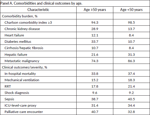
Panel B. Association of age <50 years with in-hospital mortality.
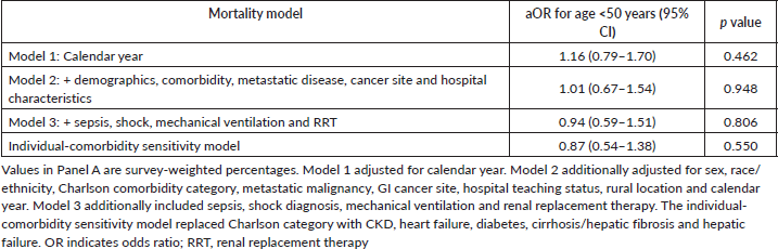
Supplementary Table S3. Principal-diagnosis TLS sensitivity analysis.
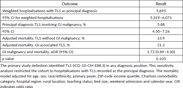
Supplementary Table S4. Disposition sensitivity analysis using all TLS hospitalisations.
Supplementary Table S5. In-hospital mortality among GI-TLS hospitalisations by cancer site and year.