Challenges to bridging the survival gap in Wilms tumour: insights from a 24-year experience from a tertiary cancer care centre in Southern India
Prasanth Srinivasan1, Gargi Das1, Balaji Thiruvengadam Kothandan1, Gautam Vydia Vedagiri2, Anand Raja3 and Venkatraman Radhakrishnan1
1Division of Paediatric Oncology , Department of Medical Oncology, Cancer Institute (W.I.A), Chennai 600036, India
2Department of Radiation Oncology, Cancer Institute (W.I.A), Chennai 600036, India
3Department of Surgical Oncology, Cancer Institute (W.I.A), Chennai 600036, India
Abstract
Wilms tumour (WT) is one of the most common paediatric renal malignancies. While the overall survival (OS) has improved above 90% in high-income countries, the outcomes remain inferior in low- and middle-income countries (LMICs) due to the healthcare system and socioeconomic barriers. This study aimed to document the demographic profile, clinical features and treatment patterns, and to evaluate the survival outcomes of children with WT managed in our institute. We retrospectively analysed patients diagnosed as WT between January 2000 and December 2023 at the Paediatric Oncology Unit of a tertiary care cancer centre in Southern India. Descriptive statistics were used to summarise the demographic profile, clinical features and treatment patterns. Event-free survival (EFS) and OS were estimated using Kaplan–Meier methods. Sixty-nine children were diagnosed with WT at our centre during the study period. The median age at diagnosis was 34 months (range: 8–159 months), with a slight male predominance (M:F ratio 1.22:1). Seventeen (25%) children without any image-defined high-risk factors underwent upfront nephrectomy while the remaining 52 (75%) children received preoperative chemotherapy followed by delayed nephrectomy. The stage distribution was as follows: Stage I–24 (35%); Stage II–15 (22%); Stage III–13 (19%); Stage 4–13 (19%); Stage 5–3 (3.5%); and Unknown–1 (1.5%). With a median follow-up of 68.5 months, the 5-year EFS and OS rates of our study cohort were 69% (95% confidence interval (CI): 56%–78%) and 78% (95% CI: 65%–87%), respectively. The most common causes of treatment failure were relapse (22%) and treatment abandonment (11%). Multivariate analysis revealed that advanced stage and upfront nephrectomy were independent predictors of inferior survival. Survival outcomes for WT in LMICs remain inferior despite multidisciplinary management. A hybrid approach – tailoring surgical timing to individualised risk – appears pragmatic. Bridging the survival gap requires addressing socioeconomic barriers and strengthening multidisciplinary care.
Keywords: child, kidney neoplasm, Wilms tumour, nephroblastoma
Correspondence to: Venkatraman Radhakrishnan
Email: venkymd@gmail.com
Published: 22/09/2026
Received: 05/05/2026
Publication costs for this article were supported by ecancer (UK Charity number 1176307).
Copyright: © the authors; licensee ecancermedicalscience. This is an Open Access article distributed under the terms of the Creative Commons Attribution License (http://creativecommons.org/licenses/by/4.0), which permits unrestricted use, distribution, and reproduction in any medium, provided the original work is properly cited.
Introduction
Paediatric renal tumours constitute 3.2%–11.1% of all childhood cancers globally [1]. Wilms tumour (WT) is the most common paediatric renal tumour [2]. Major collaborative groups, including the National Wilms Tumor Study Group/Children’s Oncology Group (NWTS/COG), the Société Internationale d’Oncologie Pédiatrique (SIOP) and the United Kingdom Children’s Cancer Study Group have played a pivotal role in advancing the understanding and management of WT [3]. As a result, the overall 3- to 5-year survival rates of WT now exceed 90% in high-income countries (HICs) [4].
The success story of WT stems from intensive multimodal treatment strategies, including surgery, chemotherapy and radiotherapy [3, 4]. Although such multimodal therapy is feasible in India, the reported overall survival (OS) rates for WT vary widely, ranging from 48% to 91%. This is due to challenges such as delayed presentation and diagnosis leading to advanced disease stage at diagnosis, treatment-related mortality, treatment abandonment, lack of uniform treatment guidelines, limited access to surgical and pathological expertise that hinders accurate staging and risk stratification and delays in the timely administration of radiotherapy [5, 6].
Since WT is one of the six index cancers identified by the World Health Organization (WHO) – Global Initiative for Childhood Cancer, efforts to bridge the survival gap between HICs and low- and middle-income countries (LMICs) require a clear understanding of the current survival rates, treatment patterns and barriers to care [7]. Although a few recent studies from India have reported the outcomes of WT, considerable heterogeneity in treatment practices and patient characteristics persists. This study aims to document our centre’s experience in managing children with WT [5, 6, 8, 9].
Methods
Study design, setting and participants
We retrospectively analysed patients evaluated for renal tumours between January 2000 and December 2023 at the Paediatric Oncology Unit, of a tertiary care cancer centre in Southern India. Our centre will be classified as setting 3 according to the SIOP – Paediatric Oncology in Developing Countries definition of resource settings necessary for diagnosing and managing children with cancer [10, 11]. This study included patients under 18 years of age with confirmed WT (typical clinical and radiological features with histopathological diagnosis of WT on biopsy and/or nephrectomy specimen) as well as probable WT (typical clinical and radiological features without histopathological diagnosis of WT on biopsy and/or nephrectomy specimen). We also included patients who had undergone upfront nephrectomy elsewhere, provided that histopathological diagnosis and staging were confirmed at our centre and essential surgical details (including information on tumour spill/rupture and lymph nodal sampling) were available. Our objective was to evaluate the demographic profiles, clinical features, treatment and survival outcomes of children with WTs managed in our unit. Prior approval and clearance were obtained from the institutional ethics committee for data analysis and publication (Ref: IEC/2025/April 16). All procedures performed in our study were in accordance with the ethical standards of the institutional and/or national research committee and with the 1964 Helsinki Declaration and its later amendments or comparable ethical standards.
Data collection
Clinical characteristics, including age, gender, presenting symptoms, duration of symptoms and sites of involvement, were abstracted from medical records. We also retrieved reports of haematological and biochemical investigations, imaging studies (X-ray, ultrasonography and computerised tomography (CT) scan) and histopathological studies from the medical records. Treatment details (chemotherapy, surgery and radiotherapy) were gathered from patients’ records. All patients in the study were followed up by medical record review until March 2025.
Initial diagnosis and evaluation
All patients presenting with a renal mass underwent contrast-enhanced CT (CECT) of the abdomen and pelvis. The clinical features and imaging characteristics were utilised to formulate a presumptive diagnosis of WT. The initial staging investigations for a child with a presumptive diagnosis of WT included chest X-ray and CT chest. Patients with characteristic lung or liver lesions were classified as having metastatic disease, whereas those without such lesions were categorised as having localised disease.
Treatment
The treatment decisions of the study participants were taken after discussion in a multidisciplinary tumour board. The resources for treatment of WT and the treatment protocol have evolved in the author’s unit during the study period; therefore, the treatment of the patients enrolled in the study is heterogeneous. The baseline CT images of the patients with presumptive diagnosis of WT were carefully evaluated for the image-defined high-risk features, namely: 1) large renal tumour crossing the midline, 2) primary tumour/lymph nodal mass infiltrating the surrounding viscera/major vessels that may warrant a multiorgan resection, 3) inferior vena cava thrombus, 4) metastatic disease and 5) bilateral WT, unilateral WT with bilateral predisposition, or WT arising from a solitary kidney/horseshoe kidney. The children presenting with any of these high-risk features were deemed unsuitable for upfront nephrectomy and were administered preoperative chemotherapy followed by delayed nephrectomy. The patients without these features underwent upfront nephrectomy.
Upfront nephrectomy (NWTS/COG approach)
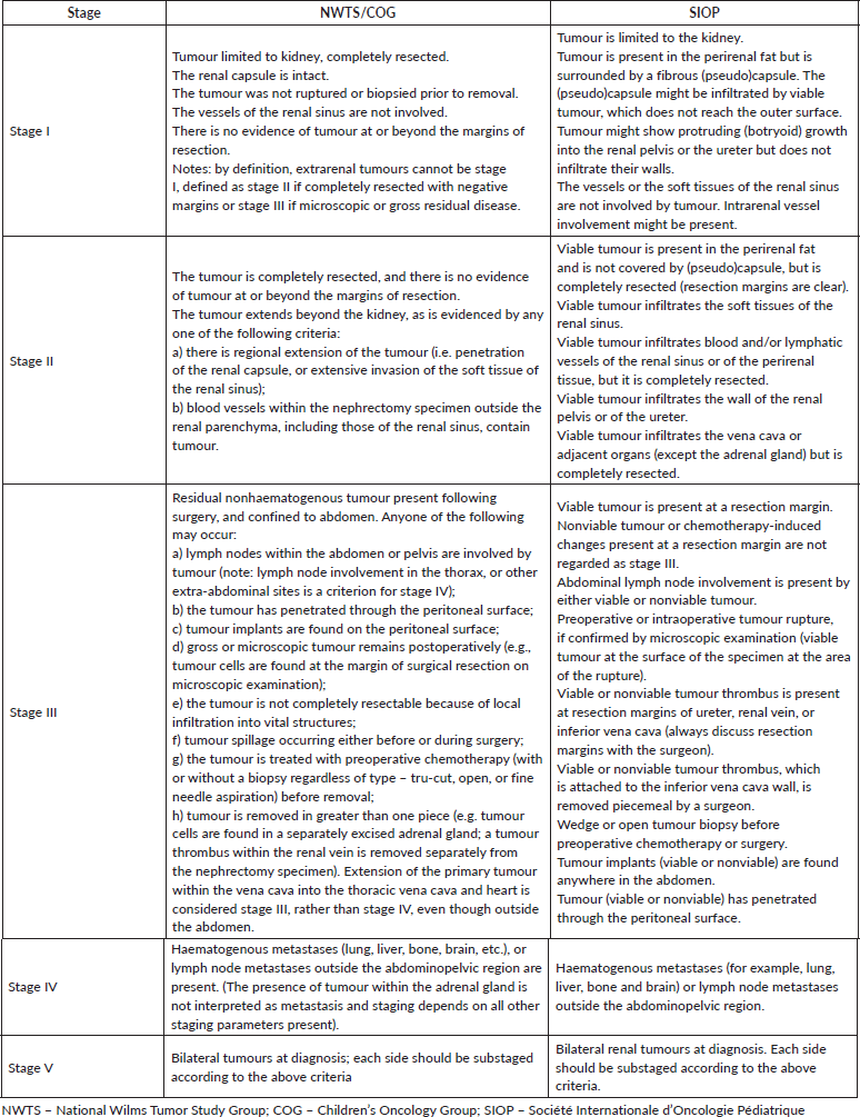
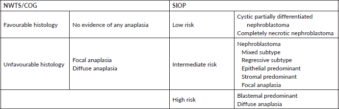
All patients deemed resectable upfront underwent open or robotic-assisted radical nephroureterectomy with standard-template retroperitoneal lymph node dissection [12]. The surgical specimens underwent histopathological examination for staging and histological subtyping, according to the NWTS/COG staging system and COG histological classification criteria, respectively [4, 13, 14] (Supplementary Tables 1 and 2). None of the patients were tested for loss of heterozygosity at 1p and 16q. Based on the assigned stage and histology, patients received adjuvant therapy, as per the NWTS-5 study protocol [15, 16].
Preoperative chemotherapy followed by delayed nephrectomy (SIOP approach)
All the patients with high-risk features precluding upfront resection would receive 4–6 weeks of preoperative chemotherapy comprising vincristine and actinomycin D with/without anthracycline (doxorubicin/epirubicin) [4, 14]. Image-guided percutaneous biopsy was reserved only for those with atypical clinical and radiological features to confirm the diagnosis of WT. Response to preoperative chemotherapy at the primary and metastatic sites, was assessed using CECT – chest, abdomen and pelvis [17].
Following preoperative chemotherapy, patients underwent open or robotic-assisted radical nephroureterectomy with standard-template retroperitoneal lymph node dissection. Nephron-sparing surgery was reserved for bilateral WT, unilateral WT with bilateral predisposition, or WT arising from a solitary kidney/horseshoe kidney [12]. The surgical specimens were examined histopathologically for stage, extent of necrosis and histological subtype. Staging was performed according to the SIOP staging system, while histological classification was based on the SIOP criteria [4, 13, 14] (Supplementary Tables 1 and 2). Based on the final stage, histological risk group and metastatic response, patients received adjuvant therapy according to the SIOP-9/SIOP-93 protocols (before 2010) and the SIOP 2001 protocol (after 2010) [18–20].
Data management and statistical considerations
A standardised case record form was designed to systematically capture the data pertinent to the study. The data were entered into Microsoft Excel 2016 (Microsoft, Redmond, CA, USA). Data analysis was performed using STATA/SE 11.2 (Stata Corp, College Station, TX, USA). Malnutrition was defined as weight for age < −2 Z score or weight for height < −2 Z score (for children <5 years) or body mass index for age < −2 Z score (for children ≥5 years). We calculated the Z-scores using the WHO Anthro software v3.2.2 and Anthro Plus software v1.0.4 for patients below 5 years and above 5 years, respectively. The median follow-up duration was calculated using the reverse Kaplan–Meier method. The Kaplan–Meier method was used to estimate event-free survival (EFS) and OS with corresponding 95% confidence intervals (CIs). An event was defined as treatment abandonment or death due to any cause or relapse or progression of the disease. EFS was calculated from the date of diagnosis to the date of the first documented event. OS was calculated from the date of diagnosis to the date of death due to any cause. The patients without an event or death were censored on the date of the last follow-up. All statistical tests were two-sided, with p < 0.05 considered statistically significant.
Results
During the 24-year study period (January 2000 to December 2023), a total of 89 children were evaluated for renal tumours at our centre. Of these, 17 children (19%) were diagnosed with non-Wilms renal tumour. Among the remaining 72 children with a provisional diagnosis of WT, 3 were excluded as they had undergone surgery elsewhere but without a confirmed histopathological diagnosis, complete staging information and essential surgical details. This manuscript reports the clinical characteristics, treatment details and survival outcomes of the remaining 69 children who were diagnosed and managed as WT at our centre during the study period (Figure 1).
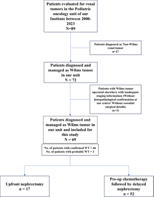
Figure 1. Distribution of children with renal tumour treated in our unit between January 2000 and December 2023.
Baseline characteristics
Among the 69 study subjects, 66 children were classified as confirmed WT while the remaining 3 were categorised as probable WT (Figure 1). The baseline characteristics of the study population are summarised in Table 1. The median age at diagnosis of the study population was 34 months (range: 8–159 months). There was a slight male predominance (M:F ratio 1.22:1) within the study cohort. About 28 out of 69 children (41%) were malnourished at the time of diagnosis. Of the 69 patients, 34 (49%) had right-sided tumours, 32 (46%) had left-sided tumours and the remaining 3 (5%) had bilateral involvement.
Table 1. Baseline characteristics of children with WT treated in our unit between January 2000 and December 2023 (N = 69).
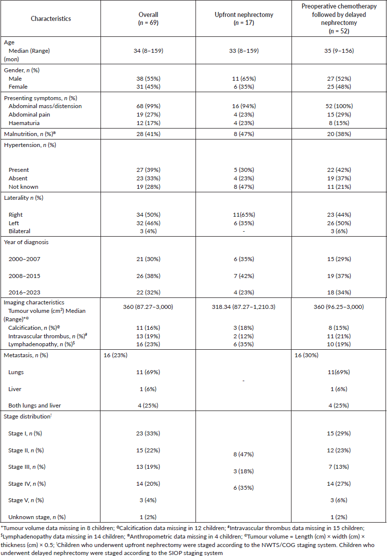
Among the 69 patients in our study cohort, 68 (99%) presented with abdominal mass or distension. In contrast, abdominal pain and haematuria were observed in only 19 (27%) and 12 (17%) patients, respectively. Hypertension at the time of diagnosis was documented in 27 (39%) children. Syndromic features and congenital anomalies were identified in nine children within our study cohort. These included undescended testes in two children, hemihypertrophy in two, horseshoe kidney in two, malrotated kidney in one, renal failure in one and developmental delay in one child. Among the study participants with available imaging details, the median tumour volume was 360 cm³ (range: 87.27–3,000 cm³). Calcifications were observed in 11 children (16%), intravascular thrombus in 13 (19%) and regional lymph node metastases in 16 (23%). Metastatic disease at presentation was documented in 16 children, of whom 11 had isolated lung metastases, 1 had isolated liver metastasis and 4 had both lung and liver involvement.
Treatment approach
Eight children who had previously undergone upfront nephrectomy elsewhere and were subsequently treated at our centre were also included in the study, as histopathological diagnosis and staging were confirmed at our institution, and essential surgical details were available. Among the remaining 61 children, 9 children who did not exhibit any of the aforementioned image-defined high-risk features underwent upfront nephrectomy. The remaining 52 children, who had one or more image-defined high-risk features, received preoperative chemotherapy followed by delayed nephrectomy. Although none of the clinical or radiological features showed statistically significant differences between the two treatment groups, children who underwent delayed nephrectomy tended to have larger tumour volumes and a higher incidence of intravascular thrombus, but a lower incidence of regional lymphadenopathy.
Upfront nephrectomy
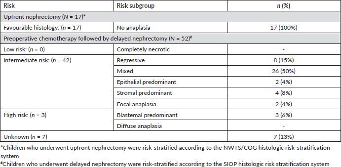
Among our study cohort, 17 children (8 – elsewhere; 9 – at our centre) underwent upfront radical nephroureterectomy. None of the patients who underwent upfront nephrectomy at our centre had either preoperative or intraoperative tumour spill, whereas four out of eight children (50%) operated upfront elsewhere had tumour spill. One child with an inferior vena cava tumour thrombus (misinterpreted as vessel compression by the mass in the preoperative imaging) who underwent upfront surgery at our institute was found to have gross residual disease postoperatively. The stage distribution among the children who underwent upfront nephrectomy was as follows: Stage I–8 (47%); Stage II–3 (18%) and Stage III–6 (35%). None of the 17 children had unfavourable histology (Table 2). Except for two children who did not receive adjuvant treatment – one due to treatment abandonment and the other due to poor general condition secondary to renal failure – the remaining 15 children received adjuvant chemotherapy: 10 with a two-drug regimen (vincristine and actinomycin D) and 5 with a three-drug regimen (vincristine, actinomycin D and doxorubicin) (Supplementary Figure 1). Additionally, 4 of the 17 children (23%) received flank irradiation.
Table 2. Histologic risk stratification of children with WT treated in our unit from January 2000 to December 2023 (N = 69).
Delayed nephrectomy
Among our study cohort, 52 children received preoperative chemotherapy, including 36 with localised WT and 16 with metastatic disease. Of these, only 25 children (48%) underwent diagnostic biopsy before treatment initiation. Among them, the biopsy findings of 22 children were concordant with the final postnephrectomy histopathological diagnosis. In the remaining three cases, nephrectomy was not performed, precluding histopathological comparison. Of the 36 children with localised disease, 35 received a two-drug preoperative chemotherapy regimen (vincristine and actinomycin D), while 1 child received a three-drug preoperative chemotherapy regimen (vincristine, actinomycin D and doxorubicin/epirubicin). Among the 16 children with metastatic disease, 9 received a two-drug preoperative chemotherapy regimen and the remaining 7 received a three-drug preoperative chemotherapy regimen (Supplementary Figure 2). Postchemotherapy tumour volume measurements were available only for 38 children, of whom 31 (81%) had a residual tumour volume of less than 500 cm³, including 20 (53%) with a volume of less than 200 cm³.
Of the 52 children who received preoperative chemotherapy, 6 did not undergo nephrectomy: 3 due to poor general condition secondary to progressive disease; 2 due to treatment abandonment and 1 child with a horseshoe kidney in whom partial nephrectomy was deemed not feasible. Among the remaining 46 children, 2 with bilateral WT and 1 with a horseshoe kidney underwent partial nephrectomy, whereas the remaining 43 underwent radical nephrectomy. Tumour rupture was observed in three children who underwent delayed nephrectomy – two had preoperative rupture and one experienced intraoperative spill.
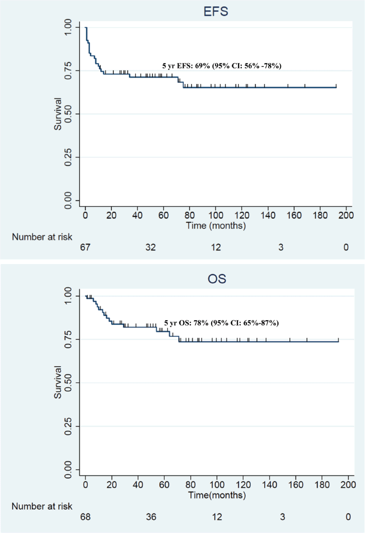
Figure 2. (a): EFS of children with WT treated in our unit between January 2000 and December 2023 (N = 69). (b): OS of children with WT treated in our unit between January 2000 and December 2023 (N = 69).
The stage distribution among the 52 children who underwent delayed nephrectomy was as follows: Stage I – 15 (29%); Stage II – 12 (23%); Stage III – 7 (13%); Stage IV – 14 (27%); Stage V – 3 (6%) and Stage not known – 1 (2%). The majority of patients (n = 42; 81%) had intermediate-risk histology, with mixed histology being the most common subtype, accounting for half of these cases (Table 2; Supplementary Figures 3 and 4). Among the 46 children who underwent delayed nephrectomy following preoperative chemotherapy, 2 patients abandoned treatment before initiating any adjuvant therapy. Of the 44 children who received adjuvant chemotherapy, 19 received a two-drug regimen (vincristine and actinomycin D), 16 received a three-drug regimen (vincristine, actinomycin D and doxorubicin/epirubicin) and the remaining 9 received a four-drug regimen (cyclophosphamide, doxorubicin, carboplatin and etoposide) (Supplementary Figure 5). Thirteen children (25%) who underwent delayed nephrectomy received radiotherapy: flank alone in nine, whole abdominal irradiation in one, flank plus whole lung irradiation (WLI) in one, WLI alone in one and WLI with liver irradiation in one child.
Survival outcomes
The median follow-up duration of the study participants who remained alive without any evidence of disease was 68.5 months (range: 11–192 months). The 5-year EFS and OS rates of the entire study cohort were 69% (95% CI: 56%–78%) and 78% (95% CI: 65%–87%), respectively. The Kaplan–Meier survival curves depicting the EFS and OS of the entire study cohort are shown in Figure 2a and b. The 5-year EFS and OS rates for patients with Stage I: 91% (95% CI: 70%–98%) and 91% (95% CI: 70%–98%); Stage II: 66% (95% CI: 37%–84%) and 63% (95% CI: 32%–83%); Stage III: 61% (95% CI: 31%–82%) and 91% (95% CI: 51%–99%); Stage IV: 52% (95% CI: 23%–75%) and 67% (95% CI: 34%–86%); and Stage V: 33% (95% CI: 9%–77%) and 100% (Figure 3a and b). The 5-year EFS and OS rates of the children who underwent upfront nephrectomy were 51% (95% CI: 25%–72%) and 60% (95% CI: 28%–82%), respectively. In comparison, those who underwent delayed nephrectomy had a 5-year EFS of 75% (95% CI: 60%–84%) and OS of 83% (95% CI: 69%–91%). However, these differences were not statistically significant for either the 5-year EFS (p = 0.2) or 5-year OS (p = 0.27) (Figure 4a and 4b). The most common cause of treatment failure among our study cohort was disease relapse (n = 15; 22%), followed by treatment abandonment (n = 8; 11%). Additionally, two children developed second malignant neoplasms: one developed papillary thyroid carcinoma 6 years after treatment completion, and another child developed Ewing sarcoma of the left tibia 2 years after treatment completion.
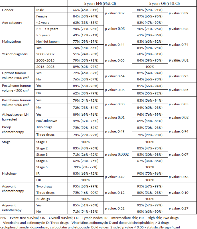
Among the entire study cohort, univariate analysis showed that the 5-year EFS and OS were significantly influenced by the year of diagnosis and the number of lymph nodes harvested (Supplementary Table 3). The patients diagnosed in more recent years (2016–2023) had significantly superior 5-year EFS and OS (86% and 95%, respectively) compared to those diagnosed in the earlier time periods. Similarly, the patients with at least seven lymph nodes harvested had markedly better 5-year EFS and OS (87% and 92%, respectively) than those with fewer lymph nodes sampled or with missing information (54% and 65%, respectively). Disease stage was significantly associated with 5-year EFS but not OS in the univariate analysis. Multivariate analysis revealed disease stage and treatment approach (upfront nephrectomy versus delayed nephrectomy) as significant independent predictors of both EFS and OS (Supplementary Table 4). Delayed nephrectomy showed lower hazards for events (HR: 0.22; 95% CI: 0.05–0.92); p = 0.04) and mortality (HR: 0.13; 95% CI: 0.02–0.85); p = 0.03). Advanced disease stage is associated with increased risk of poor EFS and OS. Univariate analyses within the upfront and delayed nephrectomy cohorts are separately summarised in Supplementary Tables 5 and 6, respectively. Multivariate Cox regression for both cohorts yielded nonconvergent models due to limited events.
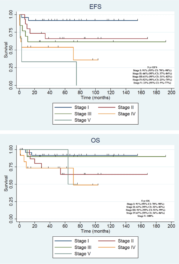
Figure 3. (a): Stage-wise EFS of children with WT treated in our unit between January 2000 and December 2023 (N = 69). (b): Stage-wise OS of children with WT treated in our unit between January 2000 and December 2023 (N = 69).
Discussion
This study reports the outcomes of children with WTs from a tertiary cancer care centre in an LMIC. With the help of multimodal therapy, we achieved a 5-year EFS of 69% and a 5-year OS of 78%. These findings underscore that, despite intensive multimodal treatment, the outcomes for WT in LMICs remain inferior to those reported in HICs.
WT constituted the majority of renal tumours (81%) in our study cohort. The slight male predominance among our study population contrasts with the global literature. This deviation likely reflects the gender disparity in seeking cancer care in India rather than any underlying biological difference [2, 21]. Among our study cohort, 41% of the children were malnourished at the time of diagnosis. This figure is comparable to the prevalence reported by Rahiman et al [22] for children with WT in North India. Among the children with available tumour volume measurements, the median tumour volume at diagnosis in our study population is less than that reported by Rahiman et al [22] (481 mL) and Joseph et al [23] (559 mL), despite having a similar stage distribution. The prevalence of metastatic disease (23%) and bilateral WT (5%) among our study cohort is consistent with the findings of various Indian and international studies [2, 5].
At our institute, we have adopted a hybrid approach for managing children with renal tumours, exploiting the advantages of both upfront and delayed nephrectomy. Among the patients with WTs in our study cohort, the majority (75%) underwent delayed nephrectomy while the remaining (25%) underwent upfront nephrectomy. In contrast, the Non-Wilms renal tumour cohort from our unit demonstrated a reverse trend, with 65% undergoing upfront nephrectomy and 35% undergoing delayed nephrectomy [24].
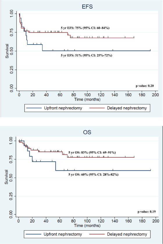
Figure 4. (a): EFS of children with WT who underwent upfront nephrectomy (n = 17) versus delayed nephrectomy (n = 52) in our unit between January 2000 and December 2023. (b): OS of children with WT who underwent upfront nephrectomy (n = 17) versus delayed nephrectomy (n = 52) in our unit between January 2000 and December 2023.
Among the nine children who underwent upfront nephrectomy at our institute, none had intraoperative tumour spill, although one child had incomplete resection. Overall, the rate of adverse surgical events among our study cohort who underwent upfront nephrectomy in-house was 3.6%, in stark contrast to 50% among the children who underwent upfront nephrectomy elsewhere (four out of eight had intraoperative tumour spill). This tailored, risk-adapted approach to decide on the timing of surgery based on imaging features has already been prospectively validated by Qureshi et al [25] who reported tumour rupture in only 1 of 19 children selected for upfront nephrectomy in the absence of high-risk imaging features. These findings underscore the importance of surgical expertise in WT management and suggest that even in high-volume tertiary centres, restricting upfront nephrectomy only to carefully selected patients at low risk for adverse surgical events would result in a significant reduction in treatment burden.
The 5-year EFS and OS rates of the entire study cohort were 69% (95% CI: 56%–78%) and 78% (95% CI: 65%–87%), respectively. These survival outcomes fall within the range of previously reported survival rates for children with WT in India [5, 6]. Notably, our results lie between the survival outcomes reported for WT in low-income countries and LMICs in Africa and those from HICs [26–28]. This gradient in survival underscores the influence of social determinants of health and the strength of the healthcare system on WT outcomes [5, 29]. Following treatment relapse (22%), treatment abandonment (11%) was the next most common cause of treatment failure. A temporal decline in treatment abandonment was observed, possibly reflecting improved social support systems in more recent years.
While the outcomes of stage I disease were comparable to global standards, the 5-year EFS and OS of stage II disease were notably inferior. Consistent with the findings of Rahiman et al [22], we also observed lower survival rates among stage II compared to stage III disease. This paradox may be attributed to inadequate lymph nodal sampling in our study cohort. Only 45% of our study subjects underwent lymph node sampling that met the recommended adequacy criteria – defined as the removal and examination of at least seven lymph nodes – with only 60% of children with stage II disease achieving this benchmark. This suboptimal lymph nodal assessment may have contributed to understaging and, consequently, undertreatment, thereby resulting in inferior outcomes. Notably, inadequate lymph node sampling was documented to be the most common protocol deviation in both resource-replete and resource-limited settings. The adverse impact of inadequate lymph nodal sampling is most pronounced among children with stage II, as documented by Kieran et al [30–32]. Furthermore, pathological staging following preoperative chemotherapy requires considerable expertise, as chemotherapy-induced changes within lymph nodes, even in the absence of viable tumour cells, constitute evidence of prior nodal metastasis and warrant classification as stage III under the SIOP staging system [33]. Failure to recognise these treatment-related changes may further contribute to understaging. Supporting this, both lymph node density and log odds of positive lymph nodes, which incorporate not only the number of positive lymph nodes but also the total number of lymph nodes harvested and the number of negative lymph nodes, have recently been identified as important prognostic factors for predicting survival outcomes in children with WT [34–36]. These metrics underscore the importance of meticulous surgical sampling and expert pathological evaluation for accurate staging and appropriate risk-adapted therapy.
Multivariate analysis identified only disease stage and treatment approach (upfront versus delayed nephrectomy) as independent prognostic factors. Compared to upfront nephrectomy, delayed nephrectomy was associated with a significantly lower risk of events and mortality, suggesting that preoperative chemotherapy may confer a survival advantage beyond its traditional role in downstaging and facilitating resection in LMIC settings. While upfront nephrectomy was limited to children with stage I–III disease, delayed nephrectomy was employed across stages I–V. By adjusting for stage as a confounding variable, multivariate analysis effectively unmasked the independent benefit of the delayed nephrectomy approach. Also in our study cohort, increased adverse surgical events associated with upfront nephrectomy performed at peripheral, less-experienced centres negated the benefit of a favourable stage distribution expected out of a tailored, risk-adapted approach to surgical timing.
Our findings emphasise the multifactorial challenges behind poorer WT outcomes in LMICs – socioeconomic constraints, healthcare system gaps and biological factors. Beyond delayed presentation and diagnostic limitations, the limited availability of surgical and pathological expertise hinders accurate staging and risk stratification. Further, additional research into the tumour biology specific to the LMIC context is essential to drive improvements in outcomes. Currently, most studies from LMICs focus predominantly on social and health system factors, with limited data on the biological and molecular characteristics of WT in these settings [29].
Strengths and limitations
This study reports the real-world experience of managing children with WT in a tertiary care centre in Southern India, highlighting the heterogeneity in management. A key strength lies in our adoption of a hybrid approach, wherein the decision between upfront and delayed nephrectomy was individualised based on multidisciplinary tumour board assessment of clinical and radiological risk factors. Notably, our study is one of the few studies from India that explicitly documents the objective criteria guiding the selection of patients for upfront nephrectomy versus delayed surgery. Our study presents one of the longest single-institution experiences in India, enabling the assessment of trends over time and survival outcomes with an adequate follow-up. By including both upfront and delayed nephrectomy cohorts, this study provides insights into the relative effectiveness and outcomes of each strategy in a real-world resource-limited setting. However, our findings must be interpreted in light of certain limitations. The retrospective study from a single centre is inherently subject to selection bias and missing data. Despite the extended study period, subgroup sizes remained small for certain clinical scenarios, limiting the power of multivariate analyses. Evolving chemotherapy regimens and supportive care practices across two decades may have introduced treatment variability, affecting outcome comparisons. Importantly, our study lacks molecular and biological characterisation of tumours, which is increasingly relevant in contemporary risk stratification. Furthermore, patient-reported outcomes, late toxicities and long-term quality of life were not captured. Nonetheless, our study reinforces the feasibility and potential advantages of a structured, multidisciplinary and individualised approach to WT management in LMICs.
Conclusion
Outcomes for WT in LMICs remain inferior to those reported in HICs, despite multimodal treatment. An individualised, risk-adapted approach to the timing of nephrectomy appears to be a pragmatic strategy in the absence of randomised evidence. Further research addressing both health system barriers and the biological characteristics of WT in LMICs is needed to bridge the survival gap.
List of abbreviations
CECT, Contrast-enhanced computed tomography; CI, Confidence incidence; COG, Children’s Oncology Group; CT, Computerised tomography; EFS, Event-free survival; HICs, High-income countries; HR, Hazard ratio; LMICs, Low- and middle-income countries; NWTS, National Wilms Tumor Study Group; OS, Overall survival; PODC, Paediatric Oncology in Developing Countries; SIOP, Société Internationale d’Oncologie Pédiatrique; WHO, World Health Organization; WLI, Whole lung irradiation; WT, Wilms tumour.
Acknowledgment
The manuscript has been read and approved by all the authors. Each author confirms that the manuscript represents honest and original work.
Conflicts of interest
The authors have no conflicts of interest to declare that are relevant to the content of this article.
Funding
The authors received no specific funding for this work.
Financial and non-financial interest declarations
The authors have no relevant financial or non-financial interests to disclose.
Consent to participate
Informed consent was waived off by the Institutional Ethics Committee of Cancer Institute (WIA), with Reg No: ECR/235/Inst/TN/2013/RR-24 (Ref No:IEC/2025/April 25) as this study was a retrospective study using anonymised data.
Consent for publication
Not applicable.
Ethical approval
Human and animal rights: This research involved only human participants.
Ethical approval: Prior approval and clearance were obtained from the Institutional Ethics Committee of Cancer Institute (WIA), with Reg No: ECR/235/Inst/TN/2013/RR-24 (Ref No:IEC/2025/April 25). All procedures performed in our study were in accordance with the ethical standards of the institutional and/or national research committee and with the 1964 Helsinki Declaration and its later amendments or comparable ethical standards.
Author contributions
VR conceived the concept, overlooked the entire study process and validated the data and manuscript; PS collected the data, performed the formal analysis and prepared the draft of the manuscript; AR verified the data and corrected the manuscript; GD, BTK and GVV corrected the manuscript.
Declaration of artificial intelligence (AI) in scientific writing
The authors declare that no AI tools were used in the preparation of this manuscript.
Clinical trial registration
Not applicable.
Data availability
The data supporting the findings of this study are available with the corresponding author.
Prior presentation
This work was previously presented as a poster at SIOP Asia, Riyadh, Saudi Arabia on April 13–15, 2025.
References
1. Libes J, Hol J, and Neto JCDA, et al (2023) Pediatric renal tumor epidemiology: global perspectives, progress, and challenges Pediatr Blood Cancer 70 e30006 https://doi.org/10.1002/pbc.30006
2. Nakata K, Colombet M, and Stiller CA, et al (2020) Incidence of childhood renal tumours: an international population‐based study Int J Cancer 147 3313–3327 https://doi.org/10.1002/ijc.33147 PMID: 32902866 PMCID: 7689773
3. Graf N, Bergeron C, and Brok J, et al (2021) Fifty years of clinical and research studies for childhood renal tumors within the International Society of Pediatric Oncology (SIOP) Ann Oncol 32 1327–1331 https://doi.org/10.1016/j.annonc.2021.08.1749 PMID: 34416363
4. Spreafico F, Fernandez CV, and Brok J, et al (2021) Wilms tumour Nat Rev Dis Primer 7 75 https://doi.org/10.1038/s41572-021-00308-8
5. Srinivasan S, Ramanathan S, and Prasad M (2023) Wilms tumor in India: a systematic review South Asian J Cancer 12 206–212 https://doi.org/10.1055/s-0042-1758567 PMID: 37969674 PMCID: 10635777
6. Siddaiahgari SR, Vaddadi S, and Bandi V, et al (2025) Patterns of care and survival of Wilms tumor in children in India: a retrospective multicentric INPHOG study Pediatr Blood Cancer 72 e31871 https://doi.org/10.1002/pbc.31871 PMID: 40611538
7. Framework C (2021) CureAll Framework: WHO Global Initiative for Childhood Cancer. Increasing Access, Advancing Quality, Saving Lives 1st edn (Geneva: World Health Organization)
8. Agrawal V, Mishra A, and Yadav SK, et al (2022) A 10-year study of the outcome of Wilms’ tumor in central India and identifying practice gaps J Indian Assoc Pediatr Surg 27 42–52 https://doi.org/10.4103/jiaps.JIAPS_314_20 PMID: 35261513 PMCID: 8853598
9. Saha S, Srinivasan S, and Nanda SS, et al (2024) Clinical profile and outcomes of childhood Wilms tumors treated in a tertiary cancer center from North India South Asian J Cancer 0044 44 [https://doi.org/10.1055/s-0044-1790223]
10. Howard SC, Davidson A, and Luna‐Fineman S, et al (2017) A framework to develop adapted treatment regimens to manage pediatric cancer in low‐ and middle‐income countries: the pediatric oncology in developing countries (PODC) committee of the international pediatric oncology society (SIOP) Pediatr Blood Cancer 64 e26879 https://doi.org/10.1002/pbc.26879
11. Israels T, Moreira C, and Scanlan T, et al (2013) SIOP PODC: clinical guidelines for the management of children with Wilms tumour in a low income setting Pediatr Blood Cancer 60 5–11 https://doi.org/10.1002/pbc.24321
12. Kumar GR, Shah AC, and Murali A, et al (2025) Surgical practices and oncological outcomes of Wilms tumor in a resource limited setting: a risk-adapted approach Pediatr Surg Int 41 105 https://doi.org/10.1007/s00383-025-05998-9 PMID: 40198412
13. Theilen TM, Braun Y, and Bochennek K, et al (2022) Multidisciplinary treatment strategies for Wilms Tumor: recent advances, technical innovations and future directions Front Pediatr 10 852185 https://doi.org/10.3389/fped.2022.852185 PMID: 35911825 PMCID: 9333359
14. Bhatnagar S (2009) Management of Wilms′ tumor: NWTS vs SIOP J Indian Assoc Pediatr Surg 14 6 https://doi.org/10.4103/0971-9261.54811
15. Dome JS, Cotton CA, and Perlman EJ, et al (2006) Treatment of anaplastic histology Wilms’ tumor: results from the Fifth National Wilms’ Tumor Study J Clin Oncol 24 2352–3258 https://doi.org/10.1200/JCO.2005.04.7852 PMID: 16710034
16. Grundy PE, Breslow NE, and Li S, et al (2005) Loss of heterozygosity for chromosomes 1p and 16q Is an adverse prognostic factor in favorable-histology Wilms tumor: a report from the national Wilms tumor study group J Clin Oncol 23 7312–7321 https://doi.org/10.1200/JCO.2005.01.2799 PMID: 16129848
17. De La Monneraye Y, Michon J, and Pacquement H, et al (2019) Indications and results of diagnostic biopsy in pediatric renal tumors: a retrospective analysis of 317 patients with critical review of SIOP guidelines Pediatr Blood Cancer 66 e27641 https://doi.org/10.1002/pbc.27641 PMID: 30746839
18. Ludwig R, Weirich A, and Pötter R, et al (1992) Präoperative chemotherapie des nephroblastoms Klin Pädiatr 204 204–213 https://doi.org/10.1055/s-2007-1025350
19. Pritchard-Jones K, Bergeron C, and De Camargo B, et al (2015) Omission of doxorubicin from the treatment of stage II–III, intermediate-risk Wilms’ tumour (SIOP WT 2001): an open-label, non-inferiority, randomised controlled trial Lancet 386 1156–1164 https://doi.org/10.1016/S0140-6736(14)62395-3 PMID: 26164096
20. Reinhard H, Semler O, and Bürger D, et al (2004) Results of the SIOP 93-01/GPOH trial and study for the treatment of patients with unilateral nonmetastatic Wilms tumor Klin Pädiatr 216 132–140 [https://doi.org/10.1055/s-2004-822625]
21. Bhatia KP, Ganguly S, and Sasi A, et al (2023) Sex disparity in childhood cancer in India: a multi-centre, individual patient data analysis Lancet Oncol 24 54–63 https://doi.org/10.1016/S1470-2045(22)00688-X
22. Rahiman EA, Trehan A, and Jain R, et al (2022) A higher tumor volume and undernutrition at diagnosis adversely affect the survival of children with Wilms tumor: a study of 200 patients Pediatr Blood Cancer 69 e29880 https://doi.org/10.1002/pbc.29880 PMID: 35841309
23. Joseph LL, Boddu D, and Srinivasan HN, et al (2022) Postchemotherapy tumor volume as a prognostic indicator in Wilms tumor: a single‐center experience from South India Pediatr Blood Cancer 69 e29454 https://doi.org/10.1002/pbc.29454
24. Srinivasan P, Das G, and Kothandan BT, et al (2025) Multidisciplinary management of children with non-Wilms renal tumor: a real-world evidence from a tertiary cancer care center in Southern India Indian J Med Paediatr Oncol 0045 [https://doi.org/10.1055/s-0045-1813008]
25. Qureshi SS, Kembhavi SA, and Bhagat M, et al (2019) Customized approach for upfront or delayed resection using radiological criteria in unilateral, nonmetastatic pediatric renal tumors: a prospective study Pediatr Blood Cancer 66 e27815 https://doi.org/10.1002/pbc.27815 PMID: 31099132
26. Jackson TJ, Al-Saadi R, and Lopez-Cortes A, et al (2023) Comparing routinely collected population level healthcare data to a prospective clinical study of Wilms Tumour in England EJC Paediatr Oncol 2 100114 https://doi.org/10.1016/j.ejcped.2023.100114
27. Fufa D, Mdoka C, and Ayalew M, et al (2024) Effectiveness of a Wilms tumour treatment guideline adapted to local circumstances in sub‐Saharan Africa: a report from Wilms Africa phase II—CANCaRe Africa Pediatr Blood Cancer 71 e31300 https://doi.org/10.1002/pbc.31300
28. Cunningham ME, Klug TD, and Nuchtern JG, et al (2020) Global disparities in Wilms tumor J Surg Res 247 34–51 https://doi.org/10.1016/j.jss.2019.10.044
29. Apple A and Lovvorn HN (2020) Wilms tumor in Sub-Saharan Africa: molecular and social determinants of a global pediatric health disparity Front Oncol 10 606380 https://doi.org/10.3389/fonc.2020.606380 PMID: 33344257 PMCID: 7746839
30. Ehrlich PF, Hamilton TE, and Gow K, et al (2016) Surgical protocol violations in children with renal tumors provides an opportunity to improve pediatric cancer care: a report from the Children’s Oncology Group Pediatr Blood Cancer 63 1905–1910 https://doi.org/10.1002/pbc.26083 PMID: 27229358 PMCID: 5030129
31. Elgendy A, Abouheba M, and Ebeid A, et al (2020) Surgical aspects, violations and outcomes of Wilms tumor—a multicenter study in a resource-limited country Egypt Pediatr Assoc Gaz 68 18 [https://doi.org/10.1186/s43054-020-00031-1]
32. Kieran K, Anderson JR, and Dome JS, et al (2012) Lymph node involvement in Wilms tumor: results from National Wilms Tumor Studies 4 and 5 J Pediatr Surg 47 700–706 https://doi.org/10.1016/j.jpedsurg.2011.08.017 PMID: 22498384 PMCID: 3976547
33. Vujanić GM, Gessler M, and Ooms AHAG, et al (2018) The UMBRELLA SIOP–RTSG 2016 Wilms tumour pathology and molecular biology protocol Nat Rev Urol 15 693–701 https://doi.org/10.1038/s41585-018-0100-3
34. You H, Yang J, and Liu Q, et al (2018) The impact of the lymph node density on overall survival in patients with Wilms’ tumor: a SEER analysis Cancer Manag Res 10 671–677 https://doi.org/10.2147/CMAR.S163514
35. Ziogas IA, Khomiak A, and Olson KE, et al (2025) The impact of lymph node ratio for children with Wilms tumors: a national cancer database analysis Cancers 17 3276 https://doi.org/10.3390/cancers17193276 PMID: 41097801 PMCID: 12523910
36. Chen S, Wan Z, and Hu S, et al (2024) Prediction values of different lymph nodes staging systems for survival of children with Wilms tumor Transl Cancer Res 13 6688–6698 https://doi.org/10.21037/tcr-24-959
Supplementary materials
Supplementary Table 1. NWTS/COG and SIOP staging systems of WT.
Supplementary Table 2. NWTS/COG and SIOP histologic risk stratification system of WT.
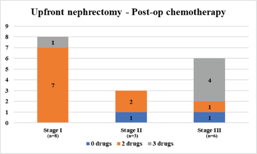
Supplementary Figure 1. Details of postop chemotherapy regimens received by children with WT, who underwent upfront nephrectomy and were treated in our unit between January 2000 and December 2023 (N = 17). Staging according to NWTS/COG staging system; 0 drug – No adjuvant postop chemotherapy received; two drugs – received vincristine and actinomycin D; three drugs – received vincristine, actinomycin D and doxorubicin.
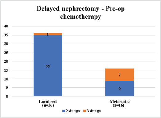
Supplementary Figure 2. Details of preop chemotherapy regimens received by children with WT treated in our unit between January 2000 and December 2023 (N = 52). Staging according to the SIOP staging system; two drugs – received vincristine and actinomycin D; three drugs – received vincristine, actinomycin D and doxorubicin/epirubicin.
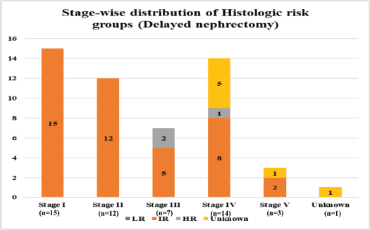
Supplementary Figure 3. Stage-wise distribution of histologic risk groups among children who underwent delayed nephrectomy in our unit between January 2000 and December 2023 (N = 52). Staging according to the SIOP staging system; Risk stratification according to SIOP risk stratification; LR – Low risk; IR – Intermediate risk; HR – High risk.
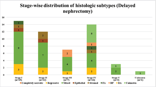
Supplementary Figure 4. Stage-wise distribution of histologic subtypes among children who underwent delayed nephrectomy in our unit between January 2000 and December 2023 (N = 52). Staging according to SIOP staging system; Histologic subtyping according to SIOP histologic classification; FA - Focal anaplasia; BP – Blastemal predominant; DA – Diffuse anaplasia.
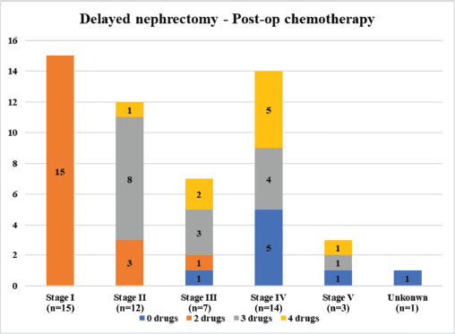
Supplementary Figure 5. Details of postop chemotherapy regimens received by children with WT, who underwent delayed nephrectomy and were treated in our unit between January 2000 and December 2023 (N = 52). Staging according to the SIOP staging system: 0 drug – No adjuvant postop chemotherapy received; two drugs – received vincristine and actinomycin D; three drugs – received vincristine, actinomycin D and doxorubicin/epirubicin; four drugs - cyclophosphamide, doxorubicin, carboplatin and etoposide.
Supplementary Table 3. Univariate analysis of factors predicting 5-year EFS and OS among children with WT treated in our unit between January 2000 and December 2023 (N = 69) using the log-rank test.
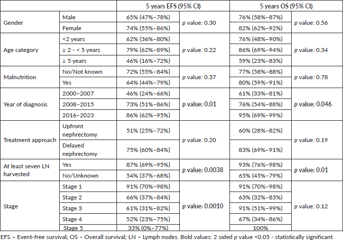
Supplementary Table 4. Multivariate analysis of factors predicting EFS and OS among children with WT treated in our unit between January 2000 and December 2023 (N = 69) using a Cox proportional hazards model.
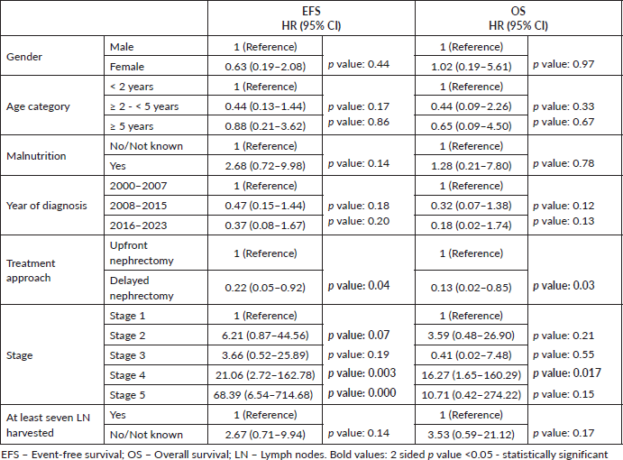
Supplementary Table 5. Univariate analysis of factors predicting 5-year EFS and OS among children with WT who underwent upfront nephrectomy in our unit between January 2000 and December 2023 (N = 17) using the log-rank test.
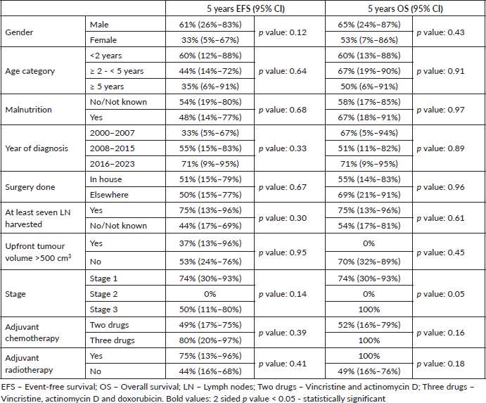
Supplementary Table 6. Univariate analysis of factors predicting 5-year EFS and OS among children with WT who underwent delayed nephrectomy in our unit between January 2000 and December 2023 (N = 52) using log-rank test.