Comprehensive genomic profiling in metastatic solid tumours: high actionability but low implementation in real-world practice
Moushumi Suryavanshi1, Prashant Mehta2, Manoj Kumar3, Saphalta Bhagmar2, Vidit Kapoor2, Dushyant Kumar3, Sweta Mishra3 and Bhawna Chauhan3
1 Department of Molecular Biology, Cytogenetics and Pathology, Amrita School of Medicine, Amrita Vishwa Vidyapeetham, Faridabad Campus, Mata Amritanandamayi Marg, Sector 88, Faridabad, Haryana 121002, India
2 Department of Hematology/Medical Oncology and BMT, Amrita School of Medicine, Amrita Vishwa Vidyapeetham, Faridabad Campus, Mata Amritanandamayi Marg, Sector 88, Faridabad, Haryana 121002, India
3 Amrita School of Medicine, Amrita Vishwa Vidyapeetham, Faridabad Campus, Mata Amritanandamayi Marg, Sector 88, Faridabad, Haryana 121002, India
Abstract
Background: Comprehensive genomic profiling (CGP) identifies actionable alterations in two-thirds of metastatic solid tumours, yet real-world treatment implementation remains unknown. We characterised both genomic yield and clinical translation barriers in 111 consecutive patients across 19 tumour types to quantify the precision oncology ‘knowing-doing gap’.
Methods: Out of 114 patients who were initially enrolled in the study, we performed retrospective analysis of 111 consecutive metastatic patients across 19 tumour types, after quality control exclusion, undergoing CGP using the Oncomine Comprehensive Assay Plus (517 genes). Genomic instability was assessed via the genomic instability metric (GIM); actionable alterations were tiered per Association for Molecular Pathology/American Society of Clinical Oncology/College of American Pathologists guidelines. Treatment implementation and clinical outcomes were tracked through October 2024 (median follow-up 11.2 months).
Results: Oncogenic alterations were detected in 109 of 111 patients (98.2%), with actionable findings in 72 patients (64.9%: Tier 1 = 32 and Tier 2 = 40). All GIM-high tumours (10/10) harboured homologous recombination repair (HRR) gene alterations. Critically, BRCA1/2 mutations were significantly enriched in GIM-high tumours (50% versus 12%, p = 0.008), whereas non-BRCA HRR genes (ATM, CHEK2, PALB2 and RAD51C/D) showed no association (50% versus 60.6%, p = 0.838). Despite 64.9% actionability, only 10 patients (13.9%) received tier-matched therapy – an 86% attrition rate attributable to clinical deterioration (22%), drug inaccessibility (19%) and financial toxicity (15%). Presumed germline variants were detected in 16.2% but confirmed in only 38.9%. Pharmacogenomic variants guided dose modifications in 29.7% of patients.
Conclusion: CGP demonstrates high diagnostic yield (98.2%) with actionable findings in two-thirds of pan-cancer patients. The differential BRCA1/2-specific association with genomic instability refines homologous recombination deficiency biology for multi-tumour Poly (ADP-ribose) polymerase inhibitor selection. An 86% attrition rate from genomic recommendation to treatment underscores urgent need for infrastructure improvements, including clinical trial access, pharmacogenomic integration and earlier CGP adoption.
Keywords: comprehensive genomic profiling, homologous recombination deficiency, precision oncology, actionable genomic alterations, treatment concordance, real-world evidence
Correspondence to: Moushumi Suryavanshi
Email: moushumisuryavanshi@fbd.amrita.edu and moushumisuryavanshi@gmail.com
Published: 01/09/2026
Received: 11/04/2026
Publication costs for this article were supported by ecancer (UK Charity number 1176307).
Copyright: © the authors; licensee ecancermedicalscience. This is an Open Access article distributed under the terms of the Creative Commons Attribution License (http://creativecommons.org/licenses/by/4.0), which permits unrestricted use, distribution, and reproduction in any medium, provided the original work is properly cited.
Introduction
The evolution of molecular diagnostics in oncology has witnessed a progressive shift from targeted single-gene testing and focussed hotspot panels to comprehensive genomic profiling (CGP), fundamentally transforming precision medicine [1, 2]. Early next-generation sequencing (NGS)-based approaches, while valuable for detecting common actionable mutations in genes such as epidermal growth factor receptor (EGFR), Kirsten rat sarcoma viral oncogene homolog and B-Raf proto-oncogene, serine/threonine kinase (BRAF) were limited by their narrow scope and inability to capture the full spectrum of clinically relevant genomic alterations. CGP platforms enable simultaneous interrogation of hundreds of cancer-related genes, detecting single nucleotide variants (SNVs), small insertions/deletions (indels), copy number variations (CNVs), gene fusions and complex genomic rearrangements from a single tumour specimen [3]. Studies demonstrate that CGP identifies actionable alterations in 30%–40% more patients compared to focussed panels, thereby expanding therapeutic options and clinical trial eligibility [4].
Beyond increased mutation detection, CGP provides critical insights into tumour biology and microenvironment. Tumour mutational burden (TMB) has emerged as a predictive biomarker for immune checkpoint inhibitor response across multiple cancer types [5], while microsatellite instability (MSI) determination provides a tissue-agnostic biomarker for immunotherapy eligibility [6]. Homologous recombination deficiency (HRD), defined by genomic instability and loss of heterozygosity (LOH) patterns, predicts Poly (ADP-ribose) polymerase (PARP) inhibitor sensitivity in ovarian cancer [7] and shows significant prevalence across breast, prostate and pancreatic tumours [8]. CGP integrates detection of homologous recombination repair (HRR) gene mutations (BRCA1/2, ATM and PALB2), copy number alterations (CNAs) and genome-wide instability signatures into a single assay, streamlining identification of patients eligible for PARP inhibitors and platinum-based therapies across tumour types.
CNV analysis enables detection of therapeutically relevant gene amplifications (ERBB2, MET, FGFR1/2 and EGFR) and homozygous deletions of tumour suppressors (CDKN2A/B and PTEN), providing both therapeutic targets and prognostic information. Categorising alterations into Tier 1 (standard-of-care) and Tier 2 (emerging/off-label) standardises CGP interpretation [9], with Tier 2 alterations expanding therapeutic options particularly through clinical trial access [10].
CGP offers dual diagnostic utility by flagging suspected germline variants in genes such as BRCA1/2 and TP53 alongside somatic mutations [11], allowing a single assay to guide targeted therapy and hereditary risk assessment. In addition, pharmacogenomic markers (DPYD and UGT1A1) identify patients at risk for severe drug toxicity, facilitating evidence-based dose adjustments per Clinical Pharmacogenetics Implementation Consortium (CPIC) guidelines [12]. Leveraging CPIC guidelines to interpret these markers facilitates evidence-based dose adjustments, thereby minimising adverse events and optimising treatment outcomes.
While recent Indian studies [13] have focussed on single disease sites, our 517-gene panel across 19 tumour types captures SNVs/indels, CNVs and fusions supporting concurrent TMB, MSI, HRD and pharmacogenomic evaluation. This pan-cancer approach enables tumour-agnostic biomarker assessment microsatellite instability-high (MSI-H/TMB-H for pembrolizumab, NTRK inhibitors) and permits cross-tumour analysis of BRCA1/2 versus non-BRCA HRR alterations with direct implications for PARP inhibitor selection.
CGP infrastructure has expanded rapidly in low- and middle-income countries (LMICs), including India, with >90% sequencing success rates achieved across major cancer centres. However, the clinical utility of this molecular infrastructure remains poorly characterised. Understanding real-world CGP translation rates in LMICs is essential for equitable deployment of genomic medicine technology and for prioritising institutional investments in precision oncology infrastructure [14, 15].
In this study, we present our institutional experience with CGP in 111 consecutive cancer patients. Our objectives were to: (1) characterise the spectrum and frequency of Tier 1 and Tier 2 targetable alterations; (2) evaluate genomic instability parameters, including HRD scores and their correlation with HRR gene mutations across tumour types; (3) assess CNVs and their therapeutic implications; (4) analyse immune biomarkers (MSI and TMB); (5) identify suspected germline variants; (6) determine prevalence of actionable pharmacogenomic variants; (7) evaluate tumour quality metrics and (8) critically assess the rate of guideline-concordant therapy administration. Through this analysis, we aim to provide insights into the clinical utility, therapeutic impact and real-world implementation of CGP in routine oncology practice.
Materials and methods
Study design and patient population
This retrospective, single-centre study included 114 consecutive patients with histologically confirmed metastatic solid tumours who underwent CGP between September 2023 and October 2024 at Amrita Institute of Medical Sciences & Research Center, Faridabad, India. The primary objective was to assess the frequency of actionable genomic alterations and the implementation rate of tier-matched therapy in routine clinical practice. Eligible patients had adequate formalin-fixed paraffin-embedded (FFPE) tumour tissue with ≥30% tumour cell content on haematoxylin–eosin (H&E)-stained slides. Macrodissection was performed when required to enrich tumour content. Clinical data, including demographics, tumour characteristics, treatment history and post-CGP therapeutic decisions, were retrieved from electronic medical records. Treatment decisions and clinical outcomes were documented through the end of the study period (October 2024). As this study was conducted at a quaternary academic trust hospital in India within a mixed public–private healthcare system, where CGP and most matched targeted therapies are not routinely covered under national healthcare schemes, most patients self-financed the testing and treatment or accessed them through private insurance support. A subset of patients received therapies through institutional clinical trial enrolment.
Appropriate pre-test and post-test counselling were performed for all patients undergoing CGP testing.
The study was approved by the Institutional Review Board (AIMS-IEC-BAS-12-25-002) and conducted according to the Declaration of Helsinki.
Comprehensive genomic profiling
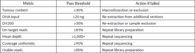
DNA was extracted from FFPE tumour sections using standard commercial kits (Thermo Fisher Scientific). Quality was assessed using the DV200 metric (≥30% required) and Qubit quantification (≥20 ng input required). CGP was performed using the Oncomine Comprehensive Assay v4 (OCA v4, 517-gene panel) on the Ion GeneStudio S5 XL System (Thermo Fisher Scientific), targeting ≥1,000× coverage depth and ≥90% uniformity. The assay simultaneously detected SNVs, insertions/deletions (indels), CNVs, gene fusions, MSI, TMB, LOH and HRD.
Samples with ≥85% on-target reads, ≥1,000× mean depth and ≥90% uniformity were considered technically successful. TMB-high was defined as ≥10 mutations/Mb. HRD status was assessed using a genomic instability metric (GIM) with a threshold of ≥16, validated against MyChoice® CDx concordance [16]. MSI status was determined using 76 microsatellite markers, with instability defined as ≥19 unstable markers. LOH-high was defined as score ≥16. DNA quality control (QC) failure was observed in 3 of the 114 samples. Therefore, subsequent testing and analysis were performed on the remaining 111 cases.
Bioinformatic analysis and variant classification
Sequence alignment to the human reference genome (hg19/GRCh37) and variant calling were performed using Torrent Suite Software v5.18.1 and Ion Reporter Software v5.20.8.0 in tumour-only analysis mode. All variants were classified according to joint Association for Molecular Pathology, American Society of Clinical Oncology and College of American Pathologists guidelines. Variants were annotated using ClinVar, COSMIC, OncoKB, CIViC and published literature.
Actionable alterations were categorised as Tier 1 (FDA-approved biomarkers with therapeutic or diagnostic significance in the specific tumour type) or Tier 2 (biomarkers with therapeutic implications based on clinical evidence in other tumour types or preclinical data). All cases were reviewed by a multidisciplinary molecular tumour board consisting of medical oncologists, pathologists and molecular geneticists to determine clinical actionability and therapeutic recommendations.
Germline pharmacogenomic variants in DPYD, CYP2D6, UGT1A1 and TPMT were identified based on variant allele frequencies (VAF; 40%–60% for heterozygous, >90% for homozygous) and classified according to CPIC guidelines. Variants suggestive of germline origin in cancer predisposition genes were flagged for confirmatory germline testing and genetic counselling based on VAF patterns and pathogenicity classification.
Treatment actionability and clinical outcomes
Clinical actionability was evaluated based on: (1) FDA-approved targeted therapies for the specific alteration and tumour type; (2) off-label therapeutic options supported by National Comprehensive Cancer Network (NCCN) guidelines or peer-reviewed evidence; (3) eligibility for biomarker-driven clinical trials and (4) immunotherapy eligibility based on MSI-H or TMB-high status.
Tier-matched therapy was defined as administration of treatment specifically targeting the identified genomic alteration according to the tier classification. Treatment implementation was tracked through medical records and reasons for non-implementation were documented when matched therapy was not administered. Actionability in this study was defined according to US FDA-approved indications. Importantly, all targeted therapies classified as actionable in the study were regulatory-approved and commercially available in India.
Clinical outcomes, including treatment response and survival status, were recorded through the study period with a median follow-up of 11.2 months from CGP testing.
Statistical analysis
Descriptive statistics summarised patient demographics, tumour characteristics, genomic findings and treatment patterns. Categorical variables were reported as frequencies and percentages and continuous variables as median with interquartile range (IQR). The frequency of alterations was calculated for the entire cohort and stratified by tumour type.
Co-occurrence and mutual exclusivity of genomic alterations were assessed using Fisher’s exact test with false discovery rate correction. Correlations between continuous variables (TMB and HRD scores) were evaluated using Spearman’s rank correlation. The association between HRD status and HRR gene mutations was assessed using the Fisher’s exact test. Associations between genomic alterations and receipt of matched therapy were evaluated using chi-square or Fisher’s exact test. All analyses were performed using R software version 4.4.2 and Python 3.x. Two-sided p-values <0.05 were considered statistically significant.
Results
Patient demographics and clinical characteristics
A total of 114 patients with metastatic solid tumours were enrolled for CGP. After QC, three cases were excluded (two for sequencing failure and one for insufficient tumour content), yielding a final cohort of 111 patients. The median age at diagnosis was 61 years (range 2–86), with 63 females (56.8%) and 48 males (43.2%). Tumour cellularity ranged from 30% to 90% (mean 60%), meeting the minimum threshold for reliable variant detection.
The cohort included diverse malignancies: ovarian cancer (n = 19, 17.1%), lung cancer (n = 13, 11.7%), prostate cancer (n = 7, 6.3%), breast cancer (n = 7, 6.3%), gallbladder (n = 6), endometrial (n = 6), hepatocellular carcinoma (n = 5), colorectal (n = 5), renal cell carcinoma (n = 4), pancreatic adenocarcinoma (n = 4) and others (n = 29).
Sequencing performance
Sequencing metrics were robust across all samples (Figure 1): median depth 2,539× (IQR 2,047×–2,922×), median on-target rate 95.0% (IQR 94.0%–96.0%) and median coverage uniformity 92.5% (IQR 90.0%–94.0%). All samples exceeded the 500× minimum depth required for variant calling.
Landscape of genomic alterations
A total of 1,006 somatic variants were identified, comprising 229 oncogenic (22.8%), 100 likely oncogenic (9.9%) and 677 variants of uncertain significance (67.3%). Oncogenic or likely oncogenic alterations were detected in 109 of 111 samples (98.2%).
The most frequently mutated genes were TP53 (n = 47, 42.3%), PIK3CA (n = 16, 14.4%), KRAS (n = 11, 9.9%), ARID1A (n = 10, 9.0%), NF1 (n = 7, 6.3%), PTEN (n = 7, 6.3%) and TERT (n = 7, 6.3%) (Figure 2). Common co-mutations included TP53+PIK3CA (n = 7, 6.3%) and TP53+KRAS (n = 6, 5.4%).
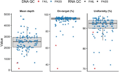
Figure 1. Sequencing QC metrics across samples. Faceted boxplots show the distribution of mean depth, on-target percentage and coverage uniformity across 114 samples. Individual points represent samples, coloured by DNA QC status and shaped by RNA QC status.
CNAs were identified in 98 samples (88.3%), frequently involving BRCA2 (n = 10), CHEK2 (n = 9), RAD51B (n = 8) and ATM/PTEN (n = 7 each). High-level amplifications (≥6 copies) occurred in 46 samples. Homozygous deletions most commonly affected TP53 (n = 39), HLA-B (n = 27), and CDKN2A (n = 26). Co-occurring loss of CDKN2A and MTAP was observed in 11 samples (9.9%) across diverse tumour types.
Gene fusions were identified in 29 samples (26.1%), representing 35 unique fusion events. Clinically actionable fusions included EML4–ALK (n = 1) and CD74–ROS1 (n = 1) in lung cancer, FGFR fusions (n = 6) across multiple tumour types and NTRK fusions (n = 2) in pancreatic cancer and glioblastoma.
Genomic biomarkers for immunotherapy and targeted therapy
All 111 samples had evaluable TMB (median 5.8 mutations/Mb, range 1.0–56.7). TMB-high (≥10 mutations/Mb) status was observed in six samples (5.4%) including endometrial (n = 2), breast (n = 1), ovarian (n = 1), lung (n = 1) and colorectal (n = 1) cancers.
MSI-H tumours were found in two samples (1.8%): one colorectal cancer carrying a pathogenic MLH1 germline variant and one endometrial cancer. All MSI-H tumours demonstrated elevated TMB (median 34.2 mutations/Mb).
HRD was quantified using the GIM, with GIM ≥16 defining GIM-high status. Ten samples (9.0%) were GIM-high. All GIM-high tumours harboured at least one HRR gene alteration (10/10, 100%), compared to 72/99 (72.7%) of GIM-low tumours (p = 0.050, Fisher’s exact test).
Notably, BRCA1/2 alterations showed a strong association with GIM-high status: 5/10 (50%) GIM-high tumours versus 12/99 (12.1%) GIM-low tumours (p = 0.008, Figure 3). In contrast, non-BRCA HRR gene alterations (ATM, CHEK2, PALB2, RAD51C and others) showed no significant association: 5/10 (50%) in GIM-high versus 60/99 (60.6%) in GIM-low tumours (p = 0.838). This distinction suggests that GIM-based HRD scoring primarily identifies BRCA-associated genomic instability.
Therapeutic actionability and clinical implementation
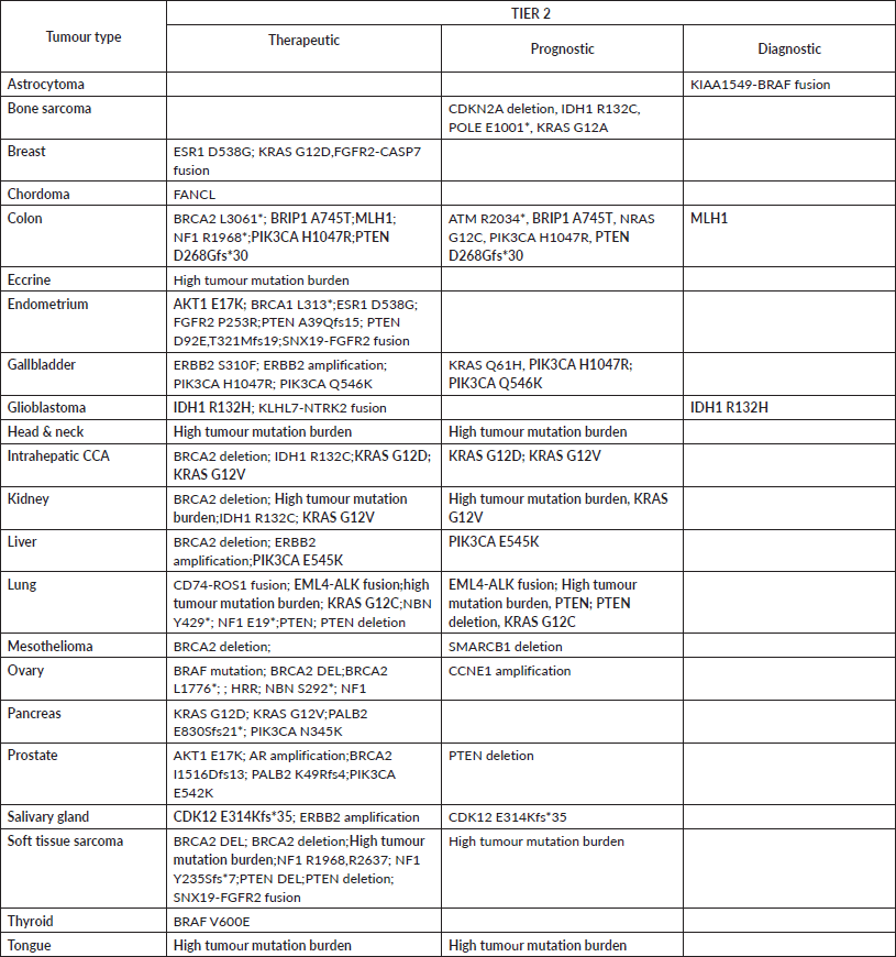
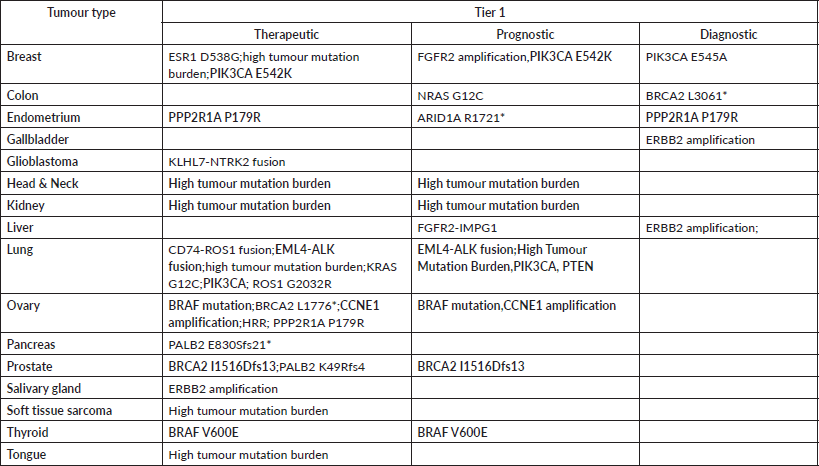
Actionable genomic alterations were identified in 72 of 111 patients (64.9%), leading to tiered therapeutic recommendations. Tier 1 recommendations (FDA-approved therapies for the specific alteration and tumour type) were made for 32 patients (28.8%), most commonly in ovarian (n = 11), lung (n = 6) and breast (n = 4) cancers. Tier 2 recommendations (clinical trial or off-label options) were made for 40 patients (36.0%). Tier-wise categorisation of biomarkers according to diagnostic, therapeutic and prognostic indications has been detailed in Supplementary Tables 1 and 2.
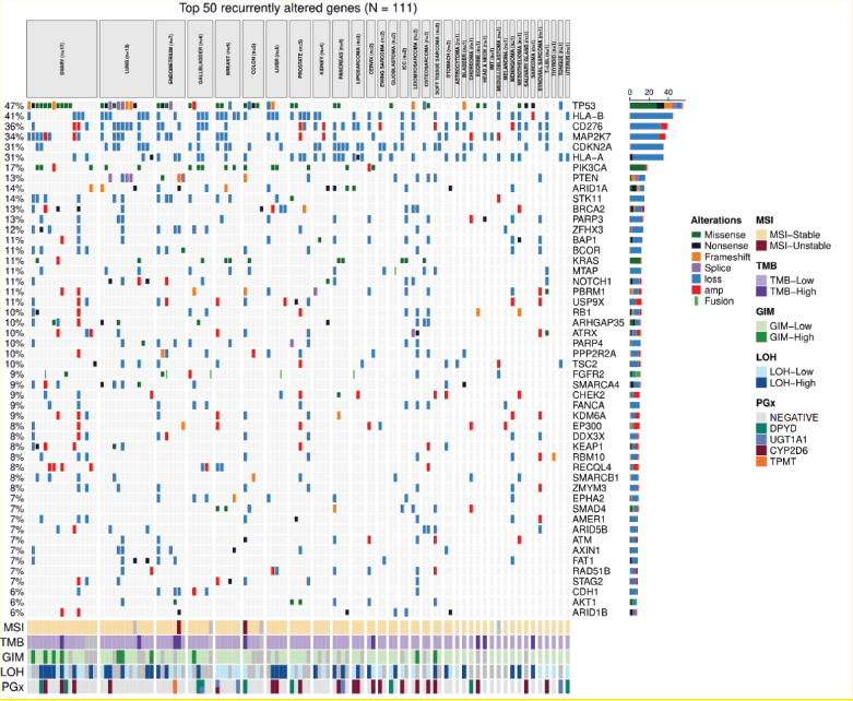
Figure 2. Genomic landscape of top 50 recurrently altered genes (N = 111). Oncoprint showing somatic alterations across 111 patients and 19 tumour types. Columns = individual patients (grouped by tumour type); rows = genes (ordered by alteration frequency, shown at right). Alteration types: green = missense, black = nonsense, orange = frameshift, purple = splice, blue = loss, red = amplification, grey = fusion. Bottom tracks: MSI, microsatellite instability; TMB, tumour mutational burden; GIM, genomic instability measure; LOH, loss of heterozygosity; PGx, pharmacogenomic variants in DPYD, UGT1A1, CYP2D6, TPMT. TP53 (47%), HLA-B (41%) and CDKN2A (31%) were most frequently altered.
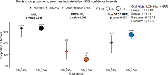
Figure 3. Genomic instability associates with BRCA1/2 but not non-BRCA HRR alterations. Proportions of tumours with HRR gene alterations by GIM status (GIM-high ≥16 versus GIM-low <16). Points show proportions with Wilson 95% confidence intervals; point size indicates sample size. Fractions denote alterations/total samples within each GIM stratum. GIM-high: n = 10; GIM-low: n = 99. All GIM-high tumours harboured ≥1 HRR alteration (10 /10, 100%) versus 72.7% of GIM-low (p = 0.050, Fisher’s exact). BRCA1/2 mutations were significantly enriched in GIM-high (50% versus 12%, p = 0.008), while non-BRCA HRR genes showed no association (50% versus 60.6%, p = 0.838). p-values by Fisher’s exact test.
Of the 72 patients with actionable recommendations, only ten (13.9% of tier recommended; 9.0% of total cohort) received therapy precisely matched to their tier recommendation: nine at Tier 1 and one at Tier 2 (Figure 4). Treatment received in the entire actionable cohort included no systemic therapy (n = 37), chemotherapy only (n = 22), multiple modalities (n = 14), targeted therapy (n = 10), immunotherapy (n = 7) and hormone therapy (n = 2).
Matched therapy cases occurred in lung (n = 2), gallbladder, liver, head and neck, kidney, tongue, ovarian and Ewing sarcoma. Actionable drivers included TMB-high (n = 4), ERBB2 amplification (n = 2), EML4–ALK fusion with ALK amplification (n = 1), CD74–ROS1 fusion with ROS1 G2032R resistance mutation (n = 1), IDH1 R132C (n = 1) and BRCA1 E23Vfs*17 (n = 1).
CGP was performed at a median of 11.2 months from diagnosis (range 0.1–23.2 months). At data cut-off, seven of ten matched therapy patients were alive (70.0%), and three had died (30.0%). Among the 62 patients who did not receive matched therapy, 35 were alive, and 27 had died. Reasons for treatment non-concordance included clinical deterioration, lack of access to targeted agents, patient refusal and alternate treatment priorities.
Germline findings and pharmacogenomic profiling
Germline testing was recommended in 18 patients (16.2%) based on tumour-only profiling suggestive of germline variants. Testing was performed in seven (38.9% of those recommended), identifying pathogenic or likely pathogenic germline variants in four patients (3.6% of total cohort; 42.9% of tested): FANCC, NBN, MLH1 and BRCA2. The 61% gap between recommendation and completion was primarily due to financial constraints (40% of non-tested), lack of genetic counselling access (35%) and patient anxiety/refusal (25%).
Thirty-three clinically relevant pharmacogenomic variants were detected involving CYP2D6 (n = 19; 15 intermediate (IM) and 4 poor metabolisers (PM)), DPYD (n = 9; all reduced activity), UGT1A1 (n = 4; 1 homozygous and 3 heterozygous) and TPMT (n = 1). These findings guided individualised dose adjustments for fluoropyrimidines, irinotecan, tamoxifen and thiopurines.
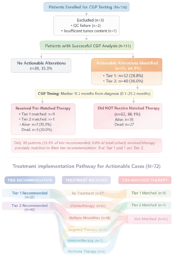
Figure 4. Flow of tier-based therapeutic recommendations to matched treatment and molecular alterations. This Sankey diagram illustrates the clinical and molecular treatment cascade across the study cohort (N = 111). Of these, 72 samples received tier-based therapeutic recommendations, including 32 Tier 1 and 40 Tier 2 recommendations. Among tier-recommended cases, chemotherapy was the most frequently administered modality (n = 28), followed by targeted therapy (n = 12), immunotherapy (n = 7) and hormone therapy (n = 2).
Tumour-type specific highlights
Key tumour-specific findings included: (1) lung cancers with actionable EML4–ALK and CD74–ROS1 fusions, both receiving matched targeted therapy; (2) ovarian cancers with frequent HRR pathway mutations (BRCA1/2 and RAD51C) and CCNE1 amplification; (3) breast cancers with PIK3CA mutations (n = 3), ESR1 D538G (n = 1) and FGFR2 amplification (n = 1); (4) endometrial cancers predominantly copy number-high (57%) with ARID1A and PPP2R1A mutations; (5) prostate cancers with DNA damage repair alterations (PALB2 and BRCA2) suitable for PARP inhibitor therapy and (6) gallbladder carcinomas with ERBB2 amplification.
Discussion
This real-world study of 111 patients with metastatic solid tumours demonstrates that CGP identifies oncogenic or likely oncogenic alterations in 98.2% of cases, with tiered actionable findings in 64.9%. However, only 13.9% of patients with actionable alterations ultimately received tier-matched therapy, highlighting a profound implementation gap between genomic discovery and therapeutic delivery. A key biological insight emerges from our HRD analysis: BRCA1/2 mutations showed significant association with genomic instability (50% in GIM-high versus 12% in GIM-low tumours, p = 0.008), while non-BRCA HRR alterations demonstrated no such correlation (p = 0.838)—a distinction with implications for PARP inhibitor patient selection and HRD biomarker interpretation. These findings underscore both the diagnostic power and practical challenges of precision oncology implementation in routine clinical practice.
Genomic landscape and biological insights
The detection of oncogenic alterations in 98.2% of profiled tumours aligns with recent pan-cancer analyses reporting actionable alterations in 59%–75% of advanced solid tumours [16]. The predominance of TP53 mutations (42.3%), PIK3CA alterations (14.4%) and KRAS variants (9.9%) reflects established driver biology across epithelial malignancies [17]. However, co-occurring alterations TP53+PIK3CA in 6.3% and TP53+KRAS in 5.4% suggest converging pathway dysregulation that may influence therapeutic response and resistance mechanisms. In ovarian cancer, TP53 mutations combined with HRD signatures predict platinum sensitivity and also identify aggressive disease requiring intensive surveillance [18].
Copy number alterations, detected in 88.3% of samples, included frequent alterations in DNA repair genes (BRCA2, CHEK2, RAD51B, ATM and PTEN). Notably, co-occurring CDKN2A/MTAP deletions (9.9%) spanned diverse tumour types and represent an emerging therapeutic vulnerability, as MTAP loss creates dependence on the methionine salvage pathway, rendering tumours selectively sensitive to PRMT5 inhibitors currently in clinical development [19]. Gene fusions identified in 26.1% of samples underscore the value of DNA-based fusion detection, though the two RNA QC failures highlight the complementary role of RNA sequencing for optimal sensitivity [20].
Contextualisation within Indian precision oncology landscape
Our oncogenic alteration detection rate (98.2%) exceeds previously reported Indian cohorts, including the Tata Memorial Hospital series (64.9% in 843 patients with 50–150 gene panels) [21] and the HCG Cancer Center cohort [22], likely attributable to our comprehensive 517-gene coverage. While our actionable finding rate (64.9%) aligns with international benchmarks, our treatment concordance rate (13.9%) more closely mirrors global real-world evidence (8%–20%) and the NOCI multicentre registry (23%) than the HCG Cancer Center’s 43% rate. This disparity reflects systematic differences in healthcare infrastructure: the HCG study involved molecular tumour board review in 44% of cases with institutional clinical trial access, whereas our consecutive cohort represents broader community practice patterns.
Our quantified implementation barriers clinical deterioration (22%), drug inaccessibility (19%) and financial toxicity (15%) validate the ‘knowing-doing gap’ identified qualitatively in prior studies and underscore that sequencing success rates exceeding 90% across all Indian studies have outpaced health system readiness for precision oncology delivery. Our study distinguishes itself through pan-cancer scope and biological depth, enabling cross-tumour biomarker validation critical for tumour-agnostic applications not captured in disease-specific cohorts.
HRD biomarker refinement: BRCA1/2 versus non-BRCA HRR alterations
A central finding is the differential association between genomic instability and HRR pathway alterations. While all GIM-high tumours harboured HRR gene alterations, BRCA1/2 mutations were significantly enriched in GIM-high cases (50% versus 12%, p = 0.008), whereas non-BRCA HRR genes (ATM, CHEK2, PALB2 and RAD51C/D) showed no association (50% versus 60.6%, p = 0.838). The differential association between BRCA1/2 mutations and GIM, but not non-BRCA HRR alterations – reflects fundamental differences in HRR pathway architecture. BRCA1/2 proteins function as rate-limiting, central coordinators of homologous recombination; biallelic loss produces profound HRD with large-scale chromosomal rearrangements, extensive LOH and characteristic ‘genomic scars’ (tandem duplications, telomeric allelic imbalance (TAI)) detectable as high GIM scores. In contrast, upstream or accessory HRR pathway components (ATM, CHEK2, PALB2 and RAD51C/D) may permit partial functional compensation or incomplete pathway shutdown, generating oncogenic alterations without equivalent genome-wide scarring patterns [23]. This hierarchy explains variable PARP inhibitor clinical responses: in the PROfound trial, BRCA1/2-mutated castration-resistant prostate cancer patients achieved superior olaparib response (33% ORR) compared to ATM-altered patients (11% ORR), despite both being HRR-pathway deficient [24]. The ARIEL3 trial similarly demonstrated that LOH-high status, a GIM surrogate, predicted rucaparib benefit in ovarian cancer, independent of BRCA mutation status [25].
This finding, unreported in prior Indian pan-cancer studies, has immediate clinical implications for expanding PARP inhibitor consideration beyond ovarian cancer to prostate, pancreatic and breast cancers, consistent with trial designs such as PROfound and POLO that restricted enrolment to BRCA1/2-altered patients despite including non-BRCA HRR genes in exploratory analyses.
Three GIM-high ovarian cancers lacked BRCA1/2 mutations, suggesting alternative mechanisms including epigenetic silencing or RAD51C/D inactivation [26]. HRR alterations were detected in breast (4/7), prostate (6/7) and pancreatic (3/4) cancers, reflecting expanding HRD assessment beyond ovarian malignancies. However, optimal HRD scoring thresholds for non-ovarian tumours require prospective validation.
Implementation gap: from genomic discovery to clinical delivery
Despite 72 patients (64.9%) receiving tiered therapeutic recommendations, only 10 (13.9%) received tier-matched therapy, an 86% attrition rate consistent with real-world CGP studies reporting treatment concordance of 8%–16% [27]. Of these, nine patients had Tier 1 alterations with FDA-approved therapies in the relevant tumour type and one patient had a Tier 2 alteration with therapeutic relevance in an alternative clinical setting. Literature identifies the commonest causes as: (1) clinical deterioration before treatment initiation (exacerbated by median CGP turnaround times of 28–63 days); (2) drug inaccessibility outside clinical trials, particularly for off-label or Tier 2 alterations and (3) financial toxicity and insurance denials [28, 29]. Notably, matched therapy was successfully delivered for alterations with robust FDA approvals: ERBB2 amplifications (2/2, 100%), TMB-high status (4/6, 67%) and ALK/ROS1 fusions (2/2, 100%), underscoring that clinical utility extends beyond variant detection to encompass reimbursement infrastructure, drug availability and payer policies. CGP was frequently performed late in the treatment course (median 11.2 months from diagnosis, range 0.1–23.2); consequently, many patients were ineligible for genomically matched therapies due to advanced disease or poor performance status at the time of result delivery.
Germline findings and cascade testing gap
Presumed germline pathogenic variants (PGPVs) were flagged in 16.2% of patients, yet confirmatory germline testing was completed in only 38.9%, a 61% gap attributable to cost, insurance barriers and patient anxiety. Among tested patients, pathogenic variants were confirmed in BRCA2, FANCC, NBN and MLH1 (Lynch syndrome), with implications for hereditary cancer syndrome diagnosis and family cascade screening. Real-world data from other CGP cohorts mirror this pattern: fewer than 20% of patients with tumour-detected PGPVs undergo genetic counselling, and cascade testing rates remain below 30% [30]. Embedding genetic counsellors within molecular tumour boards and implementing automated referral pathways for confirmed PGPVs represent actionable strategies to close this care gap.
Immunotherapy biomarkers and immune evasion
The low prevalence of MSI-H tumours (1.8%) and modest TMB-high rate (8.1%) likely reflect cohort enrichment for ovarian, lung and prostate cancer tumour types with intrinsically low MSI. Both MSI-H cases demonstrated markedly elevated TMB (median 34.2 mut/Mb), validating the linkage between mismatch repair deficiency and hypermutation [31]. Four of six TMB-high patients received immunotherapy with documented responses, supporting tumour-agnostic pembrolizumab use for TMB ≥10 mut/Mb.
Notably, HLA-A/B deletions were detected in 27% of samples – an immune evasion mechanism that may confer checkpoint inhibitor resistance by preventing tumour antigen presentation [32]. While not currently actionable, HLA genomic alterations warrant investigation as negative predictive biomarkers for immunotherapy response.
Stealth implementation of pharmacogenomics
Pharmacogenomic variants in CYP2D6 (n = 19), DPYD (n = 9) and UGT1A1 (n = 4) informed dose modifications in 29.7% of patients, preventing potentially life-threatening toxicities. DPYD deficiency (3%–5% prevalence) causes severe fluoropyrimidine toxicity mitigable through prospective dose reduction, while UGT1A1*28 homozygosity predicts grade 3–4 irinotecan toxicity [33, 34]. Despite robust evidence, pharmacogenomic testing remains underutilised (<20% implementation rates). Integration of pharmacogenomic markers into CGP workflows absent from prior Indian CGP reports, despite CPIC guideline recommendations, represents ‘stealth implementation’ that delivers actionable information without incremental cost or turnaround time. The pan-cancer context proves essential: fluoropyrimidine-based regimens span colorectal, gastric, pancreatic and breast cancers; irinotecan applies to colorectal, pancreatic and small cell lung cancer and tamoxifen metabolism affects breast cancer outcomes.
Limitations and future directions
This retrospective, single-institution study’s heterogeneous tumour distribution limits statistical power for rare malignancies. The 13.9% tier-matched therapy rate likely underestimates CGP’s clinical utility, as it excludes cases where genomic information informed clinical trial enrolment or treatment sequencing decisions. The absence of longitudinal outcome data (progression-free survival and overall survival) precludes assessment of whether matched therapy conferred survival benefit. HRD assessment relied on surrogate markers (GIM, LOH) with thresholds derived from ovarian cancer cohorts; applicability to other tumour types requires validation. Low EGFR mutation rates in lung cancer and underrepresentation of MSI-H tumours reflect institutional pre-testing algorithms, limiting generalisability. Several subgroup analyses were performed as exploratory assessments; however, the relatively small sample sizes within certain subgroups limited the statistical power to detect meaningful differences. The results of exploratory subgroup analyses should be considered hypothesis-generating and require validation in larger cohorts.
Given the heterogeneity of the cohort and the small sample size of several tumour subgroups, tumour-specific findings should be considered hypothesis-generating and require validation in larger, disease-specific cohorts.
Priority interventions to close the actionability-to-treatment gap include: accelerating turnaround times through liquid biopsy CGP, expanding clinical trial access through basket/umbrella designs, innovative reimbursement models including bundled payments for molecular-matched therapy, systematic germline follow-up through embedded genetic counselling and prospective validation of integrated HRD scoring across tumour types.
Conclusion
CGP identified clinically relevant alterations in 98.2% of metastatic solid tumours, with actionable findings in 64.9%. The strong association between BRCA1/2 mutations but not non-BRCA HRR genes and genomic instability refines HRD biology understanding and supports integrated biomarker assessment for PARP inhibitor selection. However, the 86% attrition rate from genomic recommendation to treatment delivery highlights systemic barriers, including drug access, financial toxicity and clinical deterioration. Addressing these implementation challenges through improved trial infrastructure, embedded genetic counselling and mainstreamed pharmacogenomics will be essential to realise precision oncology’s full clinical potential.
Acknowledgments
We extend our deepest gratitude to the patients and their families.
Conflicts of interest
All authors declare no financial or non-financial competing interests.
Funding
This work was conducted using institutional resources of Amrita Institute of Medical Sciences & Research Center, Faridabad, with no external funding support.
Data availability
The clinical and genomic datasets generated during this study are not publicly available due to patient privacy and institutional data protection policies, but are available from the corresponding author on reasonable request with appropriate ethics committee approval and data transfer agreements.
Code availability
This study used commercially available CGP (Oncomine™ Comprehensive Assay Plus) and standard bioinformatics analysis pipelines. No custom code was generated for this study.
Author contributions
Moushumi Suryavanshi designed the study, supervised genomic profiling analysis, performed HRD scoring, analysed data, wrote the manuscript and revised it critically. Prashant Mehta provided clinical oversight, coordinated patient enrolment and tracked treatment outcomes. Manoj Kumar performed bioinformatics and statistical analyses and generated figures. Saphalta Bhagmar and Vidit Kapoor coordinated patient recruitment, collected clinical data and documented treatment implementation. Dushyant Kumar supervised laboratory QC and sample processing workflows. Sweta Mishra and Bhawna Chauhan performed DNA extraction, library preparation and QC assessments. All authors reviewed and approved the final manuscript.
References
1. Cheng DT, Mitchell TN, and Zehir A, et al (2015) Memorial sloan kettering-integrated mutation profiling of actionable cancer targets (MSK-IMPACT): a hybridization capture-based next-generation sequencing clinical assay for solid tumor molecular oncology J Mol Diagn JMD 17(3) 251–264 https://doi.org/10.1016/j.jmoldx.2014.12.006 PMID: 25801821 PMCID: 5808190
2. Garraway LA, Verweij J, and Ballman KV (2013) Precision oncology: an overview J Clin Oncol Off J Am Soc Clin Oncol 31(15) 1803–1805 https://doi.org/10.1200/JCO.2013.49.4799
3. Zehir A, Benayed R, and Shah RH, et al (2017) Mutational landscape of metastatic cancer revealed from prospective clinical sequencing of 10,000 patients Nat Med 23(6) 703–713 https://doi.org/10.1038/nm.4333 PMID: 28481359 PMCID: 5461196
4. Sicklick JK, Kato S, and Okamura R, et al (2019) Molecular profiling of cancer patients enables personalized combination therapy: the I-PREDICT study Nat Med 25(5) 744–750 https://doi.org/10.1038/s41591-019-0407-5 PMID: 31011206 PMCID: 6553618
5. Samstein RM, Lee CH, and Shoushtari AN, et al (2019) Tumor mutational load predicts survival after immunotherapy across multiple cancer types Nat Genet 51(2) 202–206 https://doi.org/10.1038/s41588-018-0312-8 PMID: 30643254 PMCID: 6365097
6. Marcus L, Lemery SJ, and Keegan P, et al (2019) FDA approval summary: pembrolizumab for the treatment of microsatellite instability-high solid tumors Clin Cancer Res Off J Am Assoc Cancer Res 25(13) 3753–3768 https://doi.org/10.1158/1078-0432.CCR-18-4070
7. Telli ML, Timms KM, and Reid J, et al (2016) Homologous recombination deficiency (HRD) score predicts response to platinum-containing neoadjuvant chemotherapy in patients with triple-negative breast cancer Clin Cancer Res Off J Am Assoc Cancer Res 22(15) 3764–4373 https://doi.org/10.1158/1078-0432.CCR-15-2477
8. Nguyen L, Martens WMJ, and Van Hoeck A, et al (2020) Pan-cancer landscape of homologous recombination deficiency Nat Commun 11(1) 5584 https://doi.org/10.1038/s41467-020-19406-4 PMID: 33149131 PMCID: 7643118
9. Li MM, Datto M, and Duncavage EJ, et al (2017) Standards and guidelines for the interpretation and reporting of sequence variants in cancer: a joint consensus recommendation of the association for molecular pathology, American society of clinical oncology, and college of American pathologists J Mol Diagn JMD 19(1) 4–23 https://doi.org/10.1016/j.jmoldx.2016.10.002
10. Schwaederle M, Zhao M, and Lee JJ, et al (2015) Impact of precision medicine in diverse cancers: a meta-analysis of phase II clinical trials J Clin Oncol Off J Am Soc Clin Oncol 33(32) 3817–3825 https://doi.org/10.1200/JCO.2015.61.5997
11. Mandelker D, Zhang L, and Kemel Y, et al (2017) Mutation detection in patients with advanced cancer by universal sequencing of cancer-related genes in tumor and normal DNA vs guideline-based germline testing JAMA 318(9) 825–835 https://doi.org/10.1001/jama.2017.11137 PMID: 28873162 PMCID: 5611881
12. Relling MV and Evans WE (2015) Pharmacogenomics in the clinic Nature 526(7573) 343–350 https://doi.org/10.1038/nature15817 PMID: 26469045 PMCID: 4711261
13. Sharma S, Noronha V, and Yadav A, et al (2025) Comprehensive genomic profiling of indian patients with lung cancer JCO Glob Oncol 11 e2400587 https://doi.org/10.1200/GO-24-00587 PMID: 40324118
14. Marquart J, Chen EY, and Prasad V (2018) Estimation of the percentage of us patients with cancer who benefit from genome-driven oncology JAMA Oncol 4(8) 1093–1108 https://doi.org/10.1001/jamaoncol.2018.1660 PMID: 29710180 PMCID: 6143048
15. Tsimberidou AM, Fountzilas E, and Nikanjam M, et al (2020) Review of precision cancer medicine: evolution of the treatment paradigm Cancer Treat Rev 86 102019 https://doi.org/10.1016/j.ctrv.2020.102019 PMID: 32251926 PMCID: 7272286
16. Zerdes I, Filis P, and Fountoukidis G, et al (2025) Comprehensive genome profiling for treatment decisions in patients with metastatic tumors: real-world evidence meta-analysis and registry data implementation J Natl Cancer Inst 117(6) 1117–1124 https://doi.org/10.1093/jnci/djaf015 PMID: 39842854 PMCID: 12145917
17. Kandoth C, Mclellan MD, and Vandin F, et al (2013) Mutational landscape and significance across 12 major cancer types Nature 502(7471) 333–339 https://doi.org/10.1038/nature12634 PMID: 24132290 PMCID: 3927368
18. Ahmed AA, Etemadmoghadam D, and Temple J, et al (2010) Driver mutations in TP53 are ubiquitous in high grade serous carcinoma of the ovary J Pathol 221(1) 49–56 https://doi.org/10.1002/path.2696 PMID: 20229506 PMCID: 3262968
19. Kalev P, Hyer ML, and Gross S, et al (2021) MAT2A inhibition blocks the growth of MTAP-deleted cancer cells by reducing PRMT5-dependent mRNA splicing and inducing DNA damage Cancer Cell 39(2) 209–224 https://doi.org/10.1016/j.ccell.2020.12.010 PMID: 33450196
20. Abel HJ and Duncavage EJ (2013) Detection of structural DNA variation from next generation sequencing data: a review of informatic approaches Cancer Genet 206(12) 432–440 https://doi.org/10.1016/j.cancergen.2013.11.002
21. Gurav M, Epari S, and Gogte P, et al (2023) Targeted molecular profiling of solid tumours-Indian tertiary cancer centre experience J Cancer Res Clin Oncol 149(10) 7413–7425 https://doi.org/10.1007/s00432-023-04693-3 PMID: 36935431 PMCID: 11797117
22. Ghosh M, Lingaraju SM, and Krishna CR, et al (2025) Comprehensive genomic profiling reveals a unique genomic landscape in solid tumors in an Indian cancer cohort of 1000 patients: a single institutional experience Sci Rep 15(1) 12455 https://doi.org/10.1038/s41598-025-94762-z PMID: 40216820 PMCID: 11992052
23. Gulhan DC, Lee JJK, and Melloni GEM, et al (2019) Detecting the mutational signature of homologous recombination deficiency in clinical samples Nat Genet 51(5) 912–919 https://doi.org/10.1038/s41588-019-0390-2 PMID: 30988514
24. De Bono J, Mateo J, and Fizazi K, et al (2020) Olaparib for metastatic castration-resistant prostate cancer N Engl J Med 382(22) 2091–2102 https://doi.org/10.1056/NEJMoa1911440 PMID: 32343890
25. Coleman RL, Oza AM, and Lorusso D, et al (2017) Rucaparib maintenance treatment for recurrent ovarian carcinoma after response to platinum therapy (ARIEL3): a randomised, double-blind, placebo-controlled, phase 3 trial Lancet 390(10106) 1949–1961 https://doi.org/10.1016/S0140-6736(17)32440-6 PMID: 28916367 PMCID: 5901715
26. Patch AM, Christie EL, and Etemadmoghadam D, et al (2015) Whole-genome characterization of chemoresistant ovarian cancer Nature 521(7553) 489–494 https://doi.org/10.1038/nature14410 PMID: 26017449
27. Cobain EF, Wu YM, and Vats P, et al (2021) Assessment of clinical benefit of integrative genomic profiling in advanced solid tumors JAMA Oncol 7(4) 525–533 [https://doi.org/10.1001/jamaoncol.2020.7987] PMID: 33630025 PMCID: 7907987
28. Wheler JJ, Janku F, and Naing A, et al (2016) Cancer therapy directed by comprehensive genomic profiling: a single center study Cancer Res 76(13) 3690–3701 https://doi.org/10.1158/0008-5472.CAN-15-3043 PMID: 27197177
29. Marabelle A, Le DT, and Ascierto PA, et al (2020) Efficacy of pembrolizumab in patients with noncolorectal high microsatellite instability/mismatch repair-deficient cancer: results from the phase II KEYNOTE-158 study J Clin Oncol Off J Am Soc Clin Oncol 38(1) 1–10 https://doi.org/10.1200/JCO.19.02105
30. Domchek SM and Robson ME (2019) Update on genetic testing in gynecologic cancer J Clin Oncol Off J Am Soc Clin Oncol 37(27) 2501–2509 https://doi.org/10.1200/JCO.19.00363
31. Le DT, Durham JN, and Smith KN, et al (2017) Mismatch repair deficiency predicts response of solid tumors to PD-1 blockade Science 357(6349) 409–413 https://doi.org/10.1126/science.aan6733 PMID: 28596308 PMCID: 5576142
32. Mcgranahan N, Rosenthal R, and Hiley CT, et al (2017) Allele-specific HLA loss and immune escape in lung cancer evolution Cell 171(6) 1259–1271 https://doi.org/10.1016/j.cell.2017.10.001 PMID: 29107330 PMCID: 5720478
33. Gammal R, Court M, and Haidar C, et al (2016) Clinical pharmacogenetics implementation consortium (CPIC) guideline for UGT1A1 and atazanavir prescribing Clin Pharmacol Ther 99(4) 363–369 https://doi.org/10.1002/cpt.269 PMCID: 4785051
34. Lam KO, Tong CC, and Lee VHF, et al (2019) DPYD genotype-guided dose individualisation of fluoropyrimidine therapy: who and how? Lancet Oncol 20(2) e66 https://doi.org/10.1016/S1470-2045(18)30867-2 PMID: 30712799
Supplementary materials and methods
Detailed sample processing protocol
FFPE section preparation
Tumour tissue blocks were reviewed by board-certified pathologists to confirm diagnosis and assess tumour content. H&E-stained slides were prepared from each block to estimate tumour cellularity. Areas with ≥30% tumour cell content were marked. When tumour content was <30%, macrodissection was performed by scraping marked tumour areas from three to five adjacent unstained sections (5–10 μm thickness) to enrich for tumour cells.
DNA extraction and QC
Genomic DNA was extracted using the ReliaPrep™ FFPE gDNA Miniprep System and ReliaPrep™ FFPE Total RNA Miniprep System (Promega, Madison, WI, USA) according to manufacturer’s instructions. In brief, FFPE sections were deparaffinised with mineral oil, followed by protein digestion using proteinase K at 56°C for 3 hours. DNA was isolated using spin columns and eluted in 60 μL elution buffer.
DNA quantity was measured using the Qubit dsDNA HS Assay Kit on a Qubit 4 Fluorometer (Thermo Fisher Scientific). DNA quality was assessed by determining the DV200 metric (percentage of DNA fragments >200 base pairs) using the Agilent 2200 TapeStation system with Genomic DNA ScreenTape (Agilent Technologies, Santa Clara, CA, USA). Samples with ≥20 ng total DNA and DV200 ≥30% were considered suitable for library preparation. Samples failing quality metrics were re-extracted from additional sections or excluded from analysis.
Detailed NGS protocol
Library preparation
Sequencing libraries were prepared from 20 ng tumour DNA using the Ion AmpliSeq Library Kit Plus (Thermo Fisher Scientific, catalog #4488990) following the manufacturer’s protocol version 2.0. The Oncomine Comprehensive Assay v4 (OCA v4) primer pool covering 517 genes was used for multiplex PCR amplification (catalog #A43931). Amplification consisted of:
-
Initial denaturation: 99°C for 2 minutes
-
21 cycles of: 99°C for 15 seconds, 60°C for 16 minutes
Amplicons were treated with FuPa reagent to partially digest primers and phosphorylate amplicons. Adapters containing Ion Xpress Barcodes were ligated to enable sample multiplexing. Libraries were purified using Agencourt AMPure XP beads (Beckman Coulter, Indianapolis, IN, USA) at 1.2× bead:sample ratio.
Library quality and concentration were assessed using the Ion Library TaqMan Quantitation Kit (Thermo Fisher Scientific) on a QuantStudio 5 Real-Time PCR System. Libraries were normalised to 100 pM before templating.
Template preparation and sequencing
Template preparation and chip loading were automated on the Ion Chef System (Thermo Fisher Scientific) using the Ion 550 Kit-Chef (catalog #A34538). Eight barcoded libraries were pooled per Ion 550 Chip. The Ion Chef performed:
-
Emulsion PCR to clonally amplify DNA libraries on Ion Sphere Particles (ISPs)
-
Enrichment of template-positive ISPs
-
Loading of enriched ISPs onto Ion 550 Chips
Sequencing was performed on the Ion GeneStudio S5 XL System (Thermo Fisher Scientific) using Ion 540 Chips. Sequencing run parameters were set to 850 flows targeting read lengths up to 200 base pairs. Target performance metrics included:
-
Average coverage depth: ≥1,000× (range: 500–3,000×)
-
Coverage uniformity: ≥90% (percentage of amplicons covered at ≥0.2× mean depth)
-
On-target reads: ≥85%
-
Usable reads: ≥80%
-
Polyclonal reads: <30%
Detailed bioinformatic analysis pipeline
Signal processing and base calling
Raw voltage signals were converted to sequence data using Torrent Suite Software v5.18.1 (Thermo Fisher Scientific). The software performed:
-
Phase correction to account for signal decay
-
Background subtraction
-
Normalisation across chip regions
-
Base calling using flow-space algorithms
-
Quality score assignment
Alignment and variant calling
Base-called sequences were aligned to the human reference genome (hg19/GRCh37) using TMAP (Torrent Mapping Alignment Program) with default parameters optimised for amplicon sequencing. Variant calling and annotation were performed using Ion Reporter Software v5.20.8.0 with the Oncomine Comprehensive v4 workflow (tumour-only analysis mode).
Variant calling thresholds:
-
SNVs: ≥5% VAF, ≥10 variant reads, Phred quality score ≥20
-
Insertions/deletions (indels): ≥5% VAF, ≥10 variant reads
-
CNVs: ≥4 copies for amplifications, <1.5 copies for deletions, calculated using normalised coverage ratios
-
Gene fusions: ≥20 fusion reads spanning predicted breakpoint, ≥2 unique start sites
Variant filtering
Variants were filtered to remove:
-
Common germline polymorphisms (>1% allele frequency in gnomAD population database)
-
Strand-bias artifacts (Fisher Strand Score >80)
-
Low-quality variants (Phred quality <20)
-
Known sequencing artifacts in homopolymer regions
Hotspot variants in oncogenes were retained at lower VAF thresholds (≥3%) if recurrently reported in COSMIC database.
TMB calculation
TMB was calculated as the number of somatic non-synonymous mutations per megabase (mut/Mb) of coding sequence examined. The calculation included:
-
SNVs: missense, nonsense, splice site
-
Small insertions and deletions (indels) causing frameshift or in-frame changes
-
Excluded: Synonymous variants, variants in non-coding regions, known germline variants, recurrent sequencing artefacts
TMB = (Total somatic mutations) / (Coding region size in Mb)
Coding region size: 1.7 Mb (OCA v4 panel)
TMB-high threshold: ≥10 mut/Mb, based on published data correlating with immunotherapy response across tumour types.
HRD assessment
GIM calculation
HRD was assessed using a GIM derived from three components:
-
LOH score: Number of LOH events ≥15 Mb in size
-
TAI score: Number of regions with allelic imbalance extending to telomeres
-
Large-scale state transitions (LST) score: Number of chromosomal breaks between adjacent regions ≥10 Mb
GIM = LOH score + TAI score + LST score
HRD-positive threshold: GIM ≥16
Validation against MyChoice® CDx
The GIM scoring algorithm was validated using published concordance data with MyChoice® CDx (Myriad Genetics), the FDA-approved companion diagnostic for HRD assessment. Concordance rate: 92% (16).
MSI analysis
MSI status was determined using 76 microsatellite markers distributed across the genome, including:
Mononucleotide markers (primary panel):
-
BAT25, BAT26, CAT25, MON27, NR21, NR22, NR24, NR27
Dinucleotide and other repeat markers:
-
Additional 68 markers covering chromosomes 1–22, X
For each marker, the number of repeat units in tumour DNA was compared to expected germline lengths. Markers were scored as unstable if insertions/deletions were detected.
MSI classification:
-
MSI-H: ≥19 unstable markers (≥25% of markers tested)
-
MSS (Microsatellite Stable): <19 unstable markers
Pharmacogenomic variant interpretation
Germline pharmacogenomic variants were identified in genes affecting drug metabolism:
Genes analysed
-
DPYD (dihydropyrimidine dehydrogenase): 5-fluorouracil metabolism
-
CYP2D6 (cytochrome P450 2D6): tamoxifen, opioid metabolism
-
UGT1A1 (UDP glucuronosyltransferase 1A1): irinotecan metabolism
-
TPMT (thiopurine S-methyltransferase): 6-mercaptopurine, azathioprine metabolism
Germline variant identification
Variants with VAF patterns consistent with germline origin were flagged:
-
Heterozygous: VAF 40%–60%
-
Homozygous: VAF >90%
Phenotype prediction
Predicted metaboliser phenotypes based on CPIC guidelines:
-
Normal metaboliser: Wild-type or normal function alleles
-
IM: One decreased/no function allele
-
PM: Two decreased/no function alleles
-
Ultrarapid metaboliser: Gene duplication/multiplication (CYP2D6 only)
Clinical recommendations
Dosing recommendations provided according to CPIC guidelines for each metabolizer phenotype.
Germline-suspected variant identification
Variants flagged for potential germline origin in hereditary cancer predisposition genes were identified using a multi-parameter algorithm:
VAF-based criteria
-
Heterozygous pattern: VAF 40%–60%
-
Homozygous pattern: VAF >90%
-
Located in regions with copy-neutral LOH: VAF >70%
Gene-based criteria
Variants in 59 established hereditary cancer predisposition genes, including:
-
DNA repair: BRCA1, BRCA2, PALB2, ATM, CHEK2, RAD51C, RAD51D
-
Mismatch repair: MLH1, MSH2, MSH6, PMS2, EPCAM
-
Tumour suppressors: TP53, PTEN, STK11, CDH1, APC, VHL
-
Others: CDKN2A, RET, MET, BRIP1, NBN
Pathogenicity criteria
-
ClinVar classification: pathogenic or likely pathogenic
-
ACMG/AMP criteria: evidence codes supporting pathogenicity (PVS1, PS1-4, PM1-6, PP1-5)
Reporting and follow-up
Patients with germline-suspected pathogenic variants were flagged for:
-
Confirmatory germline testing from blood or saliva
-
Genetic counselling referral
-
Family cascade testing recommendation
-
Risk-reduction and surveillance recommendations per NCCN guidelines
QC metrics and troubleshooting
Sample-level QC thresholds
Failed sample management
Samples failing QC at any stage were:
-
Re-tested once using additional tissue sections
-
Excluded from analysis if repeat testing failed
-
Documented with specific failure reason
Of 114 enrolled patients, 3 were excluded: 2 for sequencing QC failure and 1 for insufficient tumour content.
Software and database versions
Analysis software
-
Torrent Suite Software: v5.18.1
-
Ion Reporter Software: v5.20.8.0
-
TMAP aligner: v5.12.3
-
Variant Caller: v5.18
Annotation databases (versions at time of analysis)
-
ClinVar: September 2024 release
-
COSMIC: v99
-
OncoKB: v4.8 (October 2024)
-
CIViC: October 2024 release dbSNP: build 156
-
gnomAD: v2.1.1
-
CPIC: Guidelines current as of October 2024
Statistical software
-
R: version 4.4.2 (R Foundation for Statistical Computing, Vienna, Austria)
-
Python: version 3.11.5
-
R packages: tidyverse (v2.0.0), ggplot2 (v3.4.4), ComplexHeatmap (v2.16.0)
-
Python packages: scipy (v1.11.3), statsmodels (v0.14.0), pandas (v2.1.1)
Data availability
Processed variant call files and clinical annotations are available upon reasonable request to the corresponding author, subject to institutional review board approval and data use agreement to protect patient confidentiality.
Supplementary Table 1. Tumour subgroups and associated biomarkers with Tier 1 clinical actionability, categorised according to therapeutic, prognostic and diagnostic relevance.
Supplementary Table 2. Tumour subgroups and associated biomarkers with Tier 2 relevance in diagnostic, prognostic and therapeutic.