Volume-replacement oncoplastic surgery using chest wall perforator flaps without Doppler mapping: surgical and patient-reported outcomes from a high-volume tertiary cancer centre in India
Ashutosh Mishra1a, Amit Kumar1b, Ajay Gogia2, Chinmay Bagla1c, Gajendra Pandit1, Jyoti Sharma3d, Jyoutishman Saikia3e, Rohan Kapoor1f, D N Sharma1, Atul Batra2g, Surendra Saini4, Supriya Mallick5, Nishkarsh Gupta6, Brajesh Ratre6, Ruchi Rathore7, Sandeep Mathur7h, Sunil Kumar1, Suryanarayana Deo8
1Department of Surgical Oncology, Dr BRA IRCH, AIIMS, New Delhi 110029, India
2Department of Medical Oncology, Dr BRA IRCH, AIIMS, New Delhi 110029, India
3Department of Surgical Oncology, NCI, AIIMS, Jhajjar, Haryana 124105, India
4Department of Radiation Oncology, Dr BRA IRCH, AIIMS, New Delhi 110029, India
5Department of Radiation Oncology, NCI, AIIMS, Jhajjar, Haryana 124105, India
6Department of Onco-anaesthesia & Palliative Medicine, Dr BRA IRCH, AIIMS, New Delhi 110029, India
7Department of Pathology, AIIMS, New Delhi 110029, India
8Department of Surgical Oncology, Indraprastha Apollo Hospital, New Delhi 110076, India
ahttps://orcid.org/0000-0002-5390-5482
bhttps://orcid.org/0009-0003-3296-1041
chttps://orcid.org/0000-0002-9838-9530
dhttps://orcid.org/0000-0001-9811-2139
ehttps://orcid.org/0000-0002-3567-6292
fhttps://orcid.org/0000-0001-8212-7547
ghttps://orcid.org/0000-0002-1934-8408
hhttps://orcid.org/0000-0002-6729-6833
Abstract
Background: Oncoplastic breast surgery seeks to preserve aesthetic outcomes without compromising oncological safety. Various flap techniques based on tumour location exist, yet limited evidence exists evaluating perforator-based oncoplastic breast reconstruction. This study evaluates surgical and patient-reported outcomes of partial breast reconstruction using chest wall perforator flaps (CWPFs) without Doppler assistance.
Methods: The study included 137 patients who underwent CWPFs post-January 2020 and completed 1 year of follow-up before April 2025. Data were analysed from a prospectively maintained database, recording demographics, perioperative events and surgical outcomes. Patient satisfaction was assessed with the BREAST-QTM questionnaire six months after radiotherapy completion.
Results: The mean age of the study population was 48.1 years. Lateral intercostal artery perforator, medial intercostal artery perforator and anterior intercostal artery perforator were performed in 59.1%, 28.5%, and 12.4%, respectively. Mean operative time for flap harvesting and insetting was 41, 23 and 25 minutes, respectively. Postoperative complications, graded using the Clavien–Dindo classification, included minor wound dehiscence (9.49%), marginal flap necrosis (2.91%), seroma (2.18%) and hematoma (0.72%). Adjuvant radiation therapy was completed by 97.08% of patients. The BREAST-QTM questionnaire, answered by 113 patients (82.4%), revealed the highest satisfaction scores for breast surgeon, medical team and office staff (median interquartile range (IQR) of 100 (0)), while sexual well-being scored lowest (median (IQR) of 72 (46)).
Conclusion: No-Doppler CWPFs offer a simple, reliable and safe solution for partial breast reconstruction, delivering high patient satisfaction without reliance on specialised imaging. This muscle-sparing, cost-effective approach reinforces the importance of anatomical precision over technology, making it a valuable addition to oncoplastic practice in diverse healthcare environments.
Keywords: breast-conserving surgery, oncoplastic reconstruction, chest wall perforator flaps, no-Doppler technique, LICAP flap, patient-reported outcomes
Correspondence to: Ashutosh Mishra
Email: drashutoshmishra11@gmail.com
Published: 01/09/2026
Received: 09/02/2026
Publication costs for this article were supported by ecancer (UK Charity number 1176307).
Copyright: © the authors; licensee ecancermedicalscience. This is an Open Access article distributed under the terms of the Creative Commons Attribution License (http://creativecommons.org/licenses/by/4.0), which permits unrestricted use, distribution, and reproduction in any medium, provided the original work is properly cited.
Introduction
Breast-conserving surgery (BCS) is standard care for early-stage breast cancer, offering oncologic safety equal to mastectomy while preserving breast aesthetics [1, 2]. However, BCS risks contour deformities in women with small- to medium-sized breasts or tumours in challenging quadrants such as lower or upper inner regions. This is particularly relevant in the Indian population, where small- to medium-sized breast volumes are common, necessitating reconstructive approaches that maintain oncologic margins [3].
Oncoplastic breast surgery integrates tumour resection with plastic surgery principles to optimise oncologic and aesthetic outcomes. Volume replacement techniques using autologous flaps have expanded reconstructive options for partial breast defects [4, 5]. Chest wall perforator flaps (CWPFs), particularly the lateral intercostal artery perforator (LICAP), medial intercostal artery perforator (MICAP) and anterior intercostal artery perforator (AICAP) flaps, have become increasingly popular because of their dependable blood supply and low donor-site complications [6, 7].
CWPFs preserve underlying muscle while providing excellent volume replacement in BCS. Traditionally, these flaps are harvested using preoperative Doppler or intraoperative imaging to identify perforators [8]. While Doppler is a useful adjunct, its routine use may be limited by operator dependency and variability in availability, particularly in high-volume or resource-constrained settings. In contrast, an anatomical, no-Doppler approach based on reliable surface landmarks offers a standardised, reproducible and cost-effective alternative, with the potential to simplify operative workflow without compromising surgical outcomes. Importantly, this approach is not intended to replace Doppler-guided techniques but rather to serve as a pragmatic alternative in appropriate clinical contexts. Despite its practical advantages, this approach remains underreported in the literature and lacks robust outcome data [9].
Preliminary reports indicate that no-Doppler CWPFs can achieve outcomes similar to those of Doppler-assisted flaps in terms of flap survival, complications and aesthetic results [10]. Patient-reported outcomes (PROs), including body image, psychosocial well-being and breast satisfaction, are now critical endpoints in breast reconstruction. The BREAST-Q tool has been validated globally to quantify patient satisfaction with surgical outcomes [11, 12].
Given the limited prospective data from South Asia and low-middle-income countries, this study aims to evaluate the surgical and PROs of no-Doppler CWPFs in a large Indian cohort.
Methods
This study was a retrospective analysis of a prospectively maintained, structured computerised breast database from a high-volume tertiary care cancer centre. This study included consecutive eligible patients who underwent BCS with immediate CWPF reconstruction between January 2020 and April 2025. All patients were followed up for a minimum duration of 1 year, with structured clinical assessments at regular intervals as per institutional protocol. The study was approved by the Institutional Ethics Committee of AIIMS, New Delhi. Written informed consent was obtained from all patients for participation, data collection and completion of PRO measures.
Patient selection criteria
-
Patients with T1–T3 tumours in whom BCS, either upfront or following neoadjuvant therapy, was anticipated to result in a significant defect (20%–50%) requiring volume replacement.
-
Patients with a moderate to poor tumour-to-breast size ratio.
-
Patients with small-to-medium-sized breasts (cup sizes A, B and C).
Surface marking the LICAP flap
For LICAP flaps, key landmarks including the inframammary fold (IMF), lateral mammary fold, mid axillary line and posterior axillary line are marked preoperatively while standing. The perforating vessels, constituting the flap mesentery, are located between the lateral mammary fold and the mid axillary line, originating from the 3rd to 7th intercostal spaces, most commonly between the 4th and 6th spaces.
The flap width is determined by the breast defect size and available donor tissue. The flap length can extend up to 30 cm without compromising vascular integrity, depending on defect requirements [13] (Figure 1).
Surface marking the MICAP flap
For MICAP flaps, the IMF is marked, and medial perforators are located 3 cm lateral to the sternal border and 1–2 cm below the IMF. The flap extends laterally with a medial base [14] (Figure 2).

Figure 1. Surface marking of LICAP flap.
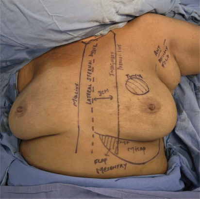
Figure 2. Surface marking of MICAP flap.
Surface marking AICAP flap
AICAP flaps are based on perforators from the rectus abdominis or oblique intercostal artery. The perforators are located at 6 o’clock, 1–2 cm below the IMF. The flap extends both sides of the perforator, with tissue at 6 o’clock forming the base [15, 16] (Figure 3).
Brief description of the surgical technique of CWPF
Tumour site and skin incision marking were done preoperatively in a standing position. For non-palpable tumours, wire localisation was used. The patient was supine, with the arm on the same side abducted at 90°. For the LICAP flap, a bolster beneath the scapula and lower back improved access to the paraspinal region for larger flaps. The surgical procedure included lumpectomy, axillary dissection, flap harvesting and flap insetting and closure. Skin incision followed preoperative marking, and breast flaps were raised above the outer lamellar fascia. A lumpectomy was performed with gross 1 cm margins. Clips were placed in the cavity for a radiotherapy boost. Axillary dissection used an axillary crease or the same incision. The flap was designed to enclose the marked perforators’ site, ensuring the donor site had enough tissue for closure without tension or vascular compromise.
The LICAP flaps can be done by turnover or as propeller flaps without twisting the perforators. If the tumour involves the skin, a lumpectomy is performed through a separate incision, removing the affected skin. The flap dimensions are adjusted to maintain a bridging skin island. Wounds were closed with sutures in layers, using drainage as needed. The patient was discharged on day 2. Postoperative complications within 1 month were documented and classified per the Clavien–Dindo classification [17]. An adjuvant treatment plan was based on histopathology results.
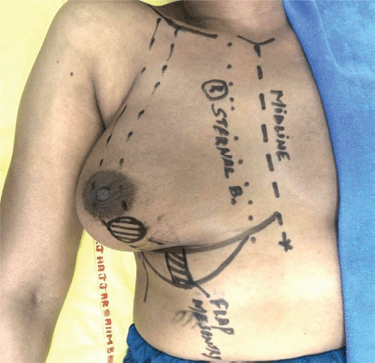
Figure 3. Surface marking of AICAP flap.
Figures 4–7 show operative pictures and postoperative outcomes of LICAP, MICAP and AICAP perforator flaps.
Data collection
Patients underwent structured follow-up with weekly clinical evaluations for the first month, followed by 3-monthly assessments as per institutional protocol, with a minimum follow-up duration of 1 year. Data collected included demographic details, clinical presentation, intraoperative findings, histopathological characteristics and postoperative complications. Complications were recorded and classified using the Clavien–Dindo classification system. PROs were assessed using the BREAST-Q™ (Breast Conserving Therapy module), administered at 6 months following completion of radiotherapy.
Statistical analysis
Continuous variables were expressed as means with standard deviation (SD) or medians with interquartile range (IQR), as appropriate. Categorical variables were presented as frequencies and percentages. Where applicable, 95% confidence intervals (CIs) were calculated for key outcome measures. Due to the retrospective design and absence of a comparator group, formal comparative statistical testing was limited; however, relevant descriptive comparisons across subgroups were performed. Statistical analysis was conducted using SPSS® version 27.
Results
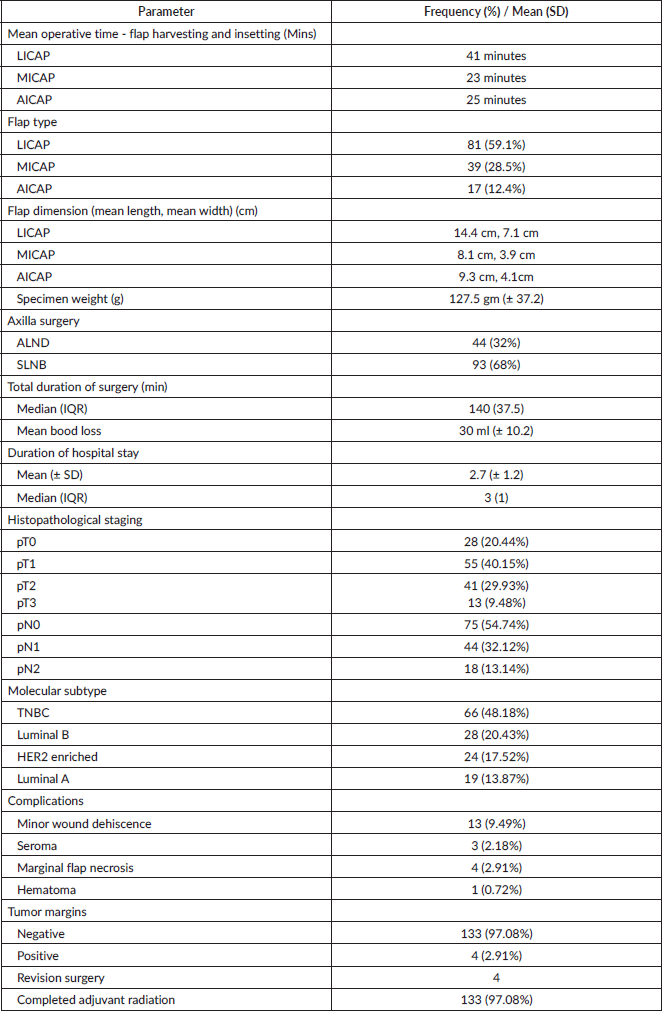
The study included 137 breast cancer patients with a mean age of 48.1 years. Eighteen patients (13.13%) were diabetic, and 11 (8.02%) had hypertension. Of note, 45.98% had right breast lumps and 54.02% had left breast lumps. Fourteen patients (10.21%) had a history of tobacco smoking. Most tumours (49.63%) were in the upper outer quadrant (UOQ). Over 70% of patients had moderate cup sizes (A and B). Fifty-five cases (40.14%) showed no ptosis, while Grade I ptosis was observed in 38 (27.73%) patients, according to the Regnault classification [18]. Preoperative staging showed 73 (53.28%) had tumours >2 to 5 cm (cT2), and 32 (23.36%) had tumours >5 cm (cT3). Sixty-nine patients (50.4%) had no nodal involvement (cN0), while 68 (49.6%) had clinically positive nodes. Additional clinical and demographic characteristics are summarised in Table 1.
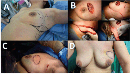
Figure 4. Operative pictures of the LICAP flap with skin island. (a): Preoperative planning and markings showing the breast lesion site and LICAP flap design. (b): Intraoperative steps: wide local excision of the cavity and LICAP flap harvesting. (c): Immediate postoperative inset of the LICAP flap into the breast defect. (d): Postoperative outcome demonstrating restored breast contour and concealed donor-site scar.
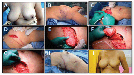
Figure 5. Operative pictures of the turnover LICAP flap. (a): Preoperative appearance of the breast with a lesion. (b): Surgical markings showing tumour excision site and LICAP flap design on the lateral chest wall. (c): Wide local excision creating a breast defect. (d): Flap incision on the lateral thoracic wall. (e): Subfascial dissection with identification of intercostal perforators. (f): LICAP flap raised on perforators and mobilised. (g): Flap transposed and inset into the breast defect. (h): Primary donor-site closure with suction drain placement. (i): Postoperative result showing satisfactory breast contour with well-concealed donor-site scar.

Figure 6. Operative pictures of the MICAP flap. (a): Marking of tumour and planned MICAP flap. (b): Raising and de-epithelialisation of the MICAP flap. (c): MICAP flap being raised for insetting at the tumour site. (d): Immediate postoperative outcome of the MICAP flap.
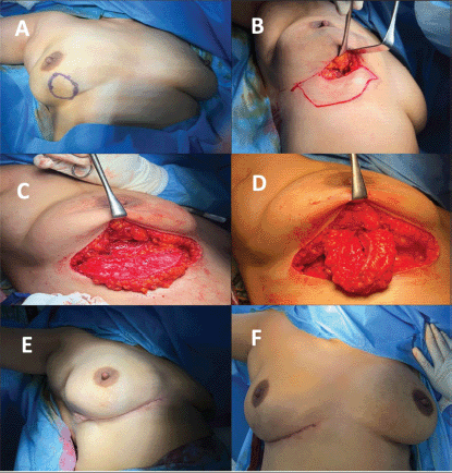
Figure 7. Operative pictures of the AICAP flap. (a): Marking of tumour in the inferior quadrant of right breast. (b): AICAP flap incision. (c): Identification of AICAP perforator at 6 o’clock position. (d): AICAP flap being raised for insetting in the cavity. (e and f): Immediate postoperative outcome of the AICAP flap.
Table 1. Baseline characteristics of the study participants.

The median total surgery duration was 140 minutes (IQR: 37.5). LICAP flaps were the most commonly performed, accounting for 81 cases (59.1%), followed by MICAP in 39 (28.5%) and AICAP in 17 (12.4%). The mean operative times for flap harvesting and insetting were 41, 23 and 25 minutes for LICAP, MICAP and AICAP flaps, respectively. Subgroup analysis based on flap type demonstrated that LICAP flaps required a longer operative time compared to MICAP and AICAP flaps. However, complication rates were comparable across all groups, with no significant variation in major outcomes. These findings highlight the versatility and safety of CWPFs across different anatomical applications. Axillary lymph node dissection (ALND) was performed in 44 cases (32%), while sentinel lymph node biopsy (SLNB) was done in 93 cases (68%).
No complications occurred in 116 patients (84.67%; 95% CI: 78.6%–90.7%). Thirteen patients (9.49%) had minor wound dehiscence, three patients (2.18%) developed seroma, four (2.91%) had marginal flap necrosis and one (0.72%) experienced hematoma. The overall complication rate was 15.32% (95% CI: 9.3%–21.3%). All complications were graded by Clavien–Dindo classification; 18 patients (13.13%) had Grade I complications, and 3 patients (2.20%) had Grade II complications. Four patients (2.91%; 95% CI: 0.1%–5.7%) had a focal positive margin and underwent margin revision surgery. Most patients (133, 97.08%; 95% CI: 94.2%–99.9%) completed adjuvant radiation therapy. Findings and complications are summarised in Table 2.
Table 2. Details of surgical procedure, histopathological report and complications.
Of the patients, 113 (82.4%) answered the postoperative questionnaire, with a follow-up time of at least 6 months postoperatively. Patient satisfaction with cosmetic outcomes after partial breast reconstruction with CWPFs was among the highest in reported literature. The BREAST Q scores for each domain are summarised in Table 3. Overall, most patients are very satisfied with post-lumpectomy breast appearance and fit, although there are minor concerns about symmetry and natural look in Figure 8.
Discussion
This study evaluated surgical and PROs of CWPFs for partial breast reconstruction after BCS. Results show that CWPF reconstructions provide high aesthetic satisfaction and low complication rates, demonstrating their value as a reliable alternative. Our findings support using CWPFs for BCS, particularly when volume replacement is needed. This is relevant as breast conservation techniques have become standard care for early-stage breast cancer, emphasising oncological outcomes while preserving aesthetics [19, 20].
CWPFs, using LICAP flaps, have gained prominence as an alternative for volume replacement in partial breast reconstruction. Studies have demonstrated the safety and efficacy of these flaps for lateral breast tumours [21].
The surgical technique using CWPFs for all breast quadrants is aligned with emerging trends in oncoplastic surgery. Our study stands out by including LICAP, MICAP and AICAP flaps. Previous research focused mainly on LICAP flaps for lateral and central breast defects, while we expanded CWPFs to cover defects across all breast quadrants. This approach allows versatile reconstruction, using MICAP and AICAP flaps for inner and lower quadrant defects [22].
Table 3. Breast-Q patient satisfaction with various domains after partial breast reconstruction with CWPFs.


Figure 8. BREAST Q ‘Satisfaction With Breast’ score after partial breast reconstruction with CWPFs.
In previous studies, the latissimus dorsi (LD) flap was the primary method for autologous reconstruction following BCS. The LD flap requires harvesting muscle, leading to higher morbidity and complications such as joint stiffness and seromas. Our study uses CWPFs, which spare shoulder muscles, reducing postoperative complications. Our findings show a lower complication rate compared to the LD flap, with 9.49% of patients experiencing minor wound dehiscence and 2.18% developing seroma. This matches previous studies reporting minimal complications with CWPF’s techniques [23].
In our study, flap design was guided by preoperative surface marking tailored to tumour location and anticipated defect size, allowing precise localisation of relevant perforators. This approach was validated by the absence of complete flap loss in the postoperative course. In contrast, handheld Doppler assessment is operator-dependent and subject to interobserver variability. Surface marking thus offers a reproducible, anatomically informed and cost-effective alternative for planning CWPFs-based reconstruction [24]. Importantly, the absence of Doppler did not necessitate a wider mesentery, as flap design followed established anatomical perforator zones, and we did not observe selection of more distant perforators or compromise in flap mobility.
The planning for the incision for harvesting the flap depends on the location and the size of the volume defect. In the earlier reports, transverse fusiform flaps were designed, but the volume replaced by these flaps was not sufficient for large volume defects. To increase the volume of flap harvest, a lazy-S/longitudinal flaps design has been adopted and published in recent research [25].
The mean tumour size in our cohort was 4.8 cm, larger than in prior studies. Despite this, the positive margin rate remained low at 2.91%, demonstrating the oncological safety of CWPFs in larger-volume resections. These findings suggest that CWPFs techniques enable adequate excision of larger tumours without compromising margin status, broadening their applicability in volume replacement during BCS [26].
Our study reported an overall complication rate of 15.32%, which is higher than in some previously published series; however, the majority of complications were minor, with 13.13% classified as Clavien–Dindo Grade I and 2.20% as Grade II. Partial flap necrosis was observed in only 3.0% of patients, and notably, no cases of complete flap loss were recorded - an outcome that has been a significant concern in traditional flap reconstructions. Furthermore, the need for revision surgery due to positive margins was limited to four cases (2.91%) of cases, substantially lower than the 10%–20% rates reported in earlier studies. These findings reinforce the safety and oncological adequacy of CWPFs, highlighting their reliability as a low-risk, effective alternative for partial breast reconstruction with a favourable morbidity profile [27].
Aesthetic outcomes are critical in oncoplastic surgery, and our study showed high patient satisfaction using BREAST-Q™. Satisfaction with breast appearance was high, with a median score of 86, supporting the favourable cosmetic results of CWPFs-based reconstruction. While satisfaction was high, lower scores in sexual well-being reflected concerns about symmetry and natural appearance. This finding aligns with studies identifying sexual well-being as a commonly affected quality-of-life domain among breast cancer survivors [28, 29].
Our study offers several strengths. First, the large sample size and prospective analysis of the database provide robust evidence regarding the surgical and aesthetic outcomes of CWPFs in partial breast reconstruction. Second, by including patients with varying tumour sizes and locations, we assessed CWPF’s utility across diverse clinical scenarios. Third, a detailed analysis of complications and patient satisfaction provides valuable insights into patient experience following CWPF-based reconstruction.
Our study highlights the safety and efficacy of one-stage immediate reconstruction using CWPFs, potentially reducing treatment burden compared to two-stage approaches. The low rate of positive margins and minimal complications demonstrates that this approach achieves favourable oncological and aesthetic outcomes.
This study has certain limitations. The relatively short follow-up period of 1 year limits assessment of long-term oncological outcomes, including disease-free and overall survival. Additionally, the absence of a comparator group restricts direct evaluation of the relative advantages of the no-Doppler technique over Doppler-guided approaches or other reconstructive methods. Multicentre studies with longer follow-up are required to validate the long-term efficacy and safety of CWPF-based reconstructions.
Initial Doppler-guided training aids understanding perforator anatomy and variability; experienced surgeons may transition to a no-Doppler approach. This technique extends anatomical expertise, not replacing foundational training.
Conclusion
No-Doppler CWPFs represent a safe and feasible option for partial breast reconstruction, offering favourable aesthetic outcomes, low complication rates and high patient satisfaction. This approach simplifies surgical planning and may be particularly useful in resource-constrained settings. However, in the absence of a comparator group, these findings should be interpreted with caution. Further comparative studies are needed to better define the role of this technique.
List of abbreviations
AICAP, Anterior intercostal artery perforator; ALND, Axillary lymph node dissection; BCS, Breast-conserving surgery; BCT, Breast conserving therapy; BMI, Body mass index; BREAST-Q™, Breast-Q patient-reported outcome measure; CWPF(s), Chest wall perforator flap(s); HER2, Human epidermal growth factor receptor 2; ICG, Indocyanine green; IMF, Inframammary fold; IQR, Interquartile range; LD flap, Latissimus dorsi flap; LICAP, Lateral intercostal artery perforator; MICAP, Medial intercostal artery perforator; NACT, Neoadjuvant chemotherapy; OBS, Oncoplastic breast surgery; PRO(s), Patient-reported outcome(s); RT, Radiotherapy; SD, Standard deviation; SLNB, Sentinel lymph node biopsy; SPSS®, Statistical Package for the Social
Sciences; TNBC, Triple-negative breast cancer; UOQ, Upper outer quadrant; UIQ, Upper inner quadrant; LOQ, Lower outer quadrant; LIQ, Lower inner quadrant.
Declaration
Portions of the technical description are derived from the authors’ previously published work. This manuscript presents a comprehensive audit of perforator flap-based reconstruction without Doppler assistance and has not been published or submitted elsewhere.
Acknowledgment
None.
Conflicts of interest
All authors declare no conflicts of interest.
Source of funding
No grant was sought or received from any funding agency for the conduct of this study. The research was conducted by using institutionally available resources.
Consent to participate
Written informed consent was obtained from all individual participants included in the study for participation, data collection and completion of patient-reported outcome measures.
Ethical approval
This study received approval from the Institutional Ethics Committee of AIIMS, New Delhi.
Data availability
The datasets used and/or analysed during the current study are available from the corresponding author on reasonable request.
Author contributions
AM and AK wrote the main manuscript text. AK prepared the figures. All authors reviewed and approved the final manuscript.
References
1. Veronesi U, Cascinelli N, and Mariani L, et al (2002) Twenty-year follow-up of a randomized study comparing breast-conserving surgery with radical mastectomy for early breast cancer N Engl J Med 347(16) 1227–1232 https://doi.org/10.1056/NEJMoa020989 PMID: 12393819
2. Van Dongen JA and Voogd AC (2000) Long-term results of a randomized trial comparing breast-conserving therapy with mastectomy: European Organization for Research and Treatment of Cancer 10801 trial J Natl Cancer Inst 92(14) 1143–1150 https://doi.org/10.1093/jnci/92.14.1143 PMID: 10904087
3. Gandhi A and Coles C (2019) Planning and provision of oncoplastic breast surgery – a national approach Breast J 25(1) 114–118
4. Clough KB and Kaufman GJ (2015) Oncoplastic surgery: pushing the limits of breast-conserving surgery Breast J 21(2) 140–146 https://doi.org/10.1111/tbj.12372 PMID: 25676776
5. Rietjens M and Urban C (2007) Oncoplastic surgery in breast cancer: a ten-year experience at the European Institute of Oncology Eur J Surg Oncol 33(2) 218–224
6. Hamdi M, Spano A, and Landuyt KV, et al (2008) The lateral intercostal artery perforators: anatomical study and clinical application in breast surgery Plast Reconstr Surg 121(2) 389–396 https://doi.org/10.1097/01.prs.0000298317.65296.cf PMID: 18300954
7. Mcculley SJ and Macmillan RD (2005) Planning and use of therapeutic mammoplasty—Nottingham approach Br J Plast Surg 58(7) 889–901 https://doi.org/10.1016/j.bjps.2005.03.008 PMID: 16043150
8. Salgarello M, Visconti G, and Barone-Adesi L (2012) Double-pedicle flap reconstruction following breast-conserving surgery Plast Reconstr Surg 130(1) 31–40 https://doi.org/10.1097/PRS.0b013e3182547d42 PMID: 22743871
9. Vijayakumar S, et al (2023) Use of anatomical landmarks for MICAP flap without preoperative Doppler: a simplified approach Indian J Surg Oncol 14(1) 110–115
10. Shanker M and Chattopadhyay R, et al (2022) Outcomes of no-Doppler chest wall perforator flap reconstruction: a single-center experience Indian J Plast Surg 55(3) 236–242
11. Pusic AL, Klassen AF, and Scott AM, et al (2009) Development of a new patient-reported outcome measure for breast surgery: the BREAST-Q Plast Reconstr Surg 124(2) 345–353 https://doi.org/10.1097/PRS.0b013e3181aee807 PMID: 19644246
12. Ward JA, Potter S, and Blazeby JM (2012) The BREAST-Q: further validation in independent clinical samples Plast Reconstr Surg 129(2) 293–302 https://doi.org/10.1097/PRS.0b013e31823aec6b
13. Mishra A, Deo SVS, and Oberoi AS, et al (2023) No Doppler single position-lateral intercostal artery perforator flap (NDSP-LICAP): a safe and versatile flap for breast oncoplasty in resource-limited setting World J Surg 47(11) 2753–2760 https://doi.org/10.1007/s00268-023-07126-x PMID: 37574468
14. Rapisarda F, Giacchino M, and Dent H, et al Medial intercostal artery perforator (MICAP) flap in oncoplastic breast conserving surgery: single centre initial experience Eur J Surgical Oncol 49(2) e99
15. Soumian S, Parmeshwar R, and Chandarana M, et al (2020) Chest wall perforator flaps for partial breast reconstruction: surgical outcomes from a multicenter study Arch Plast Surg 47(02) 153–159 https://doi.org/10.5999/aps.2019.01186 PMID: 32203992 PMCID: 7093273
16. Mishra A, Kumar A, and Deo SVS, et al (2025) No-Doppler medial intercostal artery perforator flap for partial breast reconstruction: surgical technique, feasibility, and outcomes in inner and central quadrant defects Indian J Surg Oncol https://doi.org/10.1007/s13193-025-02448-9
17. Dindo D, Demartines N, and Clavien PA (2004) Classification of surgical complications: a new proposal with evaluation in a cohort of 6336 patients and results of a survey Ann Surg 240(2) 205–213 https://doi.org/10.1097/01.sla.0000133083.54934.ae PMID: 15273542 PMCID: 1360123
18. Regnault P (1976) Breast ptosis. Definition and treatment Clin Plast Surg 3(2) 193–203 https://doi.org/10.1016/S0094-1298(20)30220-0 PMID: 1261176
19. Fisher B, Anderson S, and Bryant J, et al (2002) Twenty-year follow-up of a randomized trial comparing total mastectomy, lumpectomy, and lumpectomy plus irradiation for the treatment of invasive breast cancer N Engl J Med 347(16) 1233–1241 https://doi.org/10.1056/NEJMoa022152 PMID: 12393820
20. Agrawal SK, Mahajan S, and Ahmed R, et al (2024) Chest wall perforator flap partial breast reconstruction: a retrospective analysis of surgical, cosmetic and survival outcome Ecancermedicalscience 18 1681 https://doi.org/10.3332/ecancer.2024.1681 PMID: 38566767 PMCID: 10984835
21. Pinto R, Malata CM, and Abood A, et al (2023) Outcomes of chest wall perforator flaps in oncoplastic breast reconstruction: a multi-institutional prospective study Ann Surg Oncol 30(4) 2345–2352
22. Meybodi F, Akhavani MA, and Alvi R, et al (2019) Use of lateral intercostal artery perforator (LICAP) flaps for breast volume replacement: an evolving oncoplastic technique J Plast Reconstr Aesthet Surg 72(3) 406–412
23. Lee KT and Mun GH (2014) A systematic review of functional donor-site morbidity after latissimus dorsi muscle transfer Plast Reconstr Surg 134(2) 303–314 https://doi.org/10.1097/PRS.0000000000000365 PMID: 24732650
24. Koppiker CB, Mishra R, and Jain V, et al (2025) Chest wall perforator flaps in breast conservation: versatile, affordable, and scalable: insights from the largest single-surgeon audit from India Curr Oncol 32(3) 165 https://doi.org/10.3390/curroncol32030165 PMID: 40136369 PMCID: 11941675
25. Banys-Paluchowski M, Gruber M, and Hartmann S, et al (2023) Current indications and trends for breast reconstruction following mastectomy: an analysis of 22,079 cases Breast J 29(1) 23–30
26. Agrawal A, Malviya A, and Pandey A, et al (2023) Chest wall perforator flaps in oncoplastic breast surgery: outcomes in an Indian cohort Indian J Surg Oncol 14(1) 22–29
27. Zeeshan S, Vohra LM, and et.al. (2022) A single centre experience of local perforator flaps in oncoplastic breast surgery; a cross-sectional study Ann Med Surg (Lond) 84 104916 PMID: 36536719 PMCID: 9758369
28. Bazzarelli A, Baker L, and Petrcich W, et al (2020) Patient satisfaction following level II oncoplastic breast surgery: a comparison with mastectomy utilizing the BREAST-Q questionnaire Surg Oncol 35 556–559 https://doi.org/10.1016/j.suronc.2020.11.001 PMID: 33220630
29. Xie L, Kong X, and Lin D, et al (2025) Application of chest wall perforator flaps in oncoplastic breast-conserving surgery BMC Cancer 25(1) 328 https://doi.org/10.1186/s12885-025-13488-3 PMID: 39984927 PMCID: 11846177