Clinicopathological features, etiological factors and survival outcomes in breast cancer treated with neoadjuvant chemotherapy: a study from western Algeria
Olfa Hanaa Aissa1,2a, Aicha Rabah3, Abdelkader Bousahba3, Nawel Nassima Benchiha1,4, Boumediene Elhabachi1,5 and Lynda Addou-Klouche1,2b
1Laboratory of Nutrition, Pathology, Agro-Biotechnology and Health (LAB-NuPABS), Sidi Bel Abbes 22000, Algeria
2Department of Biology, Faculty of Life and Natural Sciences, Djillali Liabes University of Sidi Bel Abbes, BP 89, Sidi Bel Abbes 22000, Algeria
3Department of Medical Oncology, University Hospital Center of Oran, Oran 31000, Algeria
4Department of Pharmacy, Faculty of Medicine, Djillali Liabes University of Sidi-Bel-Abbes, Sidi Bel Abbes 22000, Algeria
5Department of General Surgery, Hassani Abdelkader University Hospital Center, Sidi-Bel-Abbes 22000, Algeria
ahttps://orcid.org/0000-0001-6870-4969
bhttps://orcid.org/0000-0003-4770-1050
Abstract
Breast cancer is the most frequently diagnosed malignancy and the leading cause of cancer-related death among women. It is a biologically heterogeneous disease resulting from a multifactorial process. Neoadjuvant chemotherapy (NACT) has become a key treatment for patients with locally advanced or high-risk early-stage breast cancer, with its initiation guided by clinicopathological and etiological factors reflecting tumour aggressiveness and patient profile. This study aimed to describe the distribution of these factors among breast cancer patients treated with NACT in western Algeria, focusing on breast cancer subtypes and survival outcomes. We retrospectively analysed data from 327 breast cancer patients treated with NACT between January 2020 and December 2024. Data were collected from medical records and assessed among breast cancer subtypes using the chi-square test. Survival analysis was performed using the Kaplan–Meier method. In our cohort, most patients were aged 50 years or older (68.5%) and presented with a consultation delay exceeding 3 months after symptom onset. The Luminal B (LB) subtype was predominant (48.6%). High Ki-67 expression was observed in the majority of tumours, particularly among LB and triple-negative breast cancer (TNBC) subtypes. Histologically, Scarff–Bloom–Richardson grade II tumours were the most frequent in our cohort (64.5%). T2 tumours were prevalent, and nodal involvement was predominantly N1 stage. TNBC and human epidermal growth factor receptor 2-enriched subtypes showed poorer clinical outcomes, whereas hormone receptor (HR)-positive tumours were associated with better survival. These findings underline the predominance of breast cancer among middle-aged and older women and the value of Ki-67 as a marker of tumour aggressiveness. They also confirm the prognostic relevance of HR status and emphasise the importance of early detection and personalised follow-up, supported by molecular profiling and emerging biomarkers.
Keywords: breast neoplasms, neoadjuvant therapy, biomarkers, risk factors, survival analysis
Correspondence to: Olfa Hanaa Aissa
Email: hanaa.aissa@univ-sba.dz
Published: 27/08/2026
Received: 26/01/2026
Publication costs for this article were supported by ecancer (UK Charity number 1176307).
Copyright: © the authors; licensee ecancermedicalscience. This is an Open Access article distributed under the terms of the Creative Commons Attribution License (http://creativecommons.org/licenses/by/4.0), which permits unrestricted use, distribution, and reproduction in any medium, provided the original work is properly cited.
Background
Breast cancer is the most frequently diagnosed cancer and the leading cause of cancer-related mortality among women worldwide, accounting for an estimated 2.3 million new cases and 665,684 deaths in 2022 alone [1]. In Algeria, breast cancer is also a major contributor to female cancer deaths, accounting for 41.3% of all female malignancies, with 14,601 new cases diagnosed in 2022 [2].
Breast malignancies represent a biologically diverse group of diseases that might be observed at multiple levels, such as histopathological or molecular level [3–5]. This intrinsic heterogeneity influences not only the treatment decision but also the prognosis, underscoring the importance of tailored clinical management strategies [6,7].
In many studies, triple-negative breast cancer (TNBC) subtype is reported as having the worst prognosis, as patients do not benefit from endocrine or targeted therapies, leaving chemotherapy as the main systemic treatment option [8,9]. Furthermore, it was reported that Luminal B (LB) breast cancer shows substantially worse outcomes with a more aggressive phenotype compared to Luminal A (LA) breast cancer and has a distinct profile of response to hormone therapy and chemotherapy [10].
Besides, breast cancer emerges as a multifactorial disease, resulting from a complex interplay of genetic predispositions, hormonal influences, reproductive history, environmental exposures and lifestyle factors [11,12]. These etiological factors, along with tumour-specific characteristics such as grade, stage and receptor status, contribute to disease onset, progression and treatment response. Understanding the distribution of these factors among breast cancer patients is therefore essential for developing effective prevention strategies, optimising treatment choices and improving outcomes [6].
Over the past two decades, neoadjuvant chemotherapy (NACT) administered before surgical resection has emerged as a cornerstone in the management of patients with locally advanced or high-risk early-stage breast cancer [13]. Beyond contributing to tumour downstaging, improving operability and enabling breast-conserving surgery, NACT provides an in vivo evaluation of tumour sensitivity to therapy, offering crucial prognostic and predictive insights [14–16]. The decision to initiate NACT is often based on a combination of clinicopathological and etiological factors, which reflect both tumour aggressiveness and patient profile [17,18].
In this context, this study aims to describe the distribution of clinicopathological and etiological factors among breast cancer patients treated with NACT in western Algeria, with a specific focus on breast cancer subtypes. Analysing these distributions provides valuable insights into the profiles of individuals who undergo this therapeutic approach. In addition, the study examines metastasis free survival (MFS) and overall survival (OS) patterns, offering a deeper understanding of how tumour biology influences prognosis in patients receiving NACT.
Methods
This retrospective descriptive study was carried out in the Department of Medical Oncology at the University Hospital Center of Oran (Algeria). The study included women diagnosed with breast cancer between January 2020 and December 2024.
Inclusion and exclusion criteria
A total of 383 patients were initially identified from institutional medical records. Among these, 56 patients were excluded due to incomplete or missing data. Patients diagnosed with metastatic stage (M1) were also excluded. The remaining 327 patients met all inclusion criteria and were included in the study. Inclusion criteria were as follow: Residency in the western region of Algeria; Histological confirmation of breast cancer via core needle biopsy; Treatment with NACT before surgery; Known status of oestrogen receptor (ER), progesterone receptor (PR), human epidermal growth factor receptor 2 (HER2) and Ki-67.
NACT treatment
All patients had received NACT according to their tumour subtype, stage at diagnosis, patient tolerance and the standard protocols followed in the institution. The most frequently used chemotherapy regimens included combinations of epirubicin plus cyclophosphamide (EC100) or doxorubicin plus cyclophosphamide (AC60). In some cases, taxane-based regimens were used, including docetaxel or paclitaxel, either following anthracycline-based therapy or as monotherapy. The combination of carboplatin and taxane was occasionally administered. Trastuzumab was added for patients with HER2-positive tumours according to standard clinical indications.
Immunohistochemical profiling
The expression of ER, PR, HER2, and Ki-67 proliferation index was determined by immunohistochemistry (IHC). HER2 status was confirmed by fluorescence in situ hybridisation in cases of equivocal IHC results (2+). ER and PR positivity were defined as nuclear staining in ≥1% of tumour cells. Ki-67 was considered high when the proliferation index exceeded 14%.
Breast carcinomas were classified as follows into four immunophenotypes acting as surrogates for the established molecular subtypes of breast cancer:
LA: ER+ and/or PR+, HER2- and a low Ki-67 proliferation index (≤14%).
LB: ER+ and/or PR+, HER2-, Ki-67 (>14%); ER+ and/or PR+, HER2+, any Ki-67.
HER2-enriched: ER-, PR-, HER2+.
TNBC: ER-, PR-, HER2-.
Data source
The present study was conducted in compliance with the ethical principles of the Declaration of Helsinki, as revised in 2013. Data were extracted from hospital archives and electronic medical records. Given the observational and anonymised nature of the study, formal ethical approval was not required. No direct contact with patients occurred at any stage of the study. Throughout the study, confidentiality of patient information was strictly maintained.
Statistical analysis
Statistical analyses were performed using the International Business Machines Corporation Statistical Package for the Social Sciences Statistics version 20.0. Descriptive statistics were used to analyse the distribution of clinicopathological and etiological features of the study population. Associations between these factors and breast cancer subtypes were evaluated using the chi-square test. Monte Carlo correction was applied to obtain accurate p-values in contingency tables with small expected frequencies. Survival analysis was performed using the Kaplan–Meier method. Differences between survival curves were assessed using the log-rank (Mantel-Cox) test. Median survival times and median follow-up were reported with their respective 95% confidence intervals (CI). OS was defined as the time from diagnosis to death from any cause or to the last follow-up for patients without an event. MFS was defined as the time from diagnosis to the occurrence of distant metastasis or to the last follow-up for patients without an event. Patients who died without developing distant metastasis were censored at the date of death. All statistical tests were two-sided at the 5% level of significance.
Results
Distribution of breast cancer patients and tumour characteristics among study population
Baseline patient characteristics and consultation patterns
A total of 327 female patients were included in this study. The descriptive analysis showed that most patients were aged 50 years and older at diagnosis (68.5%), with a median age of 55 years, and had a consultation delay exceeding 3 months following symptom onset (62.1%). A family history of breast cancer was reported in a subset of patients, though not predominantly (41.3%) (Table 1).
Reproductive factors
Regarding reproductive factors, the majority of patients reported menarche occurring after the age of 12 (66.1%). While most patients were parous (67%), a large proportion had never breastfed (58.7%), and the majority were postmenopausal at diagnosis (63.9%). A history of oral contraceptive (OC) use was observed in 50.8% of cases (Table 1).
Clinical and pathological tumour characteristics
In terms of tumour characteristics, the right breast was more frequently affected (48.3%), particularly in the upper outer quadrant (41.6%). Left breast was affected in 47.4% and bilateral in 4.3%. Histology showed that invasive carcinoma of no special type (NST) was the most prevalent (77.4%), with the majority of tumours classified as grade II (64.5%) followed by grade III (33.3%). Clinically, the T2 tumour stage was more frequent (46.8%), followed by the T4 stage (26.6%), and nodal involvement was predominantly N1 stage (62.7%). IHC showed that most tumours were positive for ER (65.4%) and PR (61.8%), while HER2-negative status was predominant (70.9%). A high Ki-67 proliferation index was observed in the majority of cases (59.9%) (Table 1), and LB subtype was the most represented among patients (48.6%) (Figure 1).
Table 1. Baseline clinicopathological and etiological characteristics of the study population.


Figure 1. Distribution of breast cancer subtypes in the study population. This figure illustrates the proportion of breast cancer subtypes in the study population, classified based on the immunohistochemical expression of ER, PR, HER2 and Ki-67.
Distribution of breast cancer patients and tumour characteristics by breast cancer subtypes
A comprehensive comparative analysis was conducted to evaluate the distribution of patients and tumours characteristics among each subtype of breast cancer.
Baseline patient characteristics and consultation patterns
Patients aged 50 years and older represented the majority across all subtypes (p = 0.05). Consultation delay exceeding 3 months was common in all groups, and family history of breast cancer was more frequently reported among LB and TNBC cases. However, no significant differences were observed between groups (p > 0.05) (Table 2).
Reproductive factors
Among all subtypes, most patients reported menarche after the age of 12, were parous, and postmenopausal at diagnosis. Breastfeeding was more frequently observed among patients with HER2-enriched tumours (54.5%). The use of OCs was more prevalent in the HER2-enriched (65.9%) and TNBC (52%) subtypes. Nevertheless, the distribution did not differ significantly between groups (p > 0.05) (Table 2).
Clinical and pathological tumour characteristics
Laterality differed significantly by subtype (p <0.001) with left breast involvement being more common in LA and LB (47.3% and 54.1%, respectively), whereas right-sided tumours predominated in HER2-enriched and TNBC cases (72.7% and 56% respectively). Tumour localisation was most frequently detected in the upper outer quadrant across all subtypes. Invasive carcinoma of NST represented the most common histological type across all subtypes. In parallel, Scarff–Bloom–Richardson (SBR) grade II was the most frequently observed histological grade within each subgroup, except for the HER2-enriched group, in which grade III was significantly more frequent (50%) (p = 0.046). Clinical tumour and nodal stages also varied significantly by subtype, but T2 and N1 were the most common stages observed across all of them (p <0.05). IHC showed that while nearly all LA and LB subtypes were positive for both ER and PR, HER2 positivity was detected in 32.1% of LB cases (p <0.001). A high Ki-67 index expression (>14%) was mostly observed in LB (90.6%) and TNBC subtypes (72%) (p <0.001) (Table 2).
Table 2. Distribution of clinicopathological and etiological characteristics among breast cancer subtypes.

Survival analysis
The median follow-up time was 27 months (95% CI: 25.36–28.64 months). At the end of the study, 25 (7.6%) patients had died, and up to 146 (44.6%) participants had distant disease progression.
Survival patterns among the study population
Kaplan–Meier analysis illustrated the survival distributions over time in our cohort. The median MFS was 32 months (95% CI: 27.08–36.92) (Figure 2), whereas the median OS was not reached (Figure 3).
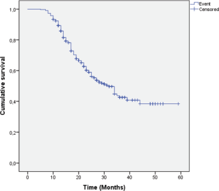
Figure 2. MFS in the study population. This figure illustrates MFS in the entire study population. Survival analysis was performed using the Kaplan–Meier method. Censored observations are indicated by tick marks.
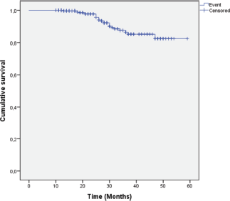
Figure 3. OS in the study population. This figure illustrates OS in the entire study population. Survival analysis was performed using the Kaplan–Meier method. Censored observations are indicated by tick marks.
Survival patterns according to breast cancer subtypes
Five-year MFS and OS rates were analysed across breast cancer subtypes. Although no statistically significant difference was observed (p = 0.464), LA tumours exhibited the most favourable MFS, while triple-negative and HER2-enriched subtypes tended to show poorer outcomes. LB cases demonstrated an intermediate survival pattern, less favourable than LA (Figure 4). OS exhibited a trend toward variation across breast cancer subtypes, approaching statistical significance (p = 0.065), with triple-negative tumours exhibiting the poorest survival, followed by HER2-enriched tumours, whereas Luminal subtypes demonstrated the most favourable outcome (Figure 5).
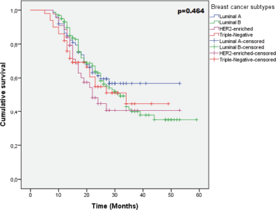
Figure 4. MFS according to breast cancer subtypes. Kaplan–Meier survival curves showing MFS according to breast cancer subtypes. The difference between survival curves was assessed using the log-rank test. The variation in MFS across breast cancer subtypes was not statistically significant (p = 0.464).
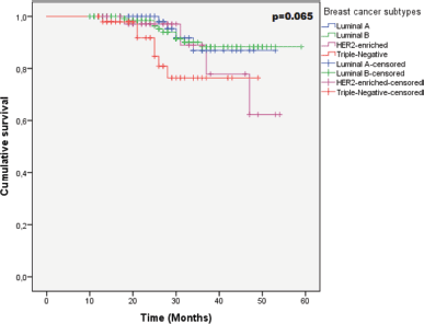
Figure 5. OS according to breast cancer subtypes. Kaplan–Meier survival curves showing OS according to breast cancer subtypes. The difference between survival curves was assessed using the log-rank test. The variation in OS across breast cancer subtypes was not statistically significant (p = 0.065).
Survival patterns according to LB subtype (HER2+ versus HER2–)
MFS analysis based on HER2 status within the LB subtype showed no statistically significant difference between the two subgroups (p = 0.165). However, LB HER2-negative subtype exhibited an earlier and more pronounced decline in MFS than LB HER2-positive subtype (Figure 6). For OS, both LB HER2-positive and HER2-negative patients maintained high survival probabilities throughout the follow-up period, yet a modestly less favourable pattern was observed among LB HER2-positive patients, with no statistically significant difference between the curves (p = 0.608) (Figure 7).

Figure 6. MFS according to LB subtype. Kaplan–Meier survival curves showing MFS according to LB HER2-negative and LB HER2-positive subtypes. Differences between subtypes were assessed using the log-rank test. The variation in MFS across LB subtypes was not statistically significant (p = 0.165).
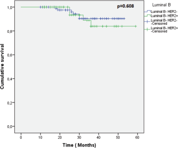
Figure 7. OS according to LB subtype. Kaplan–Meier survival curves showing OS according to LB HER2-negative and LB HER2-positive subtypes. Differences between subtypes were assessed using the log-rank test. The variation in OS across LB subtypes was not statistically significant (p = 0.608).
Survival patterns according to hormone receptors (HRs) status
Kaplan–Meier survival curves comparing HR status groups revealed a significant difference in OS with the HR-positive group showing a more favourable outcome (p = 0.015) (Figure 8). HR-positive patients also showed better MFS compared to HR-negative patients; however, the difference was not statistically significant (p = 0.156) (Figure 9).
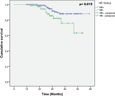
Figure 8. OS according to HR status. Kaplan–Meier survival curves showing OS according to HR status (ER/PR). Differences between groups were assessed using the log-rank test. The variation in OS across the groups was statistically significant (p = 0.015).

Figure 9. MFS according to HR status. Kaplan–Meier survival curves showing MFS according to HR status (ER/PR). Differences between groups were assessed using the log-rank test. The variation in MFS across the groups was not statistically significant (p = 0.156).
Discussion
This study analyses the clinicopathological, etiological and molecular characteristics of breast cancer in patients treated with NACT in western Algeria, with particular emphasis on their distribution among breast cancer subtypes. It also explores their association with MFS and OS, shedding light on the prognostic impact of tumour biology among these patients.
The median age of patients in our cohort was 55 years, with a predominance of women aged 50 years and older (68.5%), indicating that breast cancer is more frequent among individuals in the sixth decade of life in our study population. While these data differ from some NACT-treated series that reported a predominance of younger patients [19, 20], they align with other studies describing similar age distribution. For instance, in a large cohort of 487 patients, the median age was 52 years, with 57.9% of cases aged between 50 and 70 years [21]. These findings suggest that, in certain settings, NACT is increasingly administered in older populations.
A key finding in our study was the substantial delay in consultation, with nearly two-thirds (62.1%) of patients seeking medical assistance more than 3 months after noticing breast changes. This delay, observed across all subtypes, may be attributed to limited awareness and barriers to early detection, frequently noticed in low- and middle-income countries, including limited access to healthcare facilities, cultural taboos and socioeconomic constraints [22]. Such delay serves as a major driver for the clinical severity of the disease and may partly explain the advanced disease characteristics prevalent in our cohort, characterised by larger tumour size and higher rates of nodal involvement at presentation. Similar trends have been reported in several African studies. For instance, Salih et al [23] found that only one quarter of patients sought medical attention within the first 3 months following symptom onset. Likewise, a study conducted in Uganda reported that 89% of women presented with a delay of more than 3 months [24]. Our observations are also consistent with the conclusions of previous reports identifying consultation delays as a major contributor to advanced-stage breast cancer diagnosis [25, 26]. This underscores the need for targeted awareness strategies and earlier diagnostic pathways to reduce advanced-stage presentation that may limit the effectiveness of treatment strategies.
Reproductive characteristics, including parity and late menarche, were frequent (67% and 66.1%, respectively). Although traditionally considered protective [27–29], their predominance in our cohort may be explained by interactions with other risk factors, including genetic susceptibility, environmental exposures and lifestyle factors, which could attenuate or even offset their expected protective effect. Our findings are consistent with those from Turkish [30] and Vietnamese [31] studies, which described comparable reproductive patterns among breast cancer patients with a predominance of parous women, accounting for 86% and 92.6% of cases, respectively, and late menarche occurring among 71.5% and 95.2%, respectively. These findings suggest that traditional protective reproductive factors may not fully reflect individual risk profiles, highlighting the need for a comprehensive approach to multifactorial risk assessment.
Beyond intrinsic reproductive history, we found that OC use was more prevalent among patients with HER2-enriched and TNBC tumours in our study. These findings align with a case-control study by Bethea et al [32] in African American women, which reported prior use of OCs in more than half of TNBC cases (282/495). Moreover, meta-analytic evidence shows a significant association between OC use and TNBC [33], although the link with HER2-enriched tumours remains less clearly established. This raises the possibility that exogenous hormones may have differential effects on the development of specific breast cancer subtypes. Consequently, further research, incorporating the duration and timing of exposure, is required to better understand the underlying biological mechanisms and their role in tumour heterogeneity and aggressive clinical phenotypes, which may ultimately contribute to improved individualised risk assessment strategies.
We also observed significant associations between breast cancer subtypes and tumour characteristics. Laterality varied significantly by subtype, with left-sided tumours being more frequent in LA and LB, and right-sided tumour predominance observed in HER2-enriched and TNBC. The reasons for these differences remain unclear at this stage and warrant further investigation. Our findings differ from American and European studies, which reported aggressive subtypes occurring more frequently in the left breast [34, 35]. These discrepancies highlight that laterality-related patterns are not consistent across populations and may be influenced by ethnic variations or environmental factors.
The upper outer quadrant was the most common tumour location across all subtypes. Such predominance may reflect the higher concentration of breast glandular tissue in this region as previously reported [36]. Our finding is consistent with the pattern reported in most breast cancer studies worldwide, which show a dominance of the upper outer quadrant [37–39]. This implies that this area should remain a primary focus during imaging assessment and clinical examination due to its higher probability of harbouring malignant lesions.
In terms of clinical staging, T2 and N1 stages were the most frequently observed in the present study (46.8% and 62.7% respectively), reflecting a predominance of tumours with intermediate tumour size and early nodal involvement at diagnosis. This pattern may be influenced by the delayed presentation noted in a large proportion of patients. Our findings corroborate prior studies reporting a high prevalence of T2 and N1 stages in large NACT-treated cohorts [40, 41]. For instance, a retrospective study conducted by Cheun et al [42] in neoadjuvant settings showed that T2 and N1 stages were predominant, representing 70.2% and 43.7%, respectively. Clinically, this suggests that patients eligible for NACT are frequently diagnosed within this intermediate stage range that remains a critical period for tumour downstaging and improvement of surgical eligibility.
Histopathological examination revealed that invasive carcinoma of NST was predominant across all subtypes, in line with its well-established global predominance in breast cancer studies [20, 43]. However, the proportion of invasive lobular carcinoma (ILC), accounting for 15.9% of cases, was higher in our cohort than that reported in other NACT-treated series, such as those by Yilmaz and Cavdar [44] (7.5%) and Ghezzawi et al [45] (5.9%). This relative enrichment of ILC is particularly relevant to our earlier findings regarding age and stage, as this histological type is known to occur more frequently in older women and to be associated with a more insidious clinical presentation, often characterised by diffuse growth patterns and subtle radiological features that can delay clinical detection [46, 47]. These characteristics may have contributed to the more advanced clinical stages observed in our study.
Our investigation also revealed that SBR grade II tumours were more common in our cohort, particularly in both Luminal subtypes and TNBC, whereas SBR grade III tumours were significantly more prevalent among HER2-enriched tumours, reflecting the predominance of intermediate and high grade tumours in NACT-treated cohorts, concordant with international studies reporting similar distribution [48, 49]. This highlights the aggressive nature of tumours selected for NACT.
Immunohistochemical profiling revealed that the majority of patients had a high Ki-67 index (59.9%), in line with previous studies conducted in neoadjuvant settings [50, 51]. This reflects the predominance of highly proliferative tumours as a common feature in this context and confirms the overall aggressive tumour profile within our population. This pattern was particularly pronounced in LB and TNBC, both subtypes known to exhibit higher proliferative activity and poorer prognosis. Similar findings have been reported by a Lebanese study [52]. Ultimately, a high Ki-67 predominance underscores a rapid proliferative drive, linking aggressive biology to a higher risk of disease progression, thereby supporting the indication for NACT.
This proliferative phenotype is further supported by the predominance of the LB subtype (48.6%) within our population. Interestingly, our distribution pattern differs from several NACT-treated cohorts [53, 54]. One of these studies was conducted in Indonesia and reported a predominance of HER2-enriched subtype (32.37%) [55]. This discrepancy may be explained by differences in patient selection, particularly the inclusion of locally advanced breast cancer cases, as well as variations in tumour biology and population characteristics. Such findings point toward the heterogeneity of breast cancer subtype distribution in NACT-treated populations.
The clinical consequences of these biological patterns are reflected in survival outcomes. Although OS differences across subtypes did not reach statistical significance (p = 0.065) - possibly due to the short 27 months median follow-up - a notable trend was observed. TNBC showed the poorest OS, followed by HER2-enriched tumours, consistent with their aggressive biology. In contrast, Luminal subtypes were associated with the most favourable OS, in line with their HR positivity. This survival pattern is comparable with the one reported by Kim et al [56] and Hennigs et al [57]. These trends support the prognostic value of immunophenotypic classification and its relevance in predicting survival outcomes among breast cancer patients. This prognostic hierarchy is more clearly elucidated when focusing specifically on HR status, which emerged as the most significant indicator of outcome in our study. We found that HR-positive patients had markedly better outcomes, with a significantly prolonged OS compared to their HR-negative counterparts (p = 0.015). This reflects both the less aggressive tumour biology and treatment efficacy of hormone-sensitive tumours, emphasising the prognostic advantage conferred by HR expression. Our results align with existing literature highlighting the key role of HR status, such as the work of Ran et al [58] reporting a significantly improved OS and DFS in HR-positive patients compared to HR-negative ones [58]. This reinforces the importance of HR status assessment in guiding prognosis and therapeutic decisions.
MFS in our study also showed non-significant but consistent trends across breast cancer subtypes with LA tumours demonstrating the most favourable outcome. This may be related to intrinsic biological variations between subtypes, including the different proliferative activity, which may contribute to varying metastatic potential. These observations are in line with previously reported data, such as a study conducted in a Chinese cohort with operable breast cancer, where LA subtype had significantly higher 5-year DFS compared to other subtypes [59]. These findings highlight the prognostic heterogeneity between breast cancer subtypes, supporting the need for closer monitoring to detect and manage early metastatic progression in high-risk subtypes.
The major limitation of our study was the limited sample size, especially regarding specific breast cancer subtypes and the incomplete detailed data regarding treatment responses. This reduced the ability to perform robust subgroup analyses and explore treatment response within each subtype. As a result, potential subtype-specific associations might have been missed, highlighting the necessity of larger cohorts stratified by breast cancer subtype and treatment response in future investigations. The single-centre design of the study may also limit the generalisability of our findings. Finally, the median follow-up duration for our cohort was relatively short (27 months).
Conclusion
Taken together, our findings revealed a predominance of the LB subtype in this cohort and the poor outcome associated with TNBC and HER2-enriched subtypes, reinforcing the prognostic value of HR status in breast cancer. The frequent presentation at advanced stages highlights persistent delays in diagnosis, likely driven by sociocultural and healthcare access barriers, which continue to compromise survival outcomes. The strong expression of Ki-67 among the most aggressive subtypes underscores the role of proliferation markers in characterising tumour behaviour, particularly in NACT-treated patients. Breast cancer in this cohort appeared mainly as a disease of middle-aged and older women, with a potential greater role of non-hormonal mechanisms in tumour initiation and progression. These observations underscore the importance of early detection and timely intervention. Besides, the integration of molecular profiling, detailed pathological assessment and innovative biomarkers could enhance post-treatment surveillance by allowing earlier detection of molecular relapses and guiding personalised therapeutic strategies to improve patient outcomes.
List of abbreviations
AC60, Adriamycin (doxorubicin 60 mg/m²) and cyclophosphamide; BC, Breast cancer; CI, confidence intervals; DFS, Disease-free survival; EC100, Epirubicin (100 mg/m²) and cyclophosphamide; ER, Oestrogen receptor; HER2, Human epidermal growth factor receptor 2; HR, Hormone receptor; IHC, Immunohistochemistry; ILC, invasive lobular carcinoma; MFS, Metastasis-free survival; NACT, Neoadjuvant chemotherapy; NST, Invasive breast carcinoma of no special type; OS, Overall survival; PR, Progesterone receptor; SBR, Scarff–Bloom–Richardson; TNBC, Triple-negative breast cancer, TNM, Tumour, node, metastasis.
Acknowledgments
The authors would like to express their sincere gratitude to the Medical Oncology Department of the University Hospital Center of Oran team for their guidance and support throughout the study. The authors also sincerely thank all the women whose data were used in this study for their valuable contribution.
Conflicts of interest
The authors declare that they have no conflicts of interest.
Funding
No funding was received for this study.
Disclosure statement
Artificial intelligence-assisted tools (ChatGPT, OpenAI) were used only to improve language clarity and grammatical correctness. The authors reviewed and take full responsibility for the content.
Author contributions
OHA: Methodology, Investigation, Data curation, Formal analysis, Interpretation, Visualisation, Writing – original draft; AR, AB, BEH: Resources, Validation; NNB: Writing – review & editing; LAK: Conceptualisation, Methodology, Supervision, Writing – review & editing.
Author approval
All authors have read and approved the final manuscript and agree to its submission.
Trial registration
Not applicable.
References
1. Bray F, Laversanne M, and Sung H, et al (2024) Global cancer statistics 2022: GLOBOCAN estimates of incidence and mortality worldwide for 36 cancers in 185 countries CA Cancer J Clin 74(3) 229–263 [https://doi.org/10.3322/caac.21834] PMID: 38572751
2. Ferlay J, Ervik M, and Lam F, et al (2024) Global Cancer Observatory: Cancer Today [Internet] (Lyon: International Agency for Research on Cancer) [https://gco.iarc.who.int/media/globocan/factsheets/populations/12-algeria-fact-sheet.pdf] Date accessed: 18/08/25
3. Polyak K (2011) Heterogeneity in breast cancer J Clin Invest 121(10) 3786–3788 https://doi.org/10.1172/JCI60534 PMID: 21965334 PMCID: 3195489
4. Sørlie T, Perou CM, and Tibshirani R, et al (2001) Gene expression patterns of breast carcinomas distinguish tumor subclasses with clinical implications Proc Nat Acad Sci United States Amer 98(19) 10869–10874 https://doi.org/10.1073/pnas.191367098
5. Prat A, Pineda E, and Adamo B, et al (2015) Clinical implications of the intrinsic molecular subtypes of breast cancer Breast 24(Suppl 2) 26–S35 https://doi.org/10.1016/j.breast.2015.07.008
6. Xiong X, Zheng LW, and Ding Y, et al (2025) Breast cancer: pathogenesis and treatments Signal Transduct Target Ther 10(1) 49 https://doi.org/10.1038/s41392-024-02108-4 PMID: 39966355 PMCID: 11836418
7. Guo L, Kong D, and Liu J, et al (2023) Breast cancer heterogeneity and its implication in personalized precision therapy Exp Hematol Oncol 12(1) 3 https://doi.org/10.1186/s40164-022-00363-1 PMID: 36624542 PMCID: 9830930
8. Zagami P and Carey LA (2022) Triple negative breast cancer: pitfalls and progress Npj Breast Cancer 8 95 https://doi.org/10.1038/s41523-022-00468-0 PMID: 35987766 PMCID: 9392735
9. Cosar R, Sut N, and Ozen A, et al (2022) Breast cancer subtypes and prognosis: answers to subgroup classification questions, identifying the worst subgroup in our single-center series Breast Cancer (Dove Med Press) 14 259–280 [https://doi.org/10.2147/BCTT.S380754] PMID: 36105268 PMCID: 9467695
10. Ades F, Zardavas D, and Bozovic-Spasojevic I, et al (2014) Luminal B breast cancer: molecular characterization, clinical management, and future perspectives J Clin Oncol 32(25) 2794–2803 https://doi.org/10.1200/JCO.2013.54.1870 PMID: 25049332
11. Sha R, Kong XM, and Li XY, et al (2024) Global burden of breast cancer and attributable risk factors in 204 countries and territories, from 1990 to 2021: results from the Global Burden of Disease Study 2021 Biomarker Res 26(1) 87 https://doi.org/10.1186/s40364-024-00631-8
12. Luo J, Craver A, and Moore K, et al (2022) Etiology of breast cancer: a perspective from epidemiologic studies J Natl Cancer Cent 2(4) 195–197 [https://doi.org/10.1016/j.jncc.2022.08.004] PMID: 39036547 PMCID: 11256529
13. Bagegni NA, Davis AA, and Clifton KK, et al (2022) Targeted treatment for high-risk early-stage triple-negative breast cancer: spotlight on Pembrolizumab Breast Cancer (Dove Med Press) 14 113–123 [https://doi.org/10.2147/BCTT.S293597] PMID: 35515356 PMCID: 9064451
14. Graham PJ, Brar MS, and Foster T, et al (2015) Neoadjuvant chemotherapy for breast cancer, is practice changing? A population-based review of current surgical trends Ann Surg Oncol 22(10) 3376–3382 https://doi.org/10.1245/s10434-015-4714-x PMID: 26202561
15. Bischoff H, Espié M, and Petit T (2024) Neoadjuvant therapy: current landscape and future horizons for ER-positive/HER2-negative and triple-negative early breast cancer Curr Treat Options Oncol 25(9) 1210–1224 https://doi.org/10.1007/s11864-024-01251-y PMID: 39145854 PMCID: 11416407
16. Wang H and Mao X (2020) Evaluation of the efficacy of neoadjuvant chemotherapy for breast cancer Drug Des Devel Ther 14 2423–2433 https://doi.org/10.2147/DDDT.S253961 PMID: 32606609 PMCID: 7308147
17. Mamounas E, Poulos C, and Goertz HP, et al (2016) Neoadjuvant systemic therapy for breast cancer: factors influencing surgeons’ referrals Ann Surg Oncol 23(11) 3510–3517 https://doi.org/10.1245/s10434-016-5296-y PMID: 27283292 PMCID: 5009159
18. Gaß P, Fasching PA, and Fehm T, et al (2016) factors influencing decision-making for or against adjuvant and neoadjuvant chemotherapy in postmenopausal hormone receptor-positive breast cancer patients in the EvAluate-TM study Breast Care (Basel) 11(5) 315–322 https://doi.org/10.1159/000452468
19. Duffield JA, Blanch AJ, and Esterman A, et al (2023) The use of neoadjuvant systemic therapies in breast cancer in Australia and New Zealand ANZ J Surg 93(4) 889–895 https://doi.org/10.1111/ans.18367 PMID: 36912120
20. Chen D, Wang Q, and Dong M, et al (2023) Analysis of neoadjuvant chemotherapy for breast cancer: a 20-year retrospective analysis of patients of a single institution BMC Cancer 23(1) 984 https://doi.org/10.1186/s12885-023-11505-x PMID: 37845617 PMCID: 10577980
21. Machado MS, Santos MP, and Relvas C, et al (2026) Obesity and tolerance to neoadjuvant chemotherapy in breast cancer: a retrospective cohort study Cancers 18(6) 889 https://doi.org/10.3390/cancers18060889 PMID: 41899495 PMCID: 13024197
22. Janatolmakan M, Lashkari M, and Lehto RH, et al (2026) Factors associated with delays in breast cancer diagnosis in low- and middle-income countries: a scoping review Int J Community Based Nurs Midwifery 14(1) 22–42 [https://doi.org/10.30476/ijcbnm.2025.104511.2632] PMID: 41608531 PMCID: 12835700
23. Salih AM, Alfaki MM, and Alam-Elhuda DM, et al (2016) Factors delaying presentation of sudanese breast cancer patients: an analysis using Andersen’s model Asian Pac J Cancer Prevent APJCP 17(4) 2105–2110 https://doi.org/10.7314/APJCP.2016.17.4.2105
24. Odongo J, Makumbi T, and Kalungi S, et al (2015) Patient delay factors in women presenting with breast cancer in a low income country BMC Res Notes 8(22) 467 https://doi.org/10.1186/s13104-015-1438-8 PMID: 26395344 PMCID: 4580128
25. Tesfaw A, Demis S, and Munye T, et al (2020) Patient delay and contributing factors among breast cancer patients at two cancer referral centres in ethiopia: a cross-sectional study J Multidiscip Healthc 13 1391–1401 https://doi.org/10.2147/JMDH.S275157 PMID: 33173301 PMCID: 7646382
26. Mulugeta C, Emagneneh T, and Kumie G, et al (2024) Delayed presentation of breast cancer patients and contributing factors in East Africa: systematic review and meta-analysis PLoS One 19(11) e0309792 https://doi.org/10.1371/journal.pone.0309792 PMID: 39527621 PMCID: 11554124
27. Mao X, Omeogu C, and Karanth S, et al (2023) Association of reproductive risk factors and breast cancer molecular subtypes: a systematic review and meta-analysis BMC Cancer 23(1) 644 https://doi.org/10.1186/s12885-023-11049-0 PMID: 37430191 PMCID: 10334550
28. Travis RC and Key TJ (2003) Oestrogen exposure and breast cancer risk Breast Cancer Res 5(5) 239–247 https://doi.org/10.1186/bcr628 PMID: 12927032 PMCID: 314432
29. Clavel-Chapelon F and Group E3N (2002) Cumulative number of menstrual cycles and breast cancer risk: results from the E3N cohort study of French women Cancer Causes Control 13(9) 831–838 [https://doi.org/10.1023/A:1020684821837] PMID: 12462548 PMCID: 2001234
30. Unlu O, Kiyak D, and Caka C, et al (2017) Risk factors and histopathological features of breast cancer among women with different menopausal status and age at diagnosis J BUON 22(1) 184–191 PMID: 28365953
31. Bui OT, Tran HT, and Nguyen SM, et al (2022) Menstrual and reproductive factors in association with breast cancer risk in Vietnamese women: a case-control study Cancer Control 29 10732748221140206 https://doi.org/10.1177/10732748221140206 PMID: 36373740 PMCID: 9663618
32. Bethea TN, Rosenberg L, and Hong CC, et al (2015) A case-control analysis of oral contraceptive use and breast cancer subtypes in the African American breast cancer epidemiology and risk consortium Breast Cancer Res BCR 17(1) 22 https://doi.org/10.1186/s13058-015-0535-x PMID: 25849024 PMCID: 4358874
33. Li L, Zhong Y, and Zhang H, et al (2017) Association between oral contraceptive use as a risk factor and triple-negative breast cancer: a systematic review and meta-analysis Mol Clin Oncol 7(1) 76–80 https://doi.org/10.3892/mco.2017.1259 PMID: 28685080 PMCID: 5492671
34. Abdou Y, Gupta M, and Asaoka M, et al (2022) Left sided breast cancer is associated with aggressive biology and worse outcomes than right sided breast cancer Scientific Rep 12(1) 13377 https://doi.org/10.1038/s41598-022-16749-4
35. Barbara RC, Piotr R, and Kornel B, et al (2020) Divergent impact of breast cancer laterality on clinicopathological, angiogenic, and hemostatic profiles: a potential role of tumor localization in future outcomes J Clin Med 9(6) 1708 https://doi.org/10.3390/jcm9061708 PMID: 32498398 PMCID: 7356400
36. Lee AH (2005) Why is carcinoma of the breast more frequent in the upper outer quadrant? A case series based on needle core biopsy diagnoses Breast 14(2) 151–152 https://doi.org/10.1016/j.breast.2004.07.002 PMID: 15767185
37. Siotos C, Mccoll M, and Psoter K, et al (2018) Tumor site and breast cancer prognosis Clin Breast Cancer 18(5) 1045–1052 https://doi.org/10.1016/j.clbc.2018.05.007
38. Chan S, Chen JH, and Li S, et al (2017) Evaluation of the association between quantitative mammographic density and breast cancer occurred in different quadrants BMC Cancer 17(1) 274 https://doi.org/10.1186/s12885-017-3270-0 PMID: 28415974 PMCID: 5392962
39. Chen JH, Liao F, and Zhang Y, et al (2017) 3D MRI for quantitative analysis of quadrant percent breast density: correlation with quadrant location of breast cancer Acad Radiol 24(7) 811–817 https://doi.org/10.1016/j.acra.2016.12.016 PMID: 28131498 PMCID: 5482764
40. Hong J, Tong Y, and He J, et al (2021) Association between tumor molecular subtype, clinical stage and axillary pathological response in breast cancer patients undergoing complete pathological remission after neoadjuvant chemotherapy: potential implications for de-escalation of axillary surgery Ther Adv Med Oncol 13 1758835921996673 https://doi.org/10.1177/1758835921996673
41. Zheng C, Zheng D, and Zhang Y, et al (2025) A population based propensity score matching analysis of neoadjuvant compared to adjuvant chemotherapy in luminal breast cancer Sci Rep 15(1) 9568 https://doi.org/10.1038/s41598-025-93934-1 PMID: 40113956 PMCID: 11926370
42. Cheun JH, Kwak Y, and Kang E, et al (2024) Locoregional recurrence after neoadjuvant versus adjuvant chemotherapy based on tumor subtypes in patients with early-stage breast cancer: a multi-institutional retrospective cohort study Breast 78 103828 https://doi.org/10.1016/j.breast.2024.103828 PMID: 39515238 PMCID: 11570972
43. Wu YT, Li X, and Lu LJ, et al (2017) Effect of neoadjuvant chemotherapy on the expression of hormone receptors and Ki-67 in Chinese breast cancer patients: a retrospective study of 525 patients J Biomed Res 32(3) 191–197 [https://doi.org/10.7555/JBR.32.20170059]
44. Yilmaz C and Cavdar DK (2022) Biomarker discordances and alterations observed in breast cancer treated with neoadjuvant chemotherapy: causes, frequencies, and clinical significances Curr Oncol (Toronto Ont) 29(12) 9695–9710 https://doi.org/10.3390/curroncol29120761
45. Ghezzawi M, El Charif MH, and Lteif L, et al (2025) Patterns of response of breast cancer after neoadjuvant chemotherapy according to molecular subtype Front Oncol 15 1670594 https://doi.org/10.3389/fonc.2025.1670594 PMID: 41383511 PMCID: 12689292
46. Costantini M, Montella RA, and Fadda MP, et al (2022) Diagnostic challenge of invasive lobular carcinoma of the breast: what is the news? J Pers Med 12(6) 867 https://doi.org/10.3390/jpm12060867 PMID: 35743654 PMCID: 9224821
47. Rothschild HT, Clelland EN, and Mujir F, et al (2023) Predictors of early versus late recurrence in invasive lobular carcinoma of the breast: impact of local and systemic therapy Ann Surg Onco 30(10) 5999–6006 https://doi.org/10.1245/s10434-023-13881-x
48. Wang Y, Li L, and Liu X, et al (2021) Treatment response correlation between primary tumor and axillary lymph nodes after neoadjuvant therapy in breast cancer: a retrospective study based on real-world data Gland Surg 10(2) 656–669 https://doi.org/10.21037/gs-20-686 PMID: 33708548 PMCID: 7944072
49. Séka E, Traoré D, and Saki C, et al (2026) Pathological response to neoadjuvant chemotherapy and survival in patients with breast cancer: a multicentric retrospective study Oncol Lett 31(2) 93 https://doi.org/10.3892/ol.2026.15446 PMID: 41536517 PMCID: 12796870
50. Chen R, Ye Y, and Yang C, et al (2018) Assessment of the predictive role of pretreatment Ki-67 and Ki-67 changes in breast cancer patients receiving neoadjuvant chemotherapy according to the molecular classification: a retrospective study of 1010 patients Breast Cancer Res Treatment 170(1) 35–43 https://doi.org/10.1007/s10549-018-4730-1
51. Wang J, Sang D, and Xu B, et al (2016) Value of breast cancer molecular subtypes and Ki67 expression for the prediction of efficacy and prognosis of neoadjuvant chemotherapy in a Chinese population Med (Baltimore) 95(18) e3518 https://doi.org/10.1097/MD.0000000000003518
52. Atoui A, Bou Zerdan M, and El Mahmoud A, et al (2022) Clinical significance of breast cancer molecular subtypes and Ki67 expression as a predictive value for pathological complete response following neoadjuvant chemotherapy: experience from a tertiary care center in Lebanon Int J Breast Cancer https://doi.org/10.1155/2022/1218128 PMID: 35190777 PMCID: 8858059
53. Prat A, Fan C, and Fernández A, et al (2015) Response and survival of breast cancer intrinsic subtypes following multi-agent neoadjuvant chemotherapy BMC Med 13 303 https://doi.org/10.1186/s12916-015-0540-z PMID: 26684470 PMCID: 4683815
54. Abu El-Ela RY, Hassan M, and Kholosy RM, et al (2026) A multicenter retrospective study of neoadjuvant chemotherapy and breast conservation rate in locally advanced breast cancer Discov Oncol 17(1) 549 https://doi.org/10.1007/s12672-026-04738-2 PMID: 41863667 PMCID: 13066067
55. Prihantono and Faruk M (2023) Neoadjuvant chemotherapy response, disease-free survival, and overall survival of breast cancer in a single institution Surg Open Sci 27(15) 19–25 https://doi.org/10.1016/j.sopen.2023.07.016
56. Kim H, Park W, and Huh SJ, et al (2017) Clinical outcomes according to molecular subtypes in stage II-III breast cancer patients treated with neoadjuvant chemotherapy followed by surgery and radiotherapy Asia Pac J Clin Oncol 13(4) 329–336 https://doi.org/10.1111/ajco.12652
57. Hennigs A, Riedel F, and Gondos A, et al (2016) Prognosis of breast cancer molecular subtypes in routine clinical care: a large prospective cohort study BMC Cancer 16(1) 734 https://doi.org/10.1186/s12885-016-2766-3 PMID: 27634735 PMCID: 5024419
58. Ran R, Zhao S, and Zhou Y, et al (2024) Clinicopathological characteristics, treatment patterns and outcomes in patients with HER2-positive breast cancer based on hormone receptor status: a retrospective study BMC Cancer 24(1) 1216 https://doi.org/10.1186/s12885-024-12974-4 PMID: 39350043 PMCID: 11443648
59. Li ZS, Yao L, and Liu YQ, et al (2011) Breast cancer subtypes and survival in chinese women with operable primary breast cancer Chin J Cancer Res 23(2) 134–139 https://doi.org/10.1007/s11670-011-0134-z PMID: 23482992 PMCID: 3587543