Clinicopathological features, treatment and survival outcomes of soft tissue sarcoma in the Cuban population
Daniel F Pilco-Janeta1,2,3, Myriam De la Cruz-Puebla1,4, Daisy Guamán-Pilco1, Diego Montenegro1,5, Philipp Novotny3,6 and William Miranda7
1Latin American Institute of Precision Oncology (CancerLat), Quito 170129, Ecuador
2Department of Clinical Oncology, Hermanos Ameijeiras Clinical-Surgical Hospital, Havana 10400, Cuba
3Department of Pathology, Brigham and Women’s Hospital and Harvard Medical School, Boston, MA 02115, USA
4Department of Internal Medicine, Health Sciences Faculty, Technical University of Ambato, Ambato 180105, Ecuador
5Department of Oncology, Oncology Institute of Eastern Bolivia, Santa Cruz de la Sierra 07-0101, Bolivia
6Department of Internal Medicine, Hematology, Oncology and Rheumatology, Heidelberg University Hospital, 69120 Heidelberg, Germany
7Department of Health Services, Ministry of Public Health, Managua 14062, Nicaragua
Abstract
Introduction: Soft tissue sarcomas (STS) are rare, heterogeneous tumours with limited data from low- and middle-income countries. This study aimed to describe the clinicopathological features, treatment patterns and survival outcomes of STS in Cuba, providing an expanded real-world benchmark from the “Hermanos Ameijeiras” Clinical-Surgical Hospital, a national referral centre.
Methods: We conducted a longitudinal retrospective cohort study, including 97 adults with nongastrointestinal stromal tumour STS diagnosed from January 2006 to December 2013. Sociodemographic and clinicopathological data were obtained from medical records, and vital status was verified through the national statistics department. Five-year overall survival (OS) was estimated using Kaplan–Meier and log-rank tests. Multivariable Cox regression identified independent prognostic factors.
Results: Median age was 52 years (range, 18–82); 52/97 (53.6%) were male. Common histological subtypes were liposarcoma in 28/97 (28.9%), undifferentiated pleomorphic sarcoma in 20/97 (20.6%) and leiomyosarcoma in 13/97 (13.4%). Extremities were the most frequent site, occurring in 59/97 (60.8%). Localised disease, defined as American Joint Committee on Cancer (AJCC) stages I and II, was present in 53/97 (54.7%). Surgery—often with adjuvant therapy—was the predominant approach, used in 58/97 (59.8%). Overall, 30/97 patients (30.9%) died within 5 years after diagnosis. AJCC stage (p < 0.001), histological grade (p = 0.011) and tumour depth (p = 0.042) were associated with OS; age, subtype, tumour size and Eastern Cooperative Oncology Group status were not. Stage IV (p = 0.005) and intermediate (p = 0.011) or high (p = 0.006) grade independently predicted worse OS.
Conclusion: More than half of Cuban STS cases presented with localised disease. AJCC stage, histological grade and tumour depth were the main prognosticators of 5-year OS, underscoring the importance of early diagnosis and timely treatment within structured primary-care programmes.
Keywords: soft tissue sarcoma, Cuba, prognostic factors, overall survival, tumour grade, primary care
Correspondence to: Daniel F Pilco-Janeta
Email: dpilcojaneta@gmail.com
Published: 05/08/2026
Received: 30/11/2025
Publication costs for this article were supported by ecancer (UK Charity number 1176307).
Copyright: © the authors; licensee ecancermedicalscience. This is an Open Access article distributed under the terms of the Creative Commons Attribution License (http://creativecommons.org/licenses/by/4.0), which permits unrestricted use, distribution, and reproduction in any medium, provided the original work is properly cited.
Introduction
Soft tissue sarcomas (STS) are a rare and heterogeneous group of solid tumours originating from connective tissues and other mesenchymal cells and represent about 1% and 12% of all adult and paediatric malignancies, respectively [1, 2]. Overall, the incidence of STS ranges from 1.4 to 5.0 cases per 100,000 inhabitants [3, 4]. In 2018, 13,040 new cases with STS were diagnosed in the United States, and 5,150 patients died from this disease [5]. In Cuba, 206 new cases of soft tissue and related connective tissue cancers were reported in 2016 [6].
According to the World Health Organization (WHO) classification, nearly 100 histological subtypes of STS are described [2]. These subtypes differ in molecular characteristics, biological behavior and clinical response. The most common histological subtypes of STS include undifferentiated pleomorphic sarcoma (UPS), gastrointestinal stromal tumour (GIST), liposarcoma (LPS) and leiomyosarcoma (LMS) [7]. STS can occur in almost any anatomical site. However, 50%–75% of STS arise in the extremities [8]. The relatively low incidence, aggressiveness, several histological subtypes and different forms of clinical presentation make STS a noteworthy challenge for professionals involved in treating malignant musculoskeletal tumours.
Curative surgical resection is the cornerstone of treatment for early-stage STS [9], while Chemotherapy (e.g., Doxorubicin and Ifosfamide) alone or combined with radiotherapy is most commonly used in the management of patients with advanced STS [10]. Despite the progress made with multimodality treatments, advanced STS are still associated with high mortality rates; 30%–50% of STS patients develop metastatic disease and eventually die in 12–15 months [11, 12]. Remarkable clinicopathological factors such as disease stage, histological subtypes and tumour localisation could affect this rate.
Despite the existence of limited literature, comprehensive evaluations of STS in the Cuban population, particularly regarding clinicopathological features, treatment patterns and survival outcomes, remain scarce. This study aims to help address this knowledge gap by providing an expanded real-world cohort of adult non-GIST STS treated at the “Hermanos Ameijeiras” Clinical-Surgical Hospital, one of Cuba’s main national referral medical centres.
Methods
Study design
We conducted a longitudinal retrospective study, including 97 patients diagnosed with STS at the “Hermanos Ameijeiras” Clinical-Surgical Hospital (Havana, Cuba) from January 2006 to December 2013. As a referral centre, all cases were managed following a multidisciplinary consensus by a core team of specialists, including medical oncology, surgical oncology, pathology, radiology and orthopaedic surgery. The National Department of Statistics of the Cuban Ministry of Public Health confirmed the mortality date of the patients included in this study. The study was conducted in accordance with the principles of the Declaration of Helsinki and was reviewed and approved by the Scientific Council of the “Hermanos Ameijeiras” Clinical-Surgical Hospital on 18 June 2016. At that time, the Scientific Council was the institutional body responsible for scientific and ethical review of this type of retrospective study. No separate alphanumeric approval code was assigned in the available institutional documentation. Because anonymised retrospective clinical and pathological data were used, the requirement for informed consent was waived according to the institutional review process.
Sociodemographic and clinicopathological features
Patients’ sociodemographic variables such as age, gender and race/ethnicity as recorded in the medical records and clinicopathological variables, including main complaint, performance status (PS) Eastern Cooperative Oncology Group (ECOG/WHO score), tumour size and localisation, tumour depth, disease stage according to the American Joint Committee on Cancer (AJCC), disease recurrence, histological subtype, grade of differentiation and type of treatment, were collected from a retrospective review of medical records. It is important to note that GIST was excluded from the study due to its distinct biology and treatment approaches compared to other STS.
Pathological diagnosis
All diagnoses were established by the Pathology Department of the “Hermanos Ameijeiras” Clinical-Surgical Hospital using conventional histopathology with haematoxylin and eosin-stained sections. Immunohistochemistry was performed when clinically indicated and available, according to tumour morphology and differential diagnosis. However, in the present retrospective outcomes study, detailed marker-level immunohistochemical data were not systematically abstracted for all patients because this was not the primary objective of the analysis. Molecular testing, including next-generation sequencing, was not routinely available during the 2006–2013 study period and was not systematically performed.
Statistical analysis
We used the free R software version 4.1.2 (https://www.r-project.org/) for the statistical analysis; Figure 1 was created with BioRender.com. Overall survival (OS) was calculated from the date of diagnosis to death or last follow-up and was evaluated at 5 years. Recurrence-free survival (RFS) was defined as the time from the initiation of primary treatment to the date of the first documented disease recurrence (local or distant) or death from any cause. Univariate analysis for OS was estimated by the Kaplan–Meier method, and the log-rank test was used to calculate p values. For multivariate analyses, the Cox regression model was used to identify independent prognostic factors for OS. A p-value <0.05 was considered statistically significant.
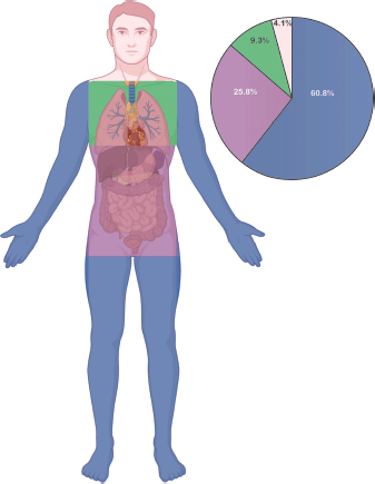
Figure 1. Anatomical distribution and frequency of STS. Illustration showing the anatomical sites affected by STS in the study population. The pie chart represents the proportion of cases according to tumour location: extremities (60.8%), abdomen and retroperitoneum (25.8%), chest wall (9.3%) and head and neck (4.1%).
Results
Patient characteristics
The sociodemographic factors of the 97 patients with STS are presented in Table 1. The mean age of patients was 51.4 years (SD = 15.9), ranging from 18 to 82 years at the time of diagnosis. In our cohort, 53.6% corresponded to male and 46.4% corresponded to female patients (ratio 1.15:1). The majority of patients were recorded as White (81.4%), followed by Mestizo (9.3%) and Black (9.3%). The main complaint of the patients was an increase in volume (60.8%), followed by pain (25.8%) and functional impotence (6.2%). According to the PS on the ECOG scale, the patients included in our study presented more frequently with ECOG 0 (33.0%) or ECOG 1 (67.0%). There were no cases with ECOG 3 or ECOG 4.
Overall, upper and lower extremities (60.8%) as well as abdomen and retroperitoneum (25.8%), followed by chest wall (9.3%) and head and neck (4.1%), were the anatomical locations of primary tumours (Figure 1). Most patients had deep tissue invasion (82.5%) and primary tumour size >5 cm (77.3%).
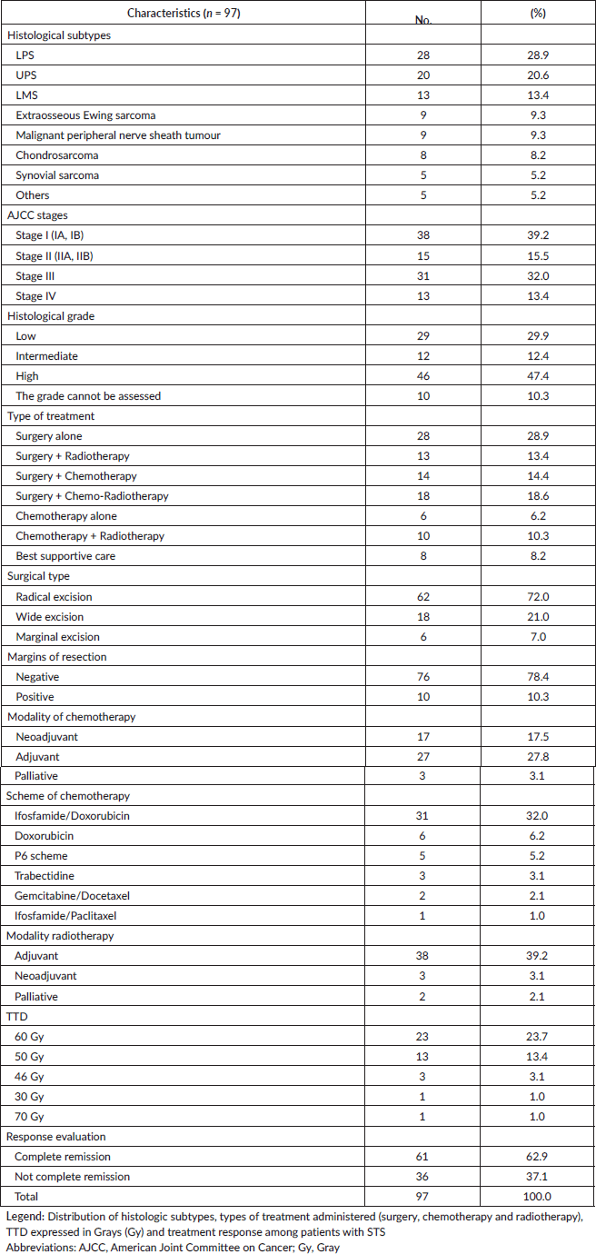
The histological analysis revealed the following distribution of the most common subtypes: LPS (28.9%), UPS (20.6%), LMS (13.4%), Extraosseous Ewing Sarcoma (9.3%), Malignant Peripheral Nerve Sheath Tumor (9.3%), Chondrosarcoma (8.2%) and Synovial Sarcoma (5.2%) (Table 2). The most frequent clinical stages were Stage I (39.2%) and Stage III (32.0%). Thirteen patients (13.4%) presented metastasis at diagnosis. Regarding histological grade, the distribution was 47.4%, 29.9% and 12.4% for high, low and intermediate, respectively.
Treatment patterns
The primary treatment modalities administered to the 97 patients are detailed in Table 2. Surgery was the most common component of therapy. Surgery alone was performed in 28 patients (28.9%). Multimodal treatment involving surgery was administered to 45 patients and included surgery with radiotherapy (13.4%), surgery with chemotherapy (14.4%) and surgery with both chemo-radiotherapy (18.6%). For the nonsurgical cohort, treatment consisted of chemotherapy alone (6.2%), combined chemotherapy and radiotherapy (10.3%) or best supportive care (8.2%). The most common drugs used as monotherapy or in combination were Ifosfamide/Doxorubicin (32.0%) and Doxorubicin (6.2%). Radiotherapy was applied in the neoadjuvant modality in 3.1%, as an adjuvant in 39.2% and as a palliative in 1.0% of the patients. Most patients received 60 Gy (23.7%) and 50 Gy (13.4%) as total tumour dose (TTD).
Response rates
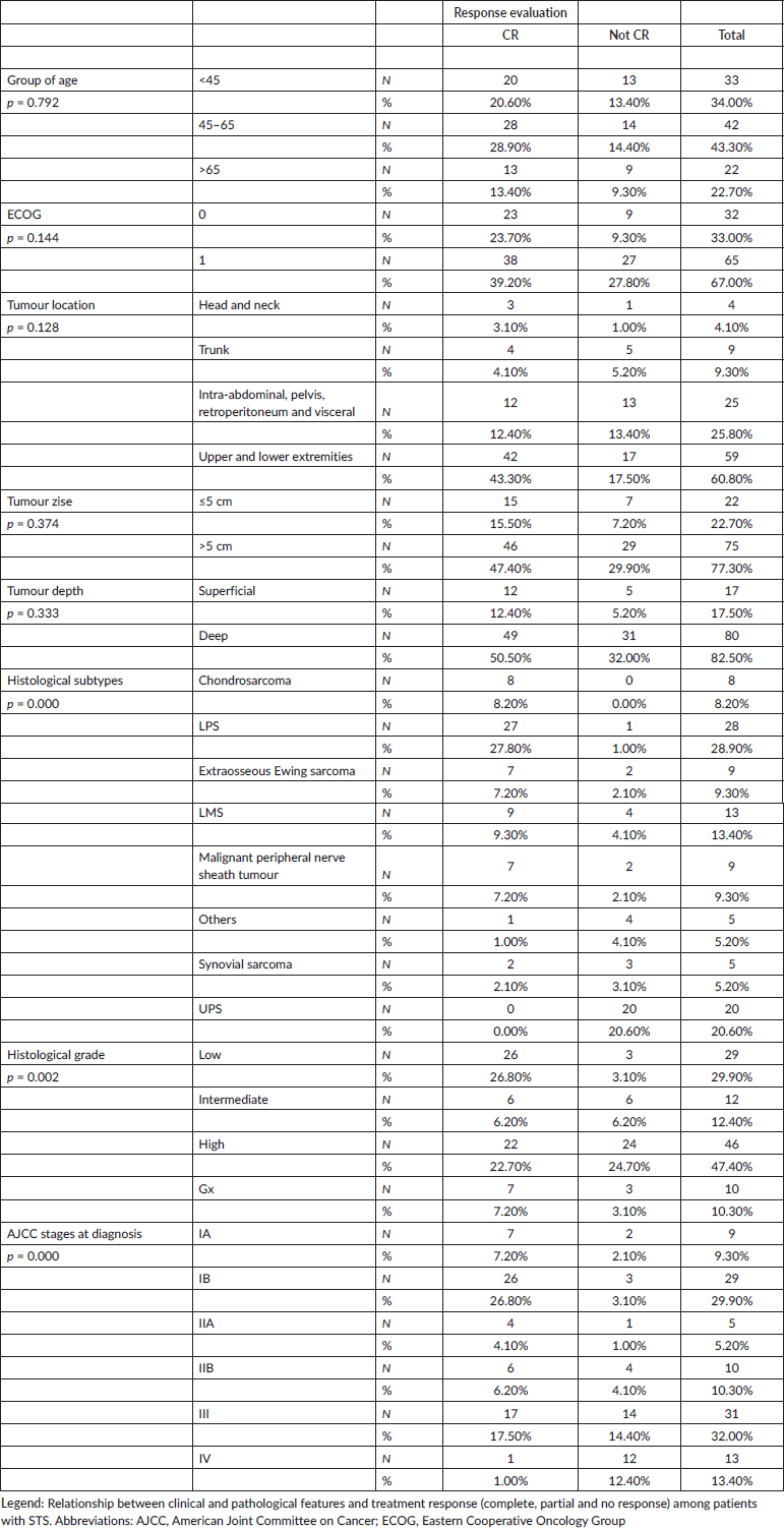
After the initial treatment, 62.9% of patients achieved a complete remission, and 37.1% did not achieve response (Table 2). Complete remission was related with: histological subtypes (p < 0.001), AJCC stages (p < 0.001) and histological grade (p = 0.002), while no association was evidenced when the patient age, tumour localisation, tumour size or ECOG were analysed (Supplementary Table S1).
Survival outcomes
The results of univariate and multivariate survival analysis are summarized in Table 3 and Figure 2. Overall, 30/97 patients (30.9%) died within 5 years after diagnosis. In the OS analysis, AJCC stage (p < 0.0001), histological grade (p = 0.011) and tumour depth (p = 0.042) were significant prognostic factors for 5-year OS (Figure 2A–C). Patient age, histological type, tumour size and ECOG were not associated with OS (Supplementary Figures S1–S4). Among these variables, clinical-stage IV (p = 0.005), intermediate (p = 0.011) and high (p = 0.006) histological grades were identified as independent risk factors for decreased OS on multivariate analysis.
The RFS for the entire cohort is presented in Supplementary Figure 5. At 5 years of follow-up, the RFS rate was 62.6% (95% CI, 52.7–74.4).
Table 1. Demographic and clinical characteristics.
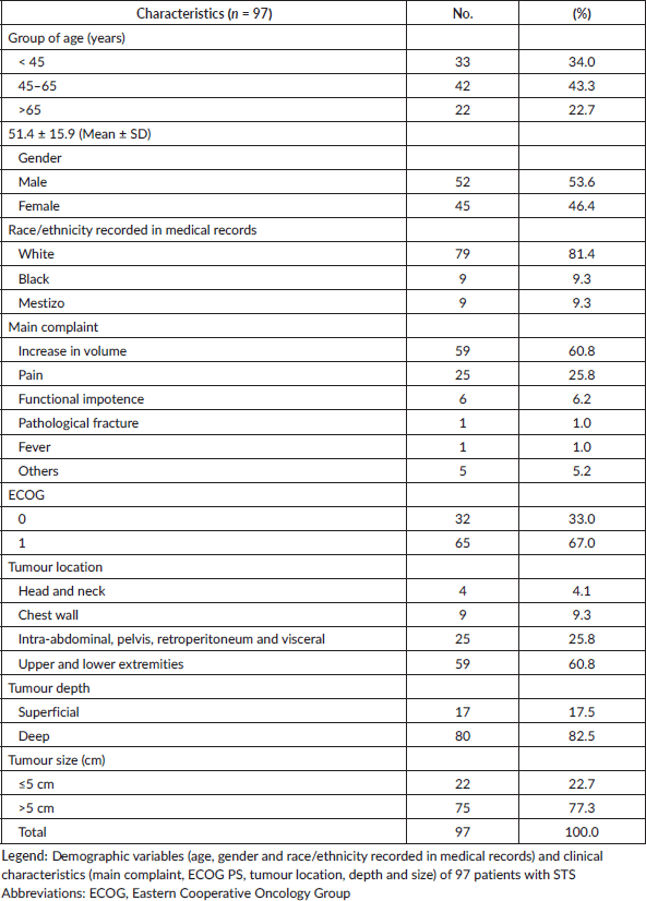
Table 2. Pathological and treatment characteristics.
Diagnostic and treatment intervals in STS
The time intervals of the patient care pathway were analysed (Figure 3). The median time from the first consultation to pathological diagnosis was 39.0 days (Interquartile range (IQR): 33.0–48.0 days) (Figure 3A). The subsequent median time from diagnosis to the initiation of primary treatment was 15.0 days (IQR: 10.0–27.0 days) (Figure 3B).
Discussion
STSs are rare and biologically heterogeneous malignancies that require coordinated clinical, radiological, surgical, pathological and oncological assessment. This expanded Cuban real-world cohort provides clinically relevant information on presentation, treatment patterns, diagnostic and treatment intervals and survival outcomes among adult patients with non-GIST STS treated at a national referral centre. The rarity and heterogeneity of STS make local data particularly relevant, especially in regions where published evidence remains limited. International guidelines emphasise that suspected STS should be assessed in experienced centres with access to multidisciplinary care, appropriate imaging, biopsy planning, pathology expertise, surgery, radiotherapy and systemic therapy when indicated [1, 13].
Table 3. Five-year OS and hazard ratios for death (multivariate analysis).
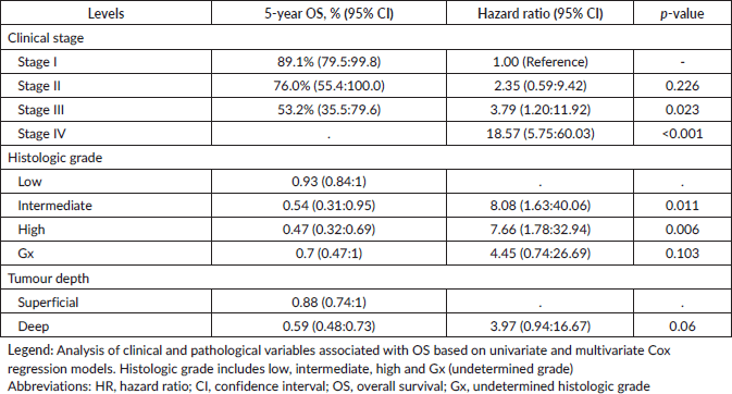
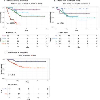
Figure 2. OS according to main prognostic factors of STS. (a): Kaplan–Meier survival curves by clinical stage (I–IV), with significant differences among stages (p < 0.0001). (b): Kaplan–Meier survival curves by histologic grade (low, intermediate, high and Gx), showing significant differences (p = 0.011). (c): Kaplan–Meier survival curves by tumour depth (superficial versus deep), with significant differences between groups (p = 0.042). The p-value for the log-rank test is shown in each plot, and the number of patients at risk at different time intervals is displayed.

Figure 3. Distribution of diagnostic and treatment intervals in patients with STS. Histograms showing the distribution of (a) time from first consultation to diagnosis (median, 39 days) and (b) time from diagnosis to treatment initiation (median, 15 days) for the cohort.
The demographic profile of this cohort was broadly comparable to international series, with a wide age distribution and slight male predominance. However, age and sex distributions in STS should be interpreted cautiously because they vary by histological subtype, anatomical site and referral pattern. European population-based data have shown that sarcomas are uncommon but highly heterogeneous, with important variation by histological subtype and primary site [4]. Similarly, studies incorporating central pathology review and molecular testing have shown that histological distribution depends strongly on diagnostic classification and ancillary testing [7]. In the present cohort, the predominance of ECOG 0–1 suggests that most patients reached the referral centre with preserved functional status, which may have facilitated eligibility for surgery and multimodality treatment.
Extremities were the most common primary tumour site, consistent with previous reports showing that a substantial proportion of adult STS arise in the limbs. Large institutional series of extremity STS have shown that tumour depth, size, grade, margin status, age, presentation status and histological subtype are clinically relevant prognostic variables [8]. From a practical clinical perspective, extremity STS may allow limb-sparing surgery when diagnosis, staging, surgical planning and radiotherapy are coordinated appropriately. However, deep-seated and large tumours remain clinically challenging because they are associated with greater surgical complexity and worse oncological outcomes.
Most tumours in this cohort were deep-seated and larger than 5 cm. The clinical importance of tumour size has been highlighted in large sarcoma cohorts, where larger tumours were associated with higher metastatic risk and poorer prognosis [14]. In the French Federation of Cancer Centers Sarcoma Group experience, tumour grade, deep location and size were key prognostic factors in adult patients with locally controlled STS [15]. These data support the clinical relevance of the variables that were significant in our cohort, particularly stage, grade and tumour depth.
The high proportion of patients with localised disease is one of the clinically relevant findings of this study. This may reflect, at least in part, the structure of the Cuban healthcare system, where primary care and referral pathways can facilitate access to specialised evaluation. The Cuban health system has been described as a national integrated model with a strong primary-care foundation [16]. The Family Doctor and Nurse Program has also been described as a central element of Cuban community-based care [17]. In our cohort, the observed diagnostic and treatment intervals suggest that, once patients entered the healthcare pathway, progression to diagnosis and treatment was relatively prompt. Nevertheless, these findings should be interpreted cautiously because the study did not capture the interval from first symptom to first medical consultation. Diagnostic intervals in sarcoma are highly variable and may be influenced by tumour biology, anatomical site, symptoms, patient factors and access to specialist assessment [18].
The predominance of surgery-based treatment in this cohort is consistent with the central role of complete surgical resection in localised STS. In clinical practice, surgery should ideally be planned within a multidisciplinary framework that integrates imaging, biopsy strategy, histological diagnosis, margin assessment, radiotherapy and systemic therapy when indicated. The fact that cases in this cohort were managed through multidisciplinary consensus is clinically important because adherence to consensus-based diagnostic and treatment recommendations has been associated with improved quality of care in adult STS [19]. In resource-constrained settings, structured referral to experienced centres may be particularly important to reduce unplanned excisions, improve staging and support risk-adapted treatment selection.
LPS was the most frequent histological subtype, followed by UPS and LMS. This distribution is clinically plausible, but it must be interpreted with caution because STS histology is not merely descriptive; it directly influences prognosis, recurrence patterns, treatment sensitivity and systemic therapy options. LPS includes biologically distinct entities, including well-differentiated, dedifferentiated, myxoid and pleomorphic subtypes, with different anatomical distributions, molecular features and clinical behaviour [20]. Therefore, the LPS group in this cohort should not be interpreted as a single homogeneous clinical entity.
Similarly, UPS remains a heterogeneous diagnostic category. In current clinical practice, classification of STS increasingly relies on integration of morphology, immunohistochemistry and molecular testing when appropriate. The 2020 WHO classification reinforced the importance of standardised pathological classification and recognition of molecularly defined entities [21]. In the present cohort, diagnoses were made according to the diagnostic resources available during 2006–2013. Immunohistochemistry was performed when clinically indicated and available, but marker-level immunohistochemical results were not systematically abstracted for all patients, and molecular testing was not routinely available. For this reason, we did not attempt retrospective molecular reclassification or detailed subtype-specific survival modelling, as doing so could have introduced misclassification and overinterpretation.
Survival analysis showed that AJCC stage, histological grade and tumour depth were associated with 5-year OS, while stage IV disease and intermediate or high histological grade independently predicted worse survival. These findings are clinically coherent and consistent with the established role of tumour burden and biological aggressiveness in STS prognosis. Stage and grade remain central to risk stratification, patient counselling, surveillance planning and selection of adjuvant or systemic treatment. Similar retrospective data from tertiary centres in resource-constrained settings have also shown that stage and treatment-related factors remain important prognostic variables in STS [22]. The lack of statistically significant association between OS and variables such as age, tumour size, ECOG status or histological subtype should not be interpreted as absence of clinical relevance. Rather, these findings likely reflect the modest sample size, the heterogeneity of histological subtypes and limited statistical power for subgroup analyses.
The RFS analysis adds clinically useful information beyond OS. Although 5-year OS was relatively favourable for patients with localised disease, recurrence remained an important outcome. Recurrence in STS may require repeated surgery, radiotherapy, systemic therapy or palliative interventions and may negatively affect function and quality of life. Current STS management increasingly emphasises histotype-specific decision-making, with treatment influenced by anatomical site, resectability, grade, histological subtype and expected sensitivity to systemic therapy [23]. Therefore, future Cuban and regional studies should aim to collect more granular pathological, treatment, recurrence and functional outcome data.
This study has several limitations. First, its retrospective, single-institution design means that the findings reflect the experience of a national referral centre and may not be fully generalisable to the entire country. Second, the outcomes are contextualised within the Cuban healthcare system, and comparisons with high-resource settings should be made cautiously given potential differences in diagnostic resources, radiotherapy access, systemic therapy availability and pathology infrastructure during the study period. Third, the retrospective nature of the study limited the availability of detailed pathological ancillary data. Although immunohistochemistry was performed when clinically indicated and available, marker-level immunohistochemical results were not systematically collected because this was not the primary objective of the study. In addition, molecular testing and next-generation sequencing were not routinely available during the 2006–2013 study period. Consequently, detailed retrospective subclassification of LPS variants, molecularly defined entities, extraosseous Ewing sarcoma and the UPS group was not feasible for the entire cohort. Fourth, the modest sample size for individual histologies limited subtype-specific survival analyses. Finally, the study did not capture the full patient interval from first symptom to first medical consultation, which limits interpretation of the complete diagnostic pathway.
In summary, this study provides an expanded real-world clinical analysis of adult non-GIST STS treated at a Cuban national referral centre. The findings support the prognostic relevance of AJCC stage, histological grade and tumour depth while also highlighting the clinical value of structured referral pathways, multidisciplinary management, and timely initiation of treatment. Future multicentre studies incorporating standardised pathology review, systematic immunohistochemistry, molecular testing where available and histology-specific outcomes would further strengthen sarcoma care and research in Cuba and comparable health systems.
List of abbreviations
AJCC, American Joint Committee on Cancer; CI, Confidence interval; ECOG, Eastern Cooperative Oncology Group; GIST, Gastrointestinal stromal tumour; IQR, Interquartile range; LMS, Leiomyosarcoma; LPS, Liposarcoma; OS, Overall survival; PS, Performance status; RFS, Recurrence-free survival; SD, Standard deviation; STS, Soft tissue sarcoma; TTD, Total tumour dose; UPS, Undifferentiated pleomorphic sarcoma; WHO, World Health Organization.
Acknowledgments
The authors thank Jorge Luis Soriano García, Marta Osorio Rodríguez, Iraida Caballero, Adriana Carr and the clinical and pathology teams of the “Hermanos Ameijeiras” Clinical-Surgical Hospital for their academic guidance, clinical mentorship and institutional support during the development of the original research project from which this work originated. These contributions are acknowledged because they supported the original institutional phase of the study; authorship of the present manuscript reflects contributions to the current analysis, manuscript preparation, final approval and accountability for the submitted work. The authors also acknowledge the support of the National Secretariat for Higher Education, Science, Technology, and Innovation of Ecuador (SENESCYT) through Ph.D. fellowships awarded to D.F.P.J. and M.D.P.
Conflicts of interest
Daniel F Pilco-Janeta has received travel grants from Pfizer, Roemmers, Roche and Bristol Myers Squibb. Jan Philipp Novotny owns stock in Onxeo. The other authors declare no potential conflicts of interest.
Funding
The authors received no financial support for this research.
Data availability
De-identified data and analysis code supporting the findings of this study are available from the corresponding author upon reasonable request.
References
1. Casali PG, Abecassis N, and Bauer S, et al (2018) Soft tissue and visceral sarcomas: ESMO–EURACAN clinical practice guidelines for diagnosis, treatment and follow-up Ann Oncol 29 iv51–iv67 https://doi.org/10.1093/annonc/mdy096
2. Fletcher CDM, Bridge JA, and Hogendoorn P, et al (2013) WHO Classification of Tumours of Soft Tissue and Bone. Fourth Edition. IARC WHO Classif Tumours (Geneva: WHO) vol 2013 no (5)
3. Wibmer C, Leithner A, and Zielonke N, et al (2010) Increasing incidence rates of soft tissue sarcomas? A population-based epidemiologic study and literature review Ann Oncol 21(5) 1106–1111 https://doi.org/10.1093/annonc/mdp415
4. Stiller CA, Trama A, and Serraino D, et al (2013) Descriptive epidemiology of sarcomas in Europe: report from the RARECARE project Eur J Cancer Oxf Engl 1990 49(3) 684–695 [https://doi.org/10.1016/j.ejca.2012.09.011]
5. Siegel RL, Miller KD, and Jemal A (2018) Cancer statistics, 2018 CA Cancer J Clin 68(1) 7–30 [https://doi.org/10.3322/caac.21442] PMID: 29313949
6. Galán Álvarez YH, Iglesias Ventura Y, and Kindelán Valerino G, et al (2020) Incidencia de cáncer en Cuba en el 2016 Rev Cubana Oncol 18(3) e46
7. Ducimetière F, Lurkin A, and Ranchère-Vince D, et al (2011) Incidence of sarcoma histotypes and molecular subtypes in a prospective epidemiological study with central pathology review and molecular testing PLoS One 6(8) e20294 https://doi.org/10.1371/journal.pone.0020294 PMID: 21826194 PMCID: 3149593
8. Weitz J, Antonescu CR, and Brennan MF (2003) Localized extremity soft tissue sarcoma: improved knowledge with unchanged survival over time J Clin Oncol Off J Am Soc Clin Oncol 21(14) 2719–2725 https://doi.org/10.1200/JCO.2003.02.026
9. Nussbaum DP, Rushing CN, and Lane WO, et al (2016) Preoperative or postoperative radiotherapy versus surgery alone for retroperitoneal sarcoma: a case-control, propensity score-matched analysis of a nationwide clinical oncology database Lancet Oncol 17(7) 966–975 https://doi.org/10.1016/S1470-2045(16)30050-X PMID: 27210906
10. Schöffski P, Cornillie J, and Wozniak A, et al (2014) Soft tissue sarcoma: an update on systemic treatment options for patients with advanced disease Oncol Res Treat 37(6) 355–362 https://doi.org/10.1159/000362631 PMID: 24903768
11. Italiano A, Mathoulin‐Pelissier S, and Cesne AL, et al (2011) Trends in survival for patients with metastatic soft-tissue sarcoma Cancer 117(5) 1049–1054 https://doi.org/10.1002/cncr.25538
12. Mastrangelo G, Coindre JM, and Ducimetière F, et al (2012) Incidence of soft tissue sarcoma and beyond: a population-based prospective study in 3 European regions Cancer 118(21) 5339–5348 https://doi.org/10.1002/cncr.27555 PMID: 22517534
13. Hayes AJ, Nixon IF, and Strauss DC, et al (2025) UK guidelines for the management of soft tissue sarcomas Br J Cancer 132(1) 11–31 https://doi.org/10.1038/s41416-024-02674-y PMCID: 11724041
14. Grimer RJ (2006) Size matters for sarcomas! Ann R Coll Surg Engl 88(6) 519–524 https://doi.org/10.1308/003588406X130651 PMID: 17059708 PMCID: 1963770
15. Coindre JM, Terrier P, and Bui NB, et al (1996) Prognostic factors in adult patients with locally controlled soft tissue sarcoma. A study of 546 patients from the French Federation of Cancer Centers Sarcoma Group J Clin Oncol Off J Am Soc Clin Oncol 14(3) 869–877 https://doi.org/10.1200/JCO.1996.14.3.869
16. Keck CW and Reed GA (2012) The curious case of Cuba Am J Public Health 102(8) e13–e22 https://doi.org/10.2105/AJPH.2012.300822 PMID: 22698011 PMCID: 3464859
17. Gorry C (2017) Cuba’s family doctor-and-nurse teams: a day in the life MEDICC Rev 19(1) 6–9 https://doi.org/10.37757/MR2017.V19.N1.2 PMID: 28225539
18. Soomers V, Husson O, and Young R, et al (2020) The sarcoma diagnostic interval: a systematic review on length, contributing factors and patient outcomes ESMO Open 5(1) 592 https://doi.org/10.1136/esmoopen-2019-000592
19. Mathoulin-Pélissier S, Chevreau C, and Bellera C, et al (2014) Adherence to consensus-based diagnosis and treatment guidelines in adult soft-tissue sarcoma patients: a French prospective population-based study Ann Oncol Off J Eur Soc Med Oncol 25(1) 225–231 https://doi.org/10.1093/annonc/mdt407
20. Lee ATJ, Thway K, and Huang PH, et al (2018) Clinical and molecular spectrum of liposarcoma J Clin Oncol Off J Am Soc Clin Oncol 36(2) 151–159 https://doi.org/10.1200/JCO.2017.74.9598
21. Sbaraglia M, Bellan E, and Dei Tos AP (2020) The 2020 WHO classification of soft tissue tumours: news and perspectives Pathologica 113(2) 70–84 https://doi.org/10.32074/1591-951X-213 PMID: 33179614 PMCID: 8167394
22. Mahyudin F, Edward M, and Basuki MH, et al (2020) Analysis of prognostic factors in soft tissue sarcoma: cancer registry from a single tertiary hospital in Indonesia. A retrospective cohort study Ann Med Surg 57 257–263 https://doi.org/10.1016/j.amsu.2020.07.053
23. Gamboa AC, Gronchi A, and Cardona K (2020) Soft-tissue sarcoma in adults: an update on the current state of histiotype-specific management in an era of personalized medicine CA Cancer J Clin 70(3) 200–229 [https://doi.org/10.3322/caac.21605] PMID: 32275330
Supplementary materials
Supplementary Table S1. Association between clinicopathological variables and treatment response.
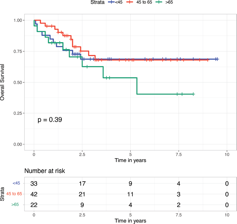
Supplementary Figure 1. OS according to age group in patients with STS. Kaplan–Meier survival curves are shown for three age groups (<45 years, 45–65 years and >65 years). OS is displayed on the vertical axis, and time in years is displayed on the horizontal axis. No statistically significant difference in survival was observed among the age groups (p = 0.39).
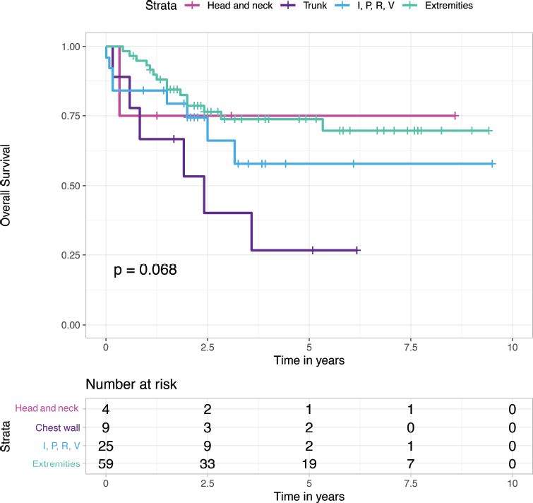
Supplementary Figure 2. OS according to tumour location in patients with STS. Kaplan–Meier survival curves are shown according to tumour location: head and neck, chest wall, intra-abdominal/pelvic/retroperitoneal/visceral (I, P, R and V) and extremities. OS is displayed on the vertical axis, and time in years is displayed on the horizontal axis. No statistically significant difference in survival was observed among the tumour locations (p = 0.068).

Supplementary Figure 3. OS according to tumour size in patients with STS. Kaplan–Meier survival curves are shown comparing tumours smaller than 5 cm versus those larger than 5 cm. OS is displayed on the vertical axis, and time is displayed in years on the horizontal axis. No statistically significant difference in survival was observed between the groups (p = 0.85).
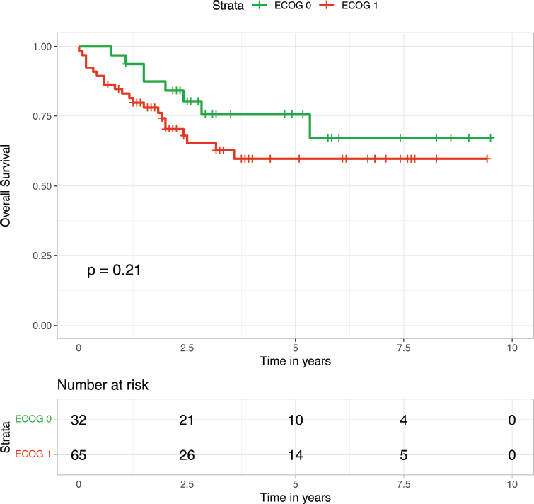
Supplementary Figure 4. OS according to ECOG PS in patients with STS. Kaplan–Meier survival curves are shown comparing patients with ECOG 0 and ECOG 1 PS. OS is displayed on the vertical axis, and time in years is displayed on the horizontal axis. No statistically significant difference in survival was observed between the groups (p = 0.21).

Supplementary Figure 5. RFS of STS. Kaplan–Meier curve showing RFS for the entire cohort (n = 97). RFS was calculated from the date of primary treatment initiation to the date of first recurrence (local or distant). The shaded area represents the 95% confidence interval. The number of patients at risk is shown below the plot.