A retrospective analysis of the clinico-epidemiological features and treatment outcomes of renal cell carcinoma at the Clinical Oncology Department at Ain Shams University in Egypt
Zizit Hamdi Mohammed El-Shahawi, Soheir Sayed Ismail, Lamia Moustafa Ahmed and Nesreen Ahmed Mosalam
Clinical Oncology Department, Faculty of Medicine, Ain Shams University, Cairo, Egypt
Abstract
Background: Renal cell carcinoma (RCC) is a biologically heterogeneous malignancy with variable clinical behaviour and survival outcomes. Real-world data from low- and middle-income countries remain limited, particularly regarding stage-specific treatment patterns and survival outcomes.
Methods: This retrospective cohort study included adult patients diagnosed with RCC and treated at the Clinical Oncology Department, Ain Shams University Hospitals, between January 2018 and December 2023. Patients were analysed as two pre-specified cohorts: (1) localised disease (Stage I–III) undergoing curative-intent surgery (n = 45) and (2) metastatic disease (Stage IV) receiving systemic therapy (n = 34). Survival outcomes were analysed using the Kaplan–Meier (KM) method and reported as medians with interquartile ranges (IQRs).
Results: A total of 79 patients were included (50 male, 63.3%; median age 57 years, IQR 50–66). In the localised cohort (n = 45), 43 underwent curative-intent surgery; disease recurrence occurred in 17 (39.5%). The 12-month disease-free survival (DFS) was 78.2%, and the 36-month DFS was 64.1%; median DFS was not reached within the observation period. In the metastatic cohort (n = 34), sunitinib was the predominant first-line agent; the KM estimated median progression-free survival (PFS) on sunitinib was 16 months (IQR 6–36), with a 6-month PFS rate of 75.4% and a 12-month PFS rate of 58.0%. The KM estimated median overall survival (OS) for Stage IV patients was 43 months from diagnosis. Disease recurrence and progression were significantly associated with inferior OS (p = 0.003 and p = 0.01, respectively).
Conclusion: Surgical resection remains the cornerstone of curative management. In the metastatic setting, sunitinib provided meaningful and durable disease control (median PFS 16 months; KM median OS 43 months) despite restricted immunotherapy access. High-risk pathological features identified in 33% of the localised cohort highlight an unmet need for adjuvant immunotherapy in resource-limited settings.
Keywords: renal cell carcinoma, nephrectomy, sunitinib, targeted therapy, survival, Kaplan–Meier, retrospective, LMIC
Correspondence to: Zizit Hamdi Mohammed El-Shahawi
Email: drzizt@gmail.com
Published: 04/08/2026
Received: 05/01/2026
Publication costs for this article were supported by ecancer (UK Charity number 1176307).
Copyright: © the authors; licensee ecancermedicalscience. This is an Open Access article distributed under the terms of the Creative Commons Attribution License (http://creativecommons.org/licenses/by/4.0), which permits unrestricted use, distribution, and reproduction in any medium, provided the original work is properly cited.
Introduction
Renal cell carcinoma (RCC) accounts for approximately 80%–85% of primary renal malignancies and represents a significant global health burden. It is the seventh most common cancer in men and the tenth in women worldwide, with clear cell RCC as the predominant histological subtype [1–3, 11, 16]. Although survival outcomes have improved due to advances in surgical techniques and systemic therapies, RCC continues to pose major challenges in advanced and metastatic stages [1, 2, 18].
In recent years, immune checkpoint inhibitor combinations have transformed the first-line treatment landscape for metastatic RCC. Despite these advances, access to novel agents remains severely limited in many low- and middle-income countries (LMICs), where sunitinib continues to represent the backbone of systemic treatment. This study aimed to retrospectively analyse the clinicoepidemiological characteristics, treatment strategies, and survival outcomes of patients with RCC treated at a tertiary oncology centre in Egypt, with separate analyses of the curative-intent surgery cohort and the metastatic/systemic therapy cohort [5–7].
Patients and methods
Study design and setting
This retrospective cohort study was conducted at the Clinical Oncology Department, Ain Shams University Hospitals, Cairo, Egypt, and included adult patients diagnosed with RCC managed between January 2018 and December 2023.
Study population
Eligible patients were identified through medical record review. Inclusion criteria comprised patients aged ≥18 years with histopathologically confirmed RCC. Two pre-specified analytical cohorts were defined: (1) Localised disease cohort - Stage I–III with curative-intent surgery (n = 45); (2) Metastatic/systemic therapy cohort - Stage IV disease (n = 34) [7–9].
Outcome measures and statistical analysis
The primary outcomes were disease-free survival (DFS) (time from curative surgery to first recurrence), progression-free survival (PFS) (time from initiation of systemic therapy to documented progression or last follow-up, with non-progressing patients censored), and overall survival (OS) (time from diagnosis to death or last follow-up). Survival outcomes were analysed using the Kaplan–Meier (KM) method with 95% confidence intervals (Greenwood method). For PFS analysis, all sunitinib-treated patients were included; patients without documented progression were treated as right-censored observations. All continuous variables are reported as median interquartile ranges (IQR). Group comparisons used the Mann–Whitney U test or log-rank test. A p-value ≤0.05 was considered statistically significant [11, 12].
Ethical considerations
This study was conducted after obtaining approval from the Research Ethics Committee, Faculty of Medicine, Ain Shams University. All patient data were anonymised prior to analysis.
Results
Patient demographics (n = 79)
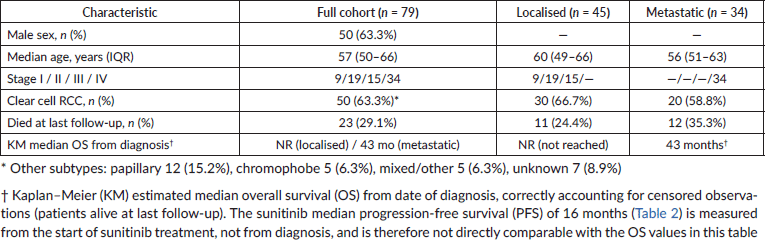
A total of 79 patients with histopathologically confirmed RCC were included. The cohort comprised 50 males (63.3%) and 29 females (36.7%). The median age at diagnosis was 57 years (IQR 50–66; range 24–84). At presentation, 34 patients (43.0%) had Stage IV metastatic disease; 15 (19.0%) had Stage III, 19 (24.1%) had Stage II and 9 (11.4%) had Stage I. Clear cell RCC was the predominant histological subtype (n = 50, 63.3%). At last follow-up, 56 patients (70.9%) were alive, and 23 (29.1%) had died (Table 1).
All OS values are KM estimates from the date of diagnosis and correctly account for censored observations (patients alive at last follow-up). KM median OS for the metastatic cohort = 43 months, consistent with Figure 1. The sunitinib median PFS of 16 months (Table 2) is measured from the start of treatment, not from diagnosis, and therefore is not directly comparable with the OS values in this table.
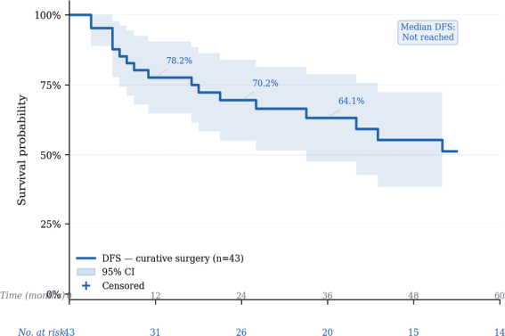
Figure 1. OS stratified by disease stage at diagnosis. KM curves with 95% CI; localised n = 45 (blue), metastatic n = 34 (red). KM median OS: localised NR; metastatic 43 months.
Table 1. Baseline patient characteristics.
Localised disease cohort: curative surgery (n = 45)
Of 45 patients with Stage I–III disease, 43 underwent surgery: radical nephrectomy n = 39 (90.7%), partial nephrectomy n = 3 (7.0%), simple nephrectomy n = 1 (2.3%). Two patients declined surgery. High-risk pathological features were present in a substantial proportion: pT3–T4 disease 15 (33.3%), perinephric fat invasion 9 (20.0%), pathological N1 5 (11.1%), nuclear grade 3–4 10 (22.2%), sarcomatoid differentiation 4 (8.9%) and lymphovascular invasion 5 (11.1%) [4, 14, 17, 23].
Disease-free survival
Disease recurrence occurred in 17 of 43 surgical patients (39.5%). The KM estimated DFS rates were as follows: 78.2% at 12 months, 70.2% at 24 months and 64.1% at 36 months (Figure 2). Median DFS was not reached within the observation period. Among relapsed patients, the median time to relapse was 11 months (IQR 6–26). No Stage I patient experienced recurrence. Disease recurrence was significantly associated with inferior OS (36-month OS: 58.0% versus 87.0%; p = 0.003; Figure 3). Lower baseline haemoglobin was significantly associated with recurrence (median 9.0 versus 12.0 g/dL; p = 0.04) [20, 27].
Table 2. Systemic therapy by line and agent.
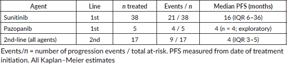
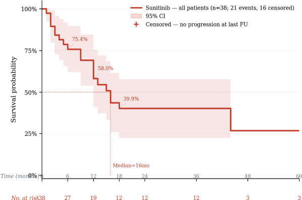
Figure 2. DFS after curative-intent surgery. KM KM curve, n = 43 surgical patients, 17 relapse events. Tick marks = censored; 95% CI as shaded band.
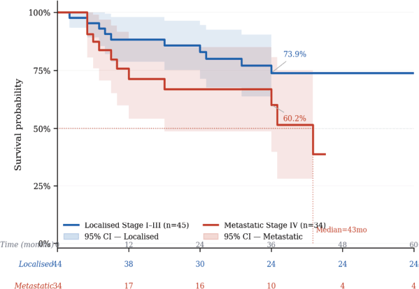
Figure 3. OS by disease recurrence status (surgical cohort). No recurrence n = 26 (teal) versus recurrence n = 17 (red). p = 0.003, log-rank test.
Metastatic/systemic therapy cohort (n = 34 at presentation)
Of 34 patients with Stage IV disease, 24 (70.6%) underwent cytoreductive nephrectomy (haematuria n = 5, loin pain n = 7, obstruction/other n = 5, selected favourable-risk n = 3). Cytoreductive surgery was performed for palliation within a multidisciplinary framework, not as routine survival-prolonging intervention, in line with CARMENA trial evidence [6].
First-line systemic therapy and PFS
Sunitinib was administered to 38 patients. KM analysis of PFS included all 38 patients (37 with follow-up time data), with 16 non-progressors treated as right-censored observations. The KM estimated median PFS was 16 months (IQR 6–36), with a 6-month PFS rate of 75.4% and a 12-month PFS rate of 58.0% (Figure 4). Pazopanib was administered to five patients (median PFS 4 months; n = 4 with events) [29, 30].
Second-line systemic therapy
In the second-line setting, 17 patients received further systemic therapy (pazopanib n = 12, everolimus n = 4, nivolumab n = 1). The median second-line PFS was 4 months (IQR 3–5; n = 9 with events) [31].
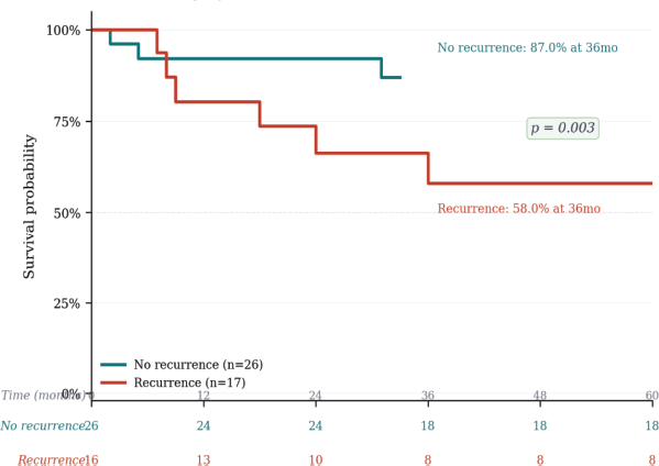
Figure 4. PFS on first-line sunitinib. All 38 treated patients (37 with follow-up data): 21 progression events, 16 censored (tick marks). Median PFS = 16 months. 95% CI as shaded band. PFS measured from start of sunitinib.
Overall survival
At last follow-up, 56 patients (70.9%) were alive, and 23 (29.1%) had died. The KM estimated median OS was not reached in the localised cohort within the observation period (36-month OS rate 73.9%; Figure 1). The KM estimated median OS for the metastatic cohort was 43 months from diagnosis (36-month OS rate 60.2%; Figure 1). Disease recurrence was significantly associated with inferior OS (36-month OS: 58.0% with recurrence versus 87.0% without; p = 0.003; Figure 3). Disease progression was similarly associated with inferior OS (p = 0.01) [21].
Discussion
This retrospective study provides real-world data on clinicoepidemiological characteristics, treatment patterns, and survival outcomes of 79 patients with RCC treated at a tertiary oncology centre in Egypt, presented within a structured two-cohort framework [12, 13].
The demographic profile is consistent with internationally published data, with a male predominance (63.3%) and a median age of 57 years (IQR 50–66). A substantial proportion (43.0%) presented with metastatic disease, reflecting delayed diagnosis characteristic of LMIC settings [12, 13, 21].
Surgical management served fundamentally different roles in the two cohorts. In the localised cohort, curative-intent nephrectomy represents the standard of care. The apparent survival advantage of surgical patients in aggregate analyses reflects stage-driven confounding rather than a direct surgical survival effect. In the metastatic cohort, 24 of 34 patients underwent cytoreductive nephrectomy for symptom palliation, within a multidisciplinary framework and in line with CARMENA trial guidance [19],which recommends against routine upfront cytoreductive surgery [6, 18].
Disease recurrence and progression were the dominant determinants of OS. Patients who relapsed had a 36-month OS of 58.0% compared with 87.0% in those who remained disease-free (p = 0.003). A substantial proportion of localised patients harboured high-risk pathological features: pT3–T4 disease (33.3%), perinephric fat invasion (20.0%), pathological N1 (11.1%), nuclear grade 3–4 (22.2%), sarcomatoid differentiation (8.9%) and lymphovascular invasion (11.1%). The KEYNOTE-564 trial [20] demonstrated that adjuvant pembrolizumab significantly improved DFS in such high-risk patients (HR 0.68; p = 0.002), with confirmed OS benefit (HR 0.62). None of our patients received adjuvant therapy due to access constraints [5, 21, 23–26].
When all 38 sunitinib-treated patients are included in the KM analysis with correct censoring of non-progressors, the median PFS was 16 months (IQR 6–36), with 75.4% of patients progression-free at 6 months and 58.0% at 12 months. The KM median OS for Stage IV patients was 43 months from diagnosis. These figures are consistent with one another: OS is measured from diagnosis and includes any pre-treatment interval, while PFS is measured from the start of sunitinib and reflects time on active therapy. The 43-month KM OS is longer than the 16-month PFS because it accounts for time after progression in patients who received subsequent therapy, and because KM correctly weights the substantial proportion of patients still alive at last follow-up. The limited use of immunotherapy reflects financial and access barriers rather than clinical preference [29–31].
The magnitude of survival improvement achievable with contemporary immunotherapy-based combinations is substantial. Four landmark trials have transformed the first-line landscape: CheckMate 214 (nivolumab + ipilimumab; OS HR 0.68), KEYNOTE-426 (pembrolizumab + axitinib; OS HR 0.53), CheckMate 9ER (nivolumab + cabozantinib; OS HR 0.60), and CLEAR (lenvatinib + pembrolizumab; PFS HR 0.39). Achieving equitable access to these regimens in LMICs requires differential pricing policies and national formulary inclusion [1, 5].
Study strengths and limitations
The strengths include real-world design, two-cohort analytical framework, comprehensive KM survival analysis with correct censoring and 95% confidence intervals. Limitations include the retrospective single-centre design, relatively small sample size, underpowered recurrence subgroup (n = 17) and absence of adjuvant immunotherapy and first-line immunotherapy access, limiting comparison with contemporary cohorts [27].
Conclusion
RCC demonstrated markedly different outcomes between the localised and metastatic cohorts in this Egyptian real-world series. Surgical resection remains the cornerstone of curative management. When analysed with correct KM methodology, sunitinib provided a median PFS of 16 months and the metastatic cohort achieved a KM median OS of 43 months from diagnosis, reflecting meaningful disease control in this resource-constrained setting. A substantial proportion of localised patients harboured high-risk features for whom adjuvant pembrolizumab is now guideline-recommended but inaccessible. Closing the immunotherapy equity gap in LMICs requires both policy intervention and advocacy.
Conflicts of interest
The authors declare no conflicts of interest.
Funding
This research received no specific grant from any funding agency in the public, commercial, or not-for-profit sectors.
Ethical approval
This study was conducted after obtaining approval from the Research Ethics Committee, Faculty of Medicine, Ain Shams University, Cairo, Egypt. All patient data were anonymised prior to analysis.
Data availability
The datasets generated and analysed during the current study are available from the corresponding author upon reasonable request.
Author contributions
Zizit Hamdi Mohammed El-shahawi: Study conception and design, data collection, analysis, manuscript drafting. Soheir Sayed Ismail: Study supervision, critical revision. Nesreen Ahmed Mosalam: Data interpretation, manuscript revision. Lamia Moustafa Ahmed: Data interpretation, manuscript revision. All authors approved the final manuscript.
References
1. Escudier B and Kataja V (2019) Renal cell carcinoma: ESMO clinical practice guidelines for diagnosis, treatment and follow-up Ann Oncol 30 706–720 https://doi.org/10.1093/annonc/mdz056 PMID: 30788497
2. Hsieh JJ, Purdue MP, and Signoretti S, et al (2017) Renal cell carcinoma Nature Rev Dis Primers 3 17009 https://doi.org/10.1038/nrdp.2017.9
3. Shuch B, Vourganti S, and Ricketts CJ, et al (2014) Defining early-onset kidney cancer: implications for germline and somatic mutation testing and clinical management J Clin Oncol 32 431–437 https://doi.org/10.1200/JCO.2013.50.8192 PMID: 24378414 PMCID: 3912328
4. Maclennan S, Imamura M, and Lapitan MC, et al (2012) Systematic review of oncological outcomes following surgical management of localised renal cancer Eur Urol 61 972–993 https://doi.org/10.1016/j.eururo.2012.02.039 PMID: 22405593
5. Méndez-Vidal MJ, Lázaro Quintela M, and Lainez-Milagro N, et al (2023) SEOM SOGUG clinical guideline for treatment of kidney cancer (2022) Clin Translational Oncol 25 2732–2748 https://doi.org/10.1007/s12094-023-03276-5
6. Bex A, Mulders P, and Jewett M, et al (2019) Comparison of immediate vs deferred cytoreductive nephrectomy in patients with synchronous metastatic renal cell carcinoma receiving Sunitinib: the SURTIME randomized clinical trial JAMA Oncol 5 164–170 https://doi.org/10.1001/jamaoncol.2018.5543 PMCID: 6439568
7. Delahunt B, Eble JN, and Samaratunga H, et al (2021) Staging of renal cell carcinoma: current progress and potential advances Pathology 53 120–128 https://doi.org/10.1016/j.pathol.2020.08.007
8. Netto GJ, Amin MB, and Berney DM, et al (2022) The 2022 World Health Organization classification of tumors of the urinary system and male genital organs — part B: prostate and urinary tract tumors Eur Urol 82 469–482 https://doi.org/10.1016/j.eururo.2022.07.002 PMID: 35965208
9. Heng DY, Xie W, and Regan MM, et al (2013) External validation and comparison with other models of the International Metastatic Renal-Cell Carcinoma Database Consortium prognostic model: a population-based study Lancet Oncol 14 141–148 https://doi.org/10.1016/S1470-2045(12)70559-4 PMID: 23312463 PMCID: 4144042
10. Das CJ (2015) Perfusion computed tomography in renal cell carcinoma World J Radiol 7 170 https://doi.org/10.4329/wjr.v7.i7.170 PMID: 26217456 PMCID: 4506935
11. Delgado A and Guddati AK (2021) Clinical endpoints in oncology - a primer Am J Cancer Res 11 1121–1131 PMID: 33948349 PMCID: 8085844
12. Rizzo M, Pezzicoli G, and Tibollo V, et al (2024) Clinical outcome predictors for metastatic renal cell carcinoma: a retrospective multicenter real-life case series BMC Cancer 24 804 https://doi.org/10.1186/s12885-024-12572-4 PMID: 38970009 PMCID: 11225140
13. Buja A, De Luca G, and Gatti M, et al (2022) Renal cell carcinoma: the population, real world, and cost-of-illness BMC Urol 22 206 https://doi.org/10.1186/s12894-022-01160-y PMID: 36536329 PMCID: 9764745
14. Karam JA (2023) Risk Factors for Recurrence After Nephrectomy (Houston: Department of Urology and Translational Molecular Pathology, The University of Texas MD Anderson Cancer Center)
15. Zisman A, Chao DH, and Pantuck AJ, et al (2002) Unclassified renal cell carcinoma: clinical features and prognostic impact of a new histological subtype J Urol 168 950–955 https://doi.org/10.1016/S0022-5347(05)64549-1 PMID: 12187197
16. Sun M (2020) Long-term outcomes after radical nephrectomy for RCC Eur Urol 78 459–467
17. Capitanio U and Montorsi F (2019) Renal cell carcinoma: epidemiology and survival trends Nature Rev Urol [https://pubmed.ncbi.nlm.nih.gov/30243799/]
18. Van Poppel H, Da Pozzo L, and Albrecht W, et al (2007) A prospective randomized EORTC intergroup phase 3 study comparing the complications of elective nephron-sparing surgery and radical nephrectomy for low-stage renal cell carcinoma Eur Urol 51 1606–1615 https://doi.org/10.1016/j.eururo.2006.11.013
19. Méjean A, Ravaud A, and Thezenas S, et al (2018) Sunitinib alone or after nephrectomy in metastatic renal-cell carcinoma N Engl J Med 379 417–427 [https://doi.org/10.1056/NEJMoa1803675]
20. Choueiri TK, Tomczak P, and Park SH, et al (2021) Adjuvant pembrolizumab after nephrectomy in renal-cell carcinoma N Engl J Med 385 683–694 [https://doi.org/10.1056/NEJMoa2106391]
21. Haas NB, Bhattacharya R, and Ogbomo AS, et al (2022) Disease-free and overall survival outcomes for localized RCC patients by disease stage J Clin Oncol 40(16_suppl) e16526 [https://doi.org/10.1200/JCO.2022.40.16_suppl.e16526]
22. Dudani S, De Velasco G, and Wells JC, et al (2021) Evaluation of clear cell, papillary, and chromophobe renal cell carcinoma metastasis sites and association with survival JAMA Netw Open 4 e2021869 https://doi.org/10.1001/jamanetworkopen.2020.21869 PMID: 33475752 PMCID: 7821027
23. Zareba P and Russo P (2019) The prognostic significance of nodal disease burden in patients with lymph node metastases from renal cell carcinoma Urol Oncol 37 302–306 https://doi.org/10.1016/j.urolonc.2019.02.006
24. Chevinsky M, Imnadze M, and Sankin A, et al (2015) Pathological stage T3a significantly increases disease recurrence across all tumor sizes in renal cell carcinoma J Urol 194 310–315 https://doi.org/10.1016/j.juro.2015.02.013 PMID: 25676433 PMCID: 4509968
25. Da Costa WH, Moniz RR, and Da Cunha IW, et al (2012) Impact of renal vein invasion and fat invasion in pT3a renal cell carcinoma BJU Int 109 544–548 https://doi.org/10.1111/j.1464-410X.2011.10366.x
26. Shah PH, Lyon TD, and Lohse CM, et al (2019) Prognostic evaluation of perinephric fat, renal sinus fat, and renal vein invasion for patients with pathological stage T3a clear-cell renal cell carcinoma BJU Int 123 270–276 https://doi.org/10.1111/bju.14523
27. Rini BI, Pal SK, and Escudier BJ, et al (2020) Tivozanib versus sorafenib in patients with advanced renal cell carcinoma (TIVO-3): a phase 3, multicentre, randomised, controlled, open-label study Lancet Oncol 21 95–104 https://doi.org/10.1016/S1470-2045(19)30735-1
28. Mozayen M, Tesfaye A, and Katato K (2012) Prognostic significance of degree of anemia in renal cell carcinoma J Clin Oncol 30 469 https://doi.org/10.1200/jco.2012.30.5_suppl.469
29. Moran M, Nickens D, and Adcock K, et al (2019) Sunitinib for metastatic renal cell carcinoma: a systematic review and meta-analysis of real-world and clinical trials data Targeted Oncol 14 405–416 https://doi.org/10.1007/s11523-019-00653-5
30. Buti S, Bersanelli M, and Massari F, et al (2021) First-line pazopanib in patients with advanced non-clear cell renal carcinoma: an Italian case series World J Clin Oncol 12 1037–1046 https://doi.org/10.5306/wjco.v12.i11.1037 PMID: 34909398 PMCID: 8641010
31. Meskawi M, Valdivieso R, and Dell’Oglio P, et al (2015) The role of everolimus in renal cell carcinoma J Kidney Cancer VHL 2 187–194 https://doi.org/10.15586/jkcvhl.2015.43 PMID: 28326273 PMCID: 5345521