Use of [18F]AlF-NOTA-Octreotide PET/CT as a diagnostic test for the detection of neuroendocrine tumours: a histopathology-referenced diagnostic accuracy study
Paulina B Crespo-Morfin1a, Rodrigo Figueroa-Mendez1b, Eleazar Ignacio-Álvarez1c, Miguel A Ávila-Rodríguez2d and Alfredo A Reza-Albarrán1e
1Instituto Nacional de Ciencias Médicas y Nutrición Salvador Zubirán, 14080 Mexico City, Mexico
2Unidad Radiofarmacia-Ciclotrón, Facultad de Medicina, Universidad Nacional Autónoma de México, 04510 Mexico City, Mexico
a https://orcid.org/0000-0003-2749-7267
b https://orcid.org/0000-0002-6064-0240
c https://orcid.org/0000-0003-0074-9508
d https://orcid.org/0000-0002-3060-410X
e https://orcid.org/0000-0002-3732-8726
Abstract
Background: Gallium-68-labeled somatostatin receptor positron emission tomography/computed tomography (PET/CT) is widely used for neuroendocrine tumour (NET) imaging, but implementation can be constrained by generator-dependent production and logistics. Fluorine-18 (18F) tracers may facilitate centralised production and broader access. We evaluated the diagnostic performance of [18F]AlF-NOTA-Octreotide (18F-OC) PET/CT for NET detection using histopathology as the reference standard.
Methods: In this cross-sectional diagnostic accuracy study, patients with suspected NET who underwent 18F-OC PET/CT and had histopathological confirmation were included. PET/CT positivity was defined a priori as standardised uptake value ≥8.3. We estimated sensitivity, specificity, predictive values, accuracy, likelihood ratios and 95% confidence intervals.
Results: Fifty-five patients were included (mean age 53.6 ± 15.1 years; 58.2% female). NET was confirmed in 39/55 (70.9%). PET/CT showed 92.3% sensitivity (36/39; 95% CI: 79.7–97.3) and 87.5% specificity (14/16; 95% CI: 64.0–96.5), with 94.7% positive predictive value, 82.4% negative predictive value and 90.9% accuracy. The positive likelihood ratio was 7.38, and the negative likelihood ratio was 0.09. False-negative cases were associated with small lesion size and/or biologically aggressive disease; false-positive cases showed mild uptake and were inflammatory on histopathology.
Conclusion: 18F-OC PET/CT demonstrated high diagnostic performance for NET detection in a histopathology-referenced cohort. Given the logistical advantages of 18F production and distribution, this tracer may support broader access to somatostatin receptor PET imaging in settings where 68Ga-based tracers are constrained.
Keywords: neuroendocrine tumours, receptors, somatostatin, positron-emission tomography and computed tomography, fluorine-18
Correspondence to: Paulina B Crespo-Morfín
Email: dra.paulinacrespom@gmail.com
Published: 21/07/2026
Received: 26/01/2026
Publication costs for this article were supported by ecancer (UK Charity number 1176307).
Copyright: © the authors; licensee ecancermedicalscience. This is an Open Access article distributed under the terms of the Creative Commons Attribution License (http://creativecommons.org/licenses/by/4.0), which permits unrestricted use, distribution, and reproduction in any medium, provided the original work is properly cited.
Introduction
Neuroendocrine tumours (NETs) are heterogeneous neoplasms arising from the diffuse endocrine system, most frequently in the gastrointestinal tract and lungs [1]. Reported incidence has increased over time, likely reflecting improved detection and heightened clinical recognition [1]. Because NETs may follow indolent courses and present with non-specific symptoms, a substantial fraction are diagnosed at advanced stages [2].
Somatostatin receptor (SSTR) imaging is central for NET localisation, staging and therapeutic decision-making [3]. While 99mTc-octreotide single-photon emission computed tomography/computed tomography (CT) was historically utilised, gallium-68-labeled somatostatin analogs now represent the gold standard due to superior sensitivity (93%–96%) and spatial resolution [4, 5]. Although [68Ga]-DOTA-SSA positron emission tomography (PET)/CT is widely used for SSTR imaging, access can be limited by generator-based production, regulatory constraints and costs, especially in resource-constrained environments [6, 7]. Fluorine-18 (18F) offers a longer half-life and supports centralised cyclotron production and distribution [8], potentially improving scalability.
[18F]AlF-NOTA-Octreotide (18F-OC) is an SSTR-targeting radiotracer with high affinity for SSTR2 and favourable biodistribution in preliminary clinical experiences [9, 10]. However, diagnostic accuracy studies anchored to histopathology remain limited. The diagnostic utility of maximum standardised uptake value (SUVmax) in 18F-OC PET/CT for NETs has been explored in recent studies. An SUVmax threshold of 8.3 has been identified as optimal for distinguishing NET lesions from non-NET ones, offering the best sensitivity–specificity balance. In the most relevant study, this cutoff yielded a sensitivity of 96.3% and a specificity of 77.8%, outperforming conventional computed tomography (CT) and magnetic resonance imaging (MRI) in the same cohort [11]. While NETs typically exhibit higher SUVmax values than benign or inflammatory lesions, overlap exists. Moreover, 18F-OC uptake tends to correlate with histologic grade and is generally higher in well-differentiated NETs (G1 and G2) [12]. We therefore aimed to evaluate the diagnostic performance of 18F-OC PET/CT for NET detection using histopathology as the reference standard, and to contextualise its performance within the published SSTR-PET literature.
Methods
Study design and participants
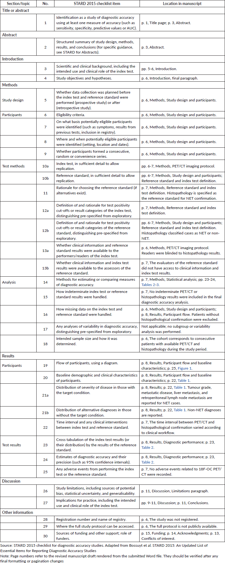
This was a retrospective cross-sectional diagnostic accuracy study of consecutive patients with suspected NET evaluated between April 2021 and February 2022 at the Instituto Nacional de Ciencias Médicas y Nutrición Salvador Zubirán (Mexico City, Mexico). During the study period, 18F-OC PET/CT was in the early stages of implementation at our institution as part of clinical diagnostic practice for patients with suspected or known NETs; it was not implemented solely for the purposes of this study. Patients were included if they underwent 18F-OC PET/CT and had histopathological confirmation (resection or biopsy). Patients without histopathology were excluded. The study was not registered, and the full protocol is not publicly available. We report the study following STARD 2015 recommendations for diagnostic accuracy reporting (Supplementary Table 1).
PET/CT imaging protocol
Whole-body PET/CT was performed 60 minutes after intravenous administration of 260 MBq of 18F-OC using a GE Discovery PET/CT 710 scanner. Low-dose CT (120 kV, 30 mAs) was acquired for attenuation correction, followed by PET acquisition (2 minutes per bed position). Images were reconstructed using ordered-subset expectation maximisation (four iterations, eight subsets). Two nuclear medicine physicians interpreted scans blinded to histopathology results.
18F-OC was produced at the Unidad Radiofarmacia-Ciclotrón (URC), Facultad de Medicina, UNAM, in a Trasis All-in-One synthesizer (Trasis, Ans, Belgium) under Good Manufacturing Practices (GMP) guidelines [13].
Reference standard and index test definition
Histopathology was used as the reference standard for NET confirmation and was performed as part of routine clinical care. Because of the retrospective design, it was not systematically recorded whether pathologists had access to PET/CT results, nor was the interval between PET/CT and histopathological confirmation consistently documented. PET/CT positivity was defined a priori as focal uptake consistent with disease with SUVmax ≥8.3, based on published work identifying this threshold as an optimal sensitivity–specificity balance for 18F-labeled octreotide analog imaging [11]. Disease extent was categorised as localised or disseminated. No adverse events related to 18F-OC PET/CT were recorded, and no indeterminate PET/CT or histopathology results were included in the final diagnostic accuracy analysis.
Statistical analysis
We computed sensitivity, specificity, positive predictive value (PPV), negative predictive value (NPV), accuracy and likelihood ratios with 95% confidence intervals. Because pretest probability varies substantially by clinical context (e.g., general population versus referral for suspected NET), post-test probability was explored using scenario-based Bayesian updating across plausible clinical pretest probabilities (see Results). Analyses were performed using MedCalc v20.0 (MedCalc Software Ltd).
Ethics
The protocol was approved by the institutional ethics committee (REG. CONBIOÉTICA-09-CEI-011-20160627). Written informed consent was obtained from all participants, including consent for publication of anonymised clinical and imaging data.
Results
Participant flow and baseline characteristics
Ninety-six patients were evaluated; 55 met inclusion criteria after excluding 41 without histopathological confirmation. The cohort had a mean age of 53.6 ± 15.1 years and 58.2% were female. NET was confirmed in 39/55 (70.9%). The participant inclusion process is shown in Figure 1. Among NET cases, most were well differentiated; metastatic disease was present in 29/39 (74.4%), most commonly involving liver and retroperitoneal lymph nodes. Non-NET diagnoses included Cushing’s disease and paraganglioma (Table 1 and Figure 2).
Diagnostic performance
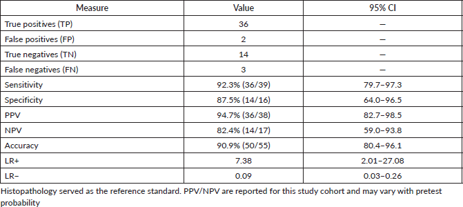
Using histopathology as the reference standard, 18F-OC PET/CT identified 36 true-positive and 14 true-negative results, with 3 false-negative and 2 false-positive findings. Sensitivity was 92.3% (36/39; 95% CI: 79.7–97.3) and specificity 87.5% (14/16; 95% CI: 64.0–96.5). The PPV was 94.7%, the NPV was 82.4% and overall accuracy was 90.9%. The positive likelihood ratio (LR+) was 7.38 and the negative likelihood ratio (LR−) was 0.09 (Table 2).
False-negative and false-positive cases
False-negative findings occurred in three situations: (i) a small (5 mm) grade 1 pancreatic NET with low uptake; (ii) a grade 3 NET with high proliferative activity (Ki-67 >20%) and (iii) a gastrinoma in a patient with impaired renal function. False-positive findings were characterised by mild uptake and corresponded to inflammatory processes on histopathology.
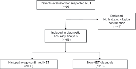
Figure 1. Participant flow diagram. Of 96 patients evaluated for suspected NET, 41 were excluded because histopathological confirmation was unavailable. The final diagnostic accuracy cohort included 55 patients, of whom 39 had histopathologically confirmed NET and 16 had non-NET diagnoses.
Table 1. Baseline characteristics and tumour features.
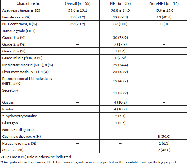
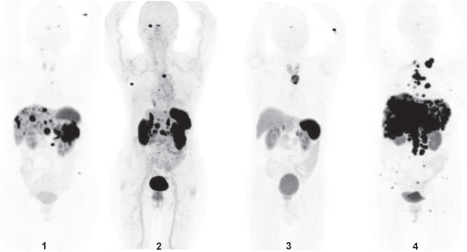
Figure 2. Representative 18F-OC PET/CT scans from four patients with NETs. Maximum intensity projection (MIP) images are shown:
1. 69-year-old female with a grade 2 NET, primary lesion located in the kidney and metastatic involvement of the liver and retroperitoneal lymph nodes.
2. 65-year-old female with a grade 1 NET located in the ileum, presenting with metastases to axillary, mediastinal and retroperitoneal lymph nodes, as well as the liver, pancreas and right orbital muscle.
3. 63-year-old male with a localised pulmonary carcinoid tumour.
4. 52-year-old female with a grade 1 NET of pancreatic origin, showing metastases to the liver, bone and retroperitoneum. The largest lesion measured 16.3 cm.
Table 2. Diagnostic performance of 18F-OC PET/CT (histopathology reference).
Scenario-based post-test probability (Bayesian interpretation)
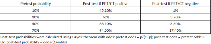
Given LR+ = 7.38 and LR− = 0.09, the post-test probability varies according to the clinical pretest probability (Table 3). Across plausible referral-setting scenarios, a positive PET/CT substantially increases the probability of NET (from 10% to 45.1% and from 70% to 94.5%), whereas a negative PET/CT markedly lowers it at lower-to-intermediate pretest probabilities (to 1.0%–8.3% for pretest values of 10%–50%). At higher pretest probability (70%), a negative result reduces the probability to 17.4%, underscoring that residual risk may remain when clinical suspicion is high and supporting the need for continued clinicopathologic correlation in selected cases. These scenario-based Bayesian effects are graphically represented in Figure 3.
Discussion
In this histopathology-referenced diagnostic accuracy study, 18F-OC PET/CT demonstrated high sensitivity and specificity for NET detection, with LR+ and LR− values consistent with a clinically useful test in referral settings. While no within-patient head-to-head comparison against [68Ga]-DOTA-SSA PET/CT was performed, the observed performance is within ranges commonly reported for SSTR-PET imaging in NET management [7]. From an implementation perspective, 18F-OC PET/CT may offer practical advantages over 68Ga-based tracers. The longer half-life of 18F enables centralised cyclotron production and regional distribution to multiple PET centers, whereas 68Ga-based imaging often depends on local generator availability and radiopharmacy infrastructure. Automated GMP-compliant production of 18F-OC has been described, supporting its technical feasibility for clinical workflows [14]. These features may reduce logistical barriers in centers without access to 68Ge/68Ga generators; however, formal cost-effectiveness analyses comparing 18F-OC with [68Ga]-DOTA-SSA tracers are still needed, particularly in lower-resource settings.
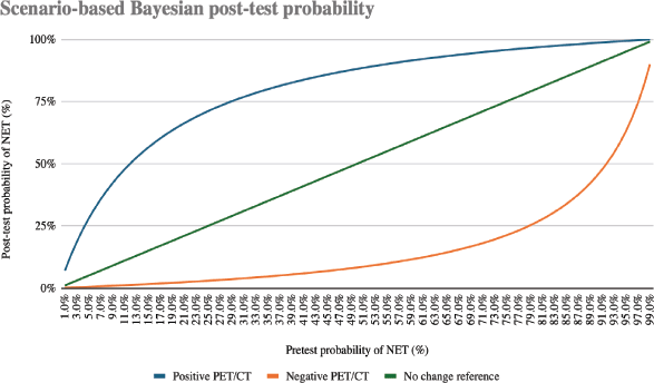
Figure 3. Scenario-based Bayesian post-test probability curves for 18F-OC PET/CT. Post-test probabilities after positive and negative PET/CT results are shown across a continuous range of clinically plausible pretest probabilities using LR+ = 7.38 and LR− = 0.09. The positive PET/CT curve illustrates the increase in post-test probability after a positive result, whereas the negative PET/CT curve illustrates the reduction in post-test probability after a negative result.
Table 3. Scenario-based post-test probability (Bayesian interpretation). (LR+ = 7.38; LR− = 0.09).
False-negative cases highlight known challenges for SSTR-targeted imaging: very small lesions, dedifferentiated or high-grade tumours with reduced SSTR expression and physiologic or patient-related factors that may alter biodistribution. In such scenarios, complementary imaging and clinicopathologic correlation remain essential [15–17]. It is also worth noting that the lower positron energy of 18F compared with 68 Ga (0.63 versus 1.9 MeV) may improve PET image quality and spatial resolution, potentially providing an advantage for the evaluation of small lesions.
False positives underscore that mild uptake can occur in inflammatory conditions and in physiologic sites of higher SSTR expression; standardised interpretation criteria and awareness of pitfalls (e.g., pancreatic uncinate uptake, splenosis and inflammatory disease) are necessary [17–19].
At our institution, 18F-OC PET/CT was incorporated into the diagnostic workflow for NET evaluation, in line with related investigations from other countries and supported by the local availability of cyclotron-based 18F production. Chen et al [11] reported that 18F-OC PET/CT improved detection and evaluation of neuroendocrine neoplasms compared with contrast-enhanced CT/MRI. Hou et al [12] characterised biodistribution and tumour uptake of 18F-OC in neuroendocrine neoplasms. In addition, Pauwels et al [20] reported superior lesion detection with 18F-OC compared with [68Ga]-DOTA-TATE/NOC in a prospective multicenter study. The major provider of PET radiopharmaceuticals in Mexico City is the URC-UNAM, which introduced 68Ga-based radiopharmaceuticals in 2013. However, the high demand for these radiopharmaceuticals, the limited activity and elevated cost of 68Ge/68Ga generators, and the relatively short half-life of 68Ga (68 minutes) prompted the shift to 18F. Additionally, the country’s current installed base of 12 cyclotron facilities and approximately 70 PET tomographs [21] supports and promotes the widespread use of 18F-based radiopharmaceuticals.
Limitations include the single-center design, modest sample size and potential selection bias introduced by excluding patients without histopathology. Additionally, without a simultaneous comparator arm ([68Ga]-DOTA-SSA), direct comparative claims cannot be made. Although Mexico has an expanding PET infrastructure, access remains concentrated in selected urban and tertiary-care centers. Therefore, PET-based diagnosis may remain restricted regardless of the radiotracer used.
Although current guidelines support SSTR-PET imaging for NET management [17, 22], 18F-OC is not yet widely incorporated as a standard tracer. Therefore, our findings should be considered supportive evidence for further validation and future multicenter, head-to-head and health-economic studies are needed to define its role in clinical guidelines.
Conclusion
18F-OC PET/CT showed high diagnostic accuracy for NET detection when referenced to histopathology. Its 18F-based production logistics may support broader access to SSTR PET imaging, particularly in settings where 68Ga-based tracers are constrained.
List of abbreviations
CI, confidence interval; LR, likelihood ratio; NET, neuroendocrine tumour; NPV, negative predictive value; PET/CT, positron emission tomography/computed tomography; PPV, positive predictive value; SSTR, somatostatin receptor; SUVmax, maximum standardised uptake value.
Acknowledgments
The authors thank the staff of the Nuclear Medicine and Radiopharmacy units at the Instituto Nacional de Ciencias Médicas y Nutrición Salvador Zubirán and the Radiopharmacy-Cyclotron Unit at Universidad Nacional Autónoma de México for their support in patient care and radiotracer production.
Conflicts of interest
The authors declare that they have no conflicts of interest.
Funding
This research received no specific grant from any funding agency in the public, commercial or not-for-profit sectors.
Author contributions
PBCM: Conceptualisation, methodology, data curation, writing (original draft). RFM: Writing (review and editing), manuscript revision, critical feedback. EIÁ: Resources, validation, visualisation. MAÁR: Supervision, project administration, radiopharmaceutical synthesis. AARA: Formal analysis, investigation, writing (review and editing).
Data availability
Data are not publicly available due to privacy and ethical restrictions, but may be available from the corresponding author upon reasonable request.
References
1. Dasari A, Shen C, and Halperin D, et al (2017) Trends in the incidence, prevalence, and survival outcomes in patients with neuroendocrine tumors in the United States JAMA Oncol 3(10) 1335–1342 https://doi.org/10.1001/jamaoncol.2017.0589 PMID: 28448665 PMCID: 5824320
2. Riihimäki M, Hemminki A, and Sundquist K, et al (2016) The epidemiology of metastases in neuroendocrine tumors Int J Cancer 139(12) 2679–2686 https://doi.org/10.1002/ijc.30400 PMID: 27553864
3. Bozkurt MF, Virgolini I, and Balogova S, et al (2017) Guideline for PET/CT imaging of neuroendocrine neoplasms with 68Ga-DOTA- conjugated somatostatin receptor targeting peptides and 18F-DOPA Eur J Nucl Med Mol Imaging 44(9) 1588–1601 https://doi.org/10.1007/s00259-017-3728-y
4. Smit Duijzentkunst DA, Kwekkeboom DJ, and Bodei L (2017) Somatostatin receptor 2-targeting compounds J Nucl Med 58(4) 54S–60S https://doi.org/10.2967/jnumed.117.191015 PMID: 28864613
5. Sadowski SM, Neychev V, and Millo C, et al (2016) Prospective study of 68Ga-DOTATATE PET/CT for detecting gastro-enteropancreatic neuroendocrine tumors and unknown primary sites J Clin Oncol 34(6) 588–596 https://doi.org/10.1200/JCO.2015.64.0987 PMCID: 4872030
6. Gabriel M, Decristoforo C, and Kendler D, et al (2007) 68Ga-DOTA-Tyr3-octreotide PET in neuroendocrine tumors: comparison with somatostatin receptor scintigraphy and CT J Nucl Med 48(4) 508–518 https://doi.org/10.2967/jnumed.106.035667 PMID: 17401086
7. Hope TA, Bergsland EK, and Bozkurt MF, et al (2018) Appropriate use criteria for somatostatin receptor PET imaging in neuroendocrine tumors J Nucl Med 59(1) 66–74 https://doi.org/10.2967/jnumed.117.202275
8. Dubash SR, Keat N, and Mapelli P, et al (2016) Clinical translation of a click-labeled 18F-octreotate radioligand for imaging neuroendocrine tumors J Nucl Med 57(8) 1207–1213 https://doi.org/10.2967/jnumed.115.169532 PMID: 27173162
9. Laverman P, Mcbride WJ, and Sharkey RM, et al (2010) A novel facile method of labeling octreotide with 18F-fluorine J Nucl Med 51(3) 454–461 https://doi.org/10.2967/jnumed.109.066902 PMID: 20150268 PMCID: 2908260
10. Xie Q, Liu T, and Ding J, et al (2021) Synthesis, preclinical evaluation, and a pilot clinical imaging study of 18F-AlF-NOTA-JR11 for neuroendocrine neoplasms compared with 68Ga-DOTA-TATE Eur J Nucl Med Mol Imag 48(10) 3129–3140 https://doi.org/10.1007/s00259-021-05249-8
11. Chen D, Yang S, and Chen J, et al (2023) Comparison of 18F-Oc PET/CT and contrast-enhanced CT/MRI in the detection and evaluation of neuroendocrine neoplasms Eur J Nucl Med Mol Imag 50(8) 2420–2431 https://doi.org/10.1007/s00259-023-06200-9
12. Hou J, Long T, and Yang N, et al (2021) Biodistribution of 18F-AlF-NOTA-octreotide in different organs and characterization of uptake in neuroendocrine neoplasms Mol Imag Biol 23(6) 827–835 https://doi.org/10.1007/s11307-021-01628-7
13. Trejo-Ballado F, Zamora-Romo E, and Gama-Romero H, et al (2023) Fully-automated GMP production of 18F-AlF-NOTA-octreotide a Trasis AiO module EJNMMI Radiopharm Chem 8(Suppl 1) PP08
14. Tshibangu T, Cawthorne C, and Serdons K, et al (2020) Automated GMP compliant production of 18F-AlF-NOTA-octreotide EJNMMI Radiopharm Chem 5(1) 4 https://doi.org/10.1186/s41181-019-0084-1 PMID: 31997090 PMCID: 6989705
15. Chan H, Moseley C, and Zhang L, et al (2019) Correlation of DOTATOC uptake and pathologic grade in neuroendocrine tumors Pancreas 48(7) 948–952 https://doi.org/10.1097/MPA.0000000000001356 PMID: 31268975
16. Yang J, Kan Y, and Ge BH, et al (2014) Diagnostic role of 68Ga-DOTATOC and 68Ga-DOTATATE PET in patients with neuroendocrine tumors: a meta-analysis Acta Radiol 55(4) 389–398 https://doi.org/10.1177/0284185113496679
17. Hope TA, Allen-Auerbach M, and Bodei L, et al (2023) SNMMI procedure standard/EANM practice guideline for SSTR PET: imaging neuroendocrine tumors J Nucl Med 64(2) 204–210 https://doi.org/10.2967/jnumed.122.264860 PMID: 36725249
18. Virgolini I, Gabriel M, and Kroiss A, et al (2016) Current knowledge on the sensitivity of 68Ga-somatostatin receptor PET and the SUVmax reference range for management of pancreatic neuroendocrine tumours Eur J Nucl Med Mol Imag 43(11) 2072–2083 https://doi.org/10.1007/s00259-016-3395-4
19. Panda A, Garg I, and Johnson GB, et al (2019) Molecular radionuclide imaging of pancreatic neoplasms Lancet Gastroenterol Hepatol 4(7) 559–570 https://doi.org/10.1016/S2468-1253(19)30081-0 PMID: 31169124
20. Pauwels E, Cleeren F, and Tshibangu T, et al (2023) 18F-AlF-NOTA-octreotide outperforms 68Ga-DOTATATE/NOC PET in neuroendocrine tumor patients: results from a prospective, multicenter study J Nucl Med 64(4) 632–638 https://doi.org/10.2967/jnumed.122.264563
21. Ávila-Rodríguez MA, Rivera Bravo B, and Kerik Rotenberg NE, et al (2019) Current status and perspectives of PET molecular imaging in Mexico Gac Med Mex 155 436–444
22. National Comprehensive Cancer Network (2026) NCCN Clinical Practice Guidelines in Oncology: Neuroendocrine and Adrenal Tumors. Version 1.2026 [Internet] (Plymouth Meeting: National Comprehensive Cancer Network) [https://www.nccn.org/guidelines/guidelines-detail?id=1448] Date accessed: 05/05/26
Supplementary Table
Supplementary Table 1. Completed STARD 2015 checklist with page and section references.