Key principles for the initial management of primary retroperitoneal tumours in adults
Walter S Nardi1, Sergio D Quildrian1, Victoria Scasso Rebzda2, Dimitri Tzanis3 and Sylvie Bonvalot3
1Sarcoma and Melanoma Unit, Department of General Surgery, British Hospital of Buenos Aires, Perdriel 74, CABA, Buenos Aires 1280AEB, Argentina
2Department of General Surgery, British Hospital of Buenos Aires, Perdriel 74, CABA, Buenos Aires 1280AEB, Argentina
3Department of Surgical Oncology, Institut Curie, 26 Rue d’Ulm, 75248 Paris, France
Abstract
Background: Primary retroperitoneal tumours (RPTs) encompass a wide range of diagnostic possibilities. Failure to recognise them appropriately may lead to suboptimal management, particularly in non-specialised centers. A structured diagnostic pathway is therefore essential to support appropriate treatment planning and timely referral to specialised teams.
Objective: To review key points of the diagnostic workup of primary RPTs, with special emphasis on those of mesenchymal origin.
Results: Primary RPTs represent a broad and heterogeneous group, with mesenchymal tumours accounting for approximately half of cases. Cross-sectional imaging plays a central role in the initial characterisation. Percutaneous core needle biopsy with histological and molecular analysis is recommended in most cases where imaging is not diagnostic. Serum tumour markers may provide valuable diagnostic clues in selected clinical settings.
Conclusion: Accurate diagnosis relies on a structured diagnostic pathway integrating imaging, biopsy when indicated and expert pathological review. Early referral to specialised multidisciplinary teams is essential to optimise management and improve oncologic outcomes.
Keywords: retroperitoneal tumour, soft tissue sarcoma, diagnosis, biopsy, imaging
Correspondence to: Walter S Nardi
Email: nardi.ws@gmail.com
Published: 20/07/2026
Received: 02/02/2026
Publication costs for this article were supported by ecancer (UK Charity number 1176307).
Copyright: © the authors; licensee ecancermedicalscience. This is an Open Access article distributed under the terms of the Creative Commons Attribution License (http://creativecommons.org/licenses/by/4.0), which permits unrestricted use, distribution, and reproduction in any medium, provided the original work is properly cited.
Introduction
Retroperitoneal tumours (RPTs) arise within the retroperitoneal space. Primary tumours originate independently of retroperitoneal organs and are predominantly of mesenchymal origin, although lymphoid neoplasms, parasympathetic tumours and extragonadal germ cell tumours may also occur. Secondary lesions are usually metastatic carcinomas [1].
According to guidelines from the European Society of Medical Oncology, National Comprehensive Cancer Network and the Trans-Atlantic Retroperitoneal Sarcoma Working Group [2–4], image-guided percutaneous core needle biopsy (CNB) is recommended in most cases where serum tumour markers and imaging do not provide a precise diagnosis. Histopathological evaluation by a certified expert pathologist is essential, as expert pathology review can modify the initial diagnosis in up to 20% of cases. Finally, a multidisciplinary tumour board (MDTB) defines the global strategy thereafter based on the results obtained.
Despite these recommendations, many patients are still initially managed in non-specialised centers. In some countries, such as France and the UK, sarcoma care is centralised in designated high-volume reference centers [5, 6]. However, this is not the standard of practice in most regions. Confusion remains in the primary care setting regarding the diagnostic algorithm and the importance of early referral of RPTs.
We discuss the initial diagnostic workup of adult patients with suspected primary RPTs, focusing on imaging evaluation, biopsy strategy and referral pathway. We also highlight key diagnostic considerations and common pitfalls that may lead to misdiagnosis and adversely impact patient care.
Methods
This narrative review is based on a non-systematic literature search performed in PubMed and supplemented by major international oncology guidelines [2–4]. Priority was given to recent clinical practice guidelines, key retrospective series and relevant review articles. The selection of studies was guided by clinical relevance and the authors’ expertise in sarcoma management.
Diagnostic approach to RPTs
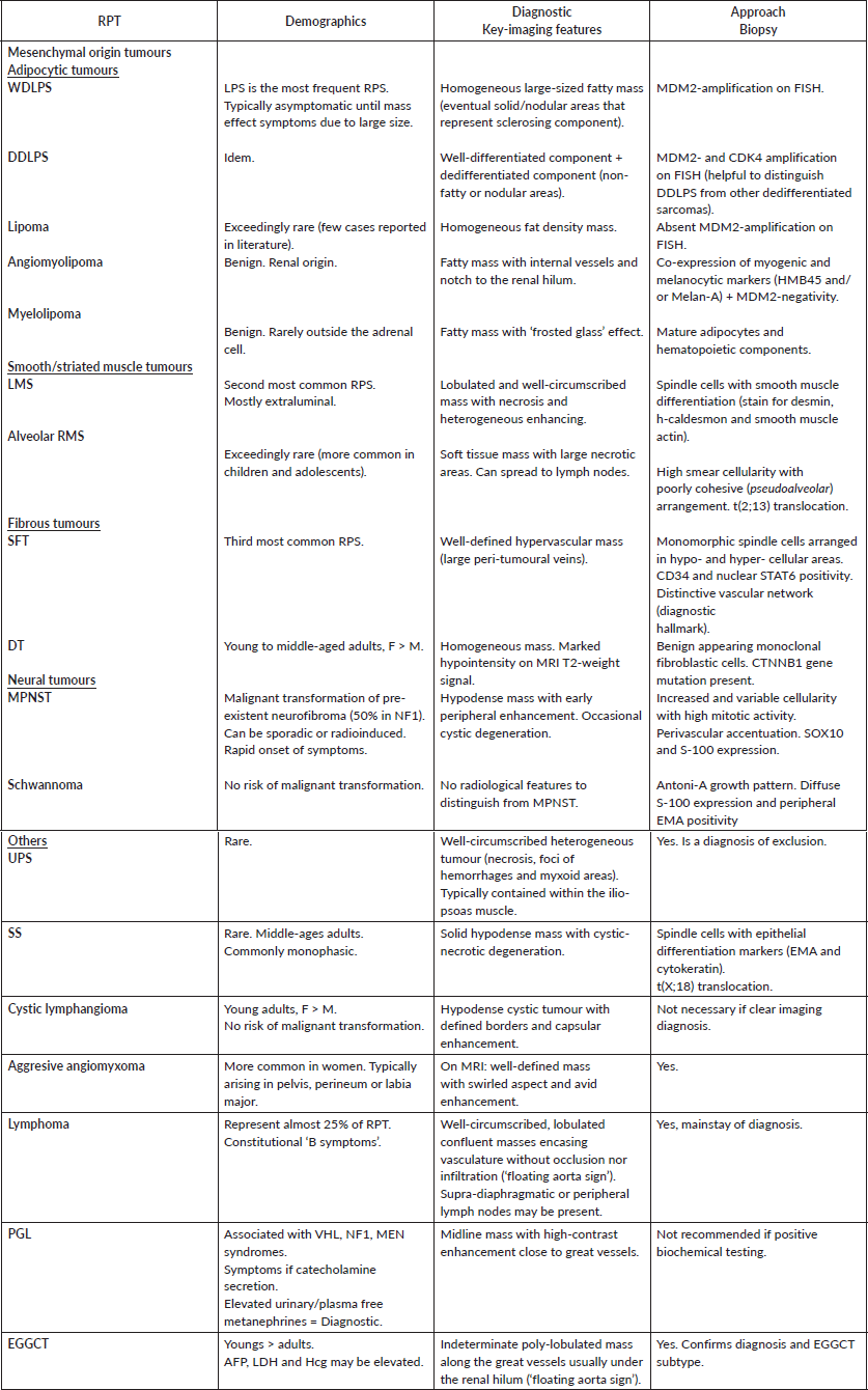
The diagnostic evaluation of RPTs should follow a structured sequence. This sequence typically progresses from initial imaging recognition to characterisation, differential diagnosis, use of serum biomarkers and histological confirmation when required. The following sections summarise key diagnostic considerations within this framework, organised by major tumour groups. Table 1 integrates imaging features, biomarker evaluation and biopsy indications and can be used as a simplified clinical algorithm.
Mesenchymal tumours
Mesenchymal tumours account for approximately half of primary RPTs. The WHO 2020 classification categorises them into different subgroups according to the tumour differentiation line and biological behaviour (benign, intermediate or malignant).
Overall, almost 30% of RPTs are sarcomas. Liposarcoma (LPS), leiomyosarcoma (LMS) and solitary fibrous tumour (SFT) account for nearly 90% of retroperitoneal (RPS) soft tissue sarcomas (STS). Generally, anterior displacement of normal anatomical structures (colon, adrenal glands, kidney and pancreas) in cross-sectional imaging is suggestive of a tumour arising from the retroperitoneal cavity.
When an RPT is identified and imaging does not establish a secure diagnosis, adequate tissue sampling is required, particularly when sarcoma is suspected, because treatment planning depends on histologic subtype and grade. Preoperative biopsy can identify patients who do not require surgery, for example, in lymphoid neoplasms. Image-guided percutaneous CNB, preferably by a ≥14–16 G needle through a posterior retroperitoneal approach and using a coaxial technique when feasible, is generally recommended in most cases. This approach provides high diagnostic accuracy, with a low rate of minor complications (3%), a reported needle track seeding of less than 0.4% and no adverse impact on oncological outcomes [7]. Once the sample is obtained, histologic and molecular biology tests are essential for a correct diagnosis and should be analysed by an expert pathologist in a reference center.
Following diagnosis, resectability should then be assessed. In RPS, staging should include a chest computed tomography (CT) to assess the presence of metastatic disease. This is particularly important for LMS as pulmonary metastases are present in 40% of cases at diagnosis [8, 9]. Assessment of liver metastasis, particularly in LMS, should also be performed on abdominal CT. Finally, following diagnosis and staging, management is typically guided by histology subtype for localised RPS once resectability is established [10]. Currently, it is recommended that specialised multidisciplinary sarcoma units carry out the treatment of sarcoma patients after MDTB meeting discussions in reference centers. Several studies have already shown the benefits of this management approach [11, 12].
Table 1. Comparative summary of main RPT.
Adipocytic tumours
Fatty tumours are the most frequent retroperitoneal histotype. Well-differentiated liposarcoma (WDLPS) and dedifferentiated liposarcoma (DDLPS) are the main malignant entities, whereas lipomas, myelolipomas and angiomyolipoma represent the benign tumours.
Contrast-enhanced CT is the imaging modality of choice to evaluate adipocytic RPTs. WDLPS typically presents as a large-sized mass with homogeneous fatty content (Figure 1a). In DDLPS, gross findings usually include the coexistence of non-fatty or nodular areas of different density (dedifferentiated component) surrounded by well-differentiated fatty mass (well-differentiated component) (Figure 1b). However, imaging overlap with benign lesions is common [13]. Therefore, although imaging findings may suggest malignancy, preoperative percutaneous biopsy is recommended. Both WDLPS and DDLPS are MDM2 amplified tumours that can be identified by fluorescence in-situ hybridisation (FISH). Biopsy should target the solid areas of the tumour systematically to avoid under-grading. This has been shown to improve sensitivity, particularly for the detection of grade 3 DDLPS [14]. Accurate diagnosis is a key step to personalise the multidisciplinary management, as WDLPS and DDLPS differ in their biological behaviour and recurrence patterns. In this setting, preoperative strategies can be considered and may include radiotherapy [15]. Currently, the STRASS 2 trial is evaluating neoadjuvant chemotherapy versus surgery alone for LMS and G3 DDLPS (ClinicalTrials.gov Identifier: NCT04031677). Its results could add new options for the peri-operative management of these patients.
Differential diagnosis between WDLPS and lipoma remains challenging. Generally, lipomas have homogeneous fat density without internal septa. Nevertheless, MDM2 gene amplification status in a biopsy specimen confirms LPS (Figure 2). Angiomyolipomas and myelolipomas are also fat-containing tumours. Angiomyolipomas show internal vessels and usually a notch or point of entrance to the renal hilum, suggesting their origin in the kidney. Myelolipomas have bone marrow within the fat (mature adipocytes), making a ‘frosted glass’ effect on enhanced CT scan. This part can be mistaken for a dedifferentiated component of an LPS [13, 16, 17]. They typically present in the adrenal lodge, rarely found outside.
Finally, surgical management principles depend on histologic subtype and are beyond the scope of this diagnostic-focused review [18, 19].
Smooth/striated muscle tumours
LMS (primary vascular sarcoma) is the second most common RPS in the adult (≅ 15%) and originates from smooth muscle cells in the wall of retroperitoneal vessels, mainly in women. It commonly arises from the inferior vena cava (IVC), but it can also originate from the renal (Figure 3), adrenal, gonadal or iliac veins. Uterine location, which is the predominant origin of all LMS, must be ruled out, especially in women with a previous history of resected uterine leiomyoma (occasionally misdiagnosed) [20]. They are mostly extraluminal and typically present as large, lobulated, well-circumscribed masses with necrotic content and heterogeneous enhancing on CT, contiguous to a vessel. Histologically, LMS shows spindle cells with smooth muscle differentiation expressing desmin, h-caldesmon and smooth muscle actin. Genetically, as well as undifferentiated pleomorphic sarcoma (UPS), LMS exhibit heterogeneous complex karyotypic abnormalities. Differential diagnosis includes benign leiomyoma (they generally do not present nuclear atypia and lack necrosis and high mitotic activity) and DDLPS in case of high-grade pleomorphic LMS. The latter can be excluded by confirming the absence of MDM2 amplification on FISH.
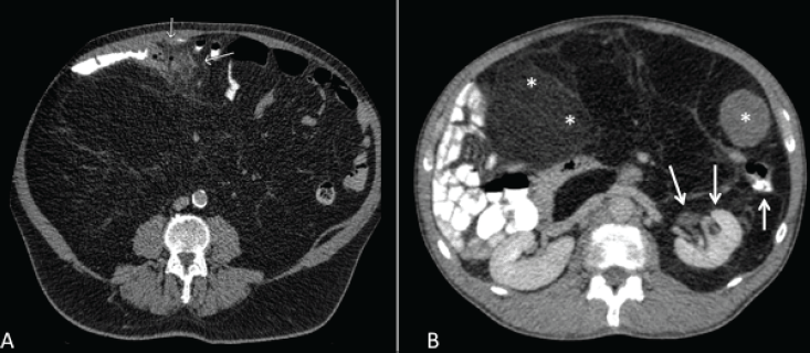
Figure 1. (a): WDLPS. (b): DDLPS. The two components of the tumour can be seen: the well-differentiated part surrounding the kidney and colon (arrows) and the dedifferentiated solid part within (asterisks).
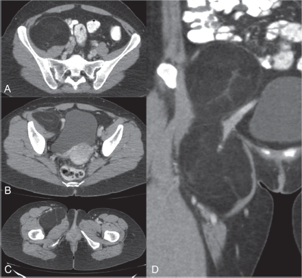
Figure 2. (a): CT scan axial-view showing a right retroperitoneal soft-tissue tumour in contact with the iliopsoas muscle and medially displacing ipsilateral iliac vessels. (b and c): Images showing herniation through inguinal canal into the thigh and displacing forwards the femoral vessels. (d): Coronal view of the tumour with a ‘dumbbell appearance’. Biopsy: lipomatous tumour without MDM2 amplification on FISH.
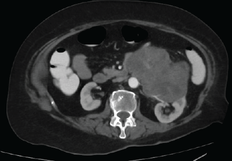
Figure 3. LMS of left renal vein.
Alveolar/Embryonal rhabdomyosarcomas (RMS) (striated muscle differentiation) are exceedingly rare in adults and common in the pediatric and adolescent population. However, pleomorphic RMS is considered a high-grade adult-type STS that can appear (although rarely) in the retroperitoneum. It has a high metastatic risk and poor prognosis. Moreover, despite sarcomas almost never metastasize to the lymph nodes, exceptionally, RMS does. Based on its chemosensitivity at other sites, neoadjuvant chemotherapy may be considered following MDTB discussion.
Fibrohistiocytic tumours
In the retroperitoneum, this group of tumours accounts mostly for desmoid tumours (DT) (unpredictable and locally aggressive potential behaviour) and SFT (variable metastatic potential).
SFT are the third most common subtype of RPS (approximately 6%). They are well-circumscribed hypervascular masses due to large peri-tumoural veins and intense contrast enhancement in delayed phases of CT scan or magnetic resonance (MRI), denoting the fibrous stroma component. Presumptive diagnosis can be confirmed with a percutaneous CNB. Histologically, they have hypocellular areas (occasionally with hypercellular components) of monomorphic spindle cells and a vascular network of thin-walled and branched vessels (this vascular pattern is called ‘stag-horn’). Immunohistochemistry shows tumoural cells with strong CD34 positivity and nuclear expression of STAT6 due to NAB2::STAT6 fusion (molecular hallmark) [21].
DT shows a wide range of T2 signal intensities on MRI [22], but usually, images do not show characteristic features that may suggest a possible diagnosis. Nonetheless, a diagnostic biopsy should be performed. Three European prospective observational trials have demonstrated that active surveillance as a first-line strategy is safe and effective for newly diagnosed DT [23–25] (Figure 4). CTNNB1 gene mutation is required to confirm the diagnosis and is present in 95% of tumours. It remains uncertain whether the mutational profile has a true impact on event-free survival (regardless of the first-line therapy chosen) [26, 27]. The DT Working Group recently published an updated consensus recommending surgery only in cases of tumour-related bowel complications (bleeding, obstruction or perforation) occurring in up to 10% of cases [28]. The main goal should be to solve the complication with resection of the tumour, but only when feasible, without excessive bowel resections that may lead to considerable morbidities. This is particularly relevant for sporadic mesenteric DTs, while for FAP-associated DTs the main objective should be symptom relief and control of the main complication rather than resection of the tumour [29, 30].
Neural tumours
Benign (schwannoma/neurofibroma) and malignant peripheral nerve sheath tumour (MPNST) are tumours arising from the nerves that can also occur in the retroperitoneum and are the most frequent entities of this subgroup.
MPNSTs can arise de novo but are more frequently seen as a malignant transformation of pre-existent plexiform neurofibromas (50% of them occurring in patients with neurofibromatosis type 1, NF-1) [31]. In fact, some studies have shown that NF-1 status is a negative predictor of overall- and distant metastasis-free survival [32, 33]. Differential diagnosis with neurofibromas or schwannomas can be challenging since there are no radiological features that reliably distinguish one from another. On CT scan, lesions appear hypodense with early peripheral enhancement after intravenous contrast and occasionally presenting cystic degeneration (Figure 5). On MRI, peritumoural edema, intratumoural heterogeneous masses or invasive margins are suggestive, although not specific, of MPNST. Moreover, rapid onset of neurologic pain (which is a classic presenting symptom) or interval tumour growth may suggest malignancy. Microscopically, increased and variable cellularity with high mitotic activity and perivascular accentuation is a typical morphologic feature. Instead, schwannomas exhibit a characteristic Antoni-A growth pattern with diffuse S-100 expression and peripheral epithelial membrane antigen (EMA) positivity on perineural cells. Schwannomas do not have a risk of malignant transformation (Figure 6).
Ganglioneuroma is a rare, benign, differentiated neurogenic tumour that arises in neural crest cells. The retroperitoneum is the second most common site after the posterior mediastinum and typically presents in children or young adults. Characteristically, they present well-defined, rounded, homogeneous masses with fibrous shapes, low pre-contrast attenuation on CT scan and occasional punctate calcifications (Figure 7). Definitive diagnosis is made by histopathologic examination. Features include mature ganglion cells admixed with clusters of spindle cells (positive for S-100). Active surveillance is an option [34].
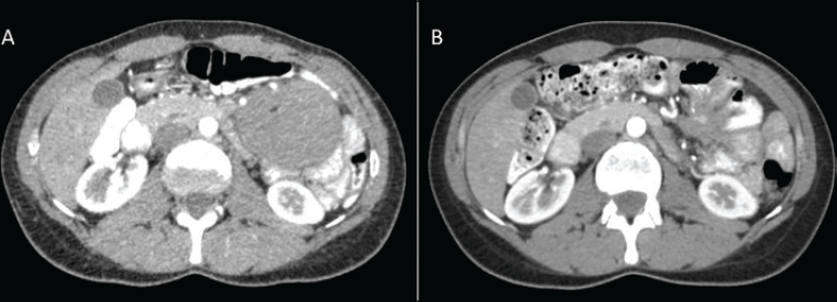
Figure 4. (a): Intra-abdominal asymptomatic mass measuring 9 cm. Biopsy confirmed DT and active surveillance (AS) was decided after multidisciplinary discussion. (b): CT scan after 3 years of AS.
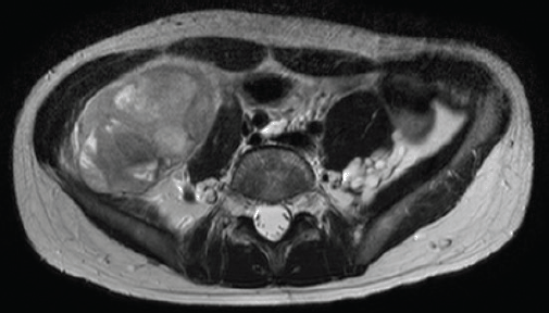
Figure 5. (a and b): MPNST originating in the right crural nerve territory and displacing medially the psoas muscle in a NF-1 patient. Neurofibromas (arrows).
Vascular tumours
Cystic lymphangiomas are benign tumours originating from the endothelium of lymphatic vessels with no risk of malignant transformation. They predominantly occur in young patients, mostly females. They most frequently present in the head and neck region or mediastinum, but also can appear in retroperitoneum, mesentery or greater/lesser omentum. Diagnosis is based on imaging findings showing a typical hypodense cystic tumour with defined borders and capsular enhancement on CT scan (Figure 8), and does not require biopsy. They may enlarge slowly until they become symptomatic lesions due to compression of surrounding structures or even encase adjacent organs, which makes surgical resection more difficult.
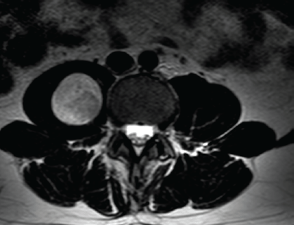
Figure 6. (a and b): Schwannoma with cystic degeneration contained within the left psoas muscle. Percutaneous biopsy showed areas of densely populated spindle cells (Antoni-A) positive for S100 and SOX10 on immunohistochemistry confirming nerve sheath differentiation.
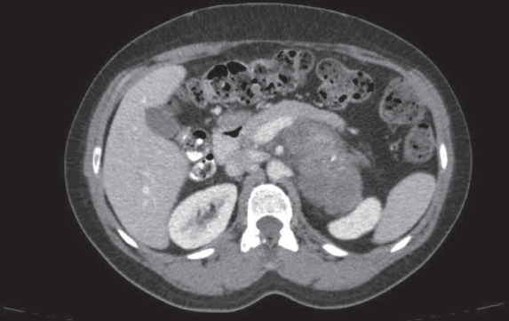
Figure 7. Ganglioneuroma. Well-defined retroperitoneal mass with punctate calcifications. Diagnosis was established by biopsy, as ganglioneuromas lack pathognomonic imaging features.

Figure 8. Cystic lymphangioma.
Primary aortic involvement is very rare. It is usually affected by adjacent tumours with a secondary wide encasement or partial infiltration [35]. However, it can also be affected by primary malignant vascular tumours: high-grade angiosarcoma and intimal sarcomas (that generally grow into the aortic lumen). Retroperitoneal angiosarcoma has a dismal prognosis and mortality is usually related to local complications, mainly thrombo-embolic events.
Extra-skeletal osseous/cartilaginous tumours
They are rare in the retroperitoneum. Extra-skeletal Ewing’s sarcoma, known for its chemosensitivity, benefits from an already established neoadjuvant chemotherapy. Because the site of origin (bone versus soft tissue) determines the therapeutic strategy, accurate diagnosis through imaging and biopsy is essential to guide therapy.
Others
UPS accounts for approximately 10% of sarcomas in the adult and is rare in the retroperitoneum. It is typically a fast-growing mass contained within the musculature (ilio-psoas muscle), but direct invasion into adjacent organs may be present. Images show a well-circumscribed heterogeneous tumour with necrosis, foci of hemorrhages and myxoid areas. Morphologic, immunohistochemical and molecular findings are non-specific; therefore, UPS remains a diagnosis of exclusion. Cellularity is marked by an extensive pleomorphism without an identified line of differentiation. High mitotic count and abundant necrosis are usually present. Before classifying a tumour as UPS, pleomorphic DDLPS must be ruled out [30].
Perivascular epithelioid cell tumours (PEComas) are a family of mesenchymal tumours with myomelanocytic differentiation, sharing a specific cell type, the PEComas. They exhibit different biologic potential: from benign behaviour (angiomyolipoma), intermediate course (sclerosing PEComa) to malignant behaviour with higher rates of recurrence and mortality (PEComa NOS – not otherwise specified). The latter is composed of a nest of epithelioid and spindle cells intimately arranged around blood vessels walls, expressing both melanocytic and smooth muscle markers. Most are sporadic but can be associated with tuberous sclerosis. They are often large (> 5 cm) heterogeneous infiltrative masses with strong arterial enhancement and small calcifications. However, there are no pathognomonic imaging features, and a biopsy is needed for diagnosis. Most PEComas do not recur after complete surgical resection, but a subset (malignant subtype) can evolve during follow-up with locally aggressive recurrences or distant metastasis. These scenarios entail a poor prognosis with little or no effective therapy described so far [36].
Synovial sarcoma (SS) is rare. It appears as solid hypodense masses with cystic-necrotic degeneration on CT scan that can be confused with MPNST or LMS. Microscopically, SS are commonly monophasic comprised only of spindle cells, with expression of epithelial differentiation markers (EMA and cytokeratin). Its unique t(X;18) translocation confirms the diagnosis.
Aggressive angiomyxoma is a highly vascularised tumour with intermediate differentiation, usually affecting women. Commonly arising from the perineum (often clinically as a perineal mass) or labium major, can also appear in the sub-peritoneal space. If suspected, an MRI scan should be performed showing a well-defined mass with a swirled aspect and avid enhancement after contrast administration on T2-weighted images (Figure 9). It is a benign lesion with no propensity to metastasize but a historically reported high rate of local recurrence after surgery. After conservative surgery in reference centers, particularly in young patients, a good local control can be achieved [37].
Lymphoid tumours
Lymphomas, represent almost 25% of primary RPTs. A wide variety of subtypes exist and each one of them has its own prognostic and therapeutic considerations. Lymphomas are considered clinic-pathologic syndromes with variable presentations. Constitutional ‘B symptoms’ (low-grade fever, night sweats, weight loss) as well as multiple peripheral lymph nodes are not always present. On imaging, lymphomas present as well-circumscribed, lobulated, confluent masses along great vessels. These masses typically encase the vasculature without occlusion nor infiltration (Figure 10). Noncontiguous spread (multiple enlarged lymph nodes) along iliac vessels can also be present and favours a diagnosis of lymphoma.
Diffuse large B-cell lymphoma is the most frequent in adults (35% of all non-Hodgkin’s lymphomas) and the most frequently associated with HIV infection and immunosuppression. Pathological assessment through CNB remains the mainstay of diagnosis and chemotherapy is the standard of care in this disease [38, 39].

Figure 9. Aggressive perineal angiomyxoma. Coronal (a): and axial (b): views.
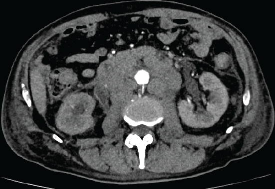
Figure 10. Retroperitoneal well circumscribed confluent and lobulated mass encasing but not infiltrating the abdominal aorta and displacing it away from the vertebral column (‘floating aorta sign’) most commonly suggests a lymphoma diagnosis.
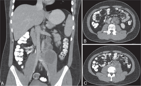
Figure 11. CT scan coronal (a): and axial views (b and c): of an EGGCT in a 24 year-old male with elevated serum tumour markers (mainly β-human chorionic gonadotropin). Percutaneous biopsy confirmed choriocarcinoma.
Retroperitoneal paraganglioma (PGL) and adrenal tumours
PGL is a rare catecholamine-secreting neuroendocrine tumour arising from the sympathetic and para-aortic retroperitoneal sympathetic ganglia. Pheochromocytoma is called when tumour arises in the adrenal gland (by definition, strictly not a primary RPT). Many of them are sporadic but some can be associated with genetic inheritable mutations (mostly MEN2A, Von-Hippel Lindau disease or NF-1). Particular scenarios should raise awareness to ask for genetic counseling [40]. Up to 40% of PGL patients present with symptoms due to active catecholamine secretion such as paroxysmal hypertension, palpitations, hyperglycemia and/or flushing. On imaging, midline masses with high contrast enhancement close to the aorta or IVC with intratumoural vessels are characteristic. If suspected, biochemical testing should be performed. Marked elevation of plasma-free or urinary fractionated metanephrines (2 or 3 times above the upper limit of normal values) is diagnostic [41]. Biopsy is not recommended as it can produce a sudden release of hormones with potential life-threatening hypertension. Definitive treatment for PGL is complete surgical resection. Preoperative alpha-adrenergic blockade is recommended to prevent perioperative hemodynamic instability.
Sometimes, because of the size, tumours arising in the adrenal lodge can raise doubts of the true origin and be confused with a primary RPT. Adrenocortical carcinoma must be suspected if the adrenal gland is not visible.
Germ cell tumours
Primary germ cell tumours originate from the testicles or ovaries. Retroperitoneal lymphatic spread can be present at diagnosis or as disease progression after treatment during follow-up. Extra-gonadal germ cell tumours (EGGCTs) are a heterogeneous group composed by malignant germ cells arising in extra-gonadal locations without the presence of a gonadal primary tumour. EGGCTs are uncommon neoplasms with an overall incidence of 1.8 to 3.4/1 million and retroperitoneum is a common location following the brain and mediastinum [42].
Altogether (primary or secondary), germ cell tumours account for approximately one quarter of all RPT and are an important differential diagnosis when assessing a retroperitoneal mass. On imaging, an indeterminate poly-lobulated mass along the great vessels, usually under the renal hilia may be observed. In the diagnostic process, specific serum tumour markers (α-fetoprotein, β-human chorionic gonadotropin, lactate dehydrogenase) are frequently elevated in EGGCTs [43]. A previous history of disease in a young male patient is highly evocative and if not, a testicular ultrasound should be considered. Percutaneous CNB is encouraged to confirm diagnosis and type of EGGCT (Figure 11). Morphological and immunohistochemical characteristics of gonadal germ cell tumours are the same as the extra-gonadal counterparts. However, a precise diagnosis is important to determine the therapeutic algorithm that often includes chemotherapy, followed or not by surgical resection.
Diagnostic pitfalls and common errors
Awareness of common diagnostic pitfalls is essential to avoid mismanagement and inappropriate therapeutic decisions.
Misinterpretation of imaging findings is common, especially in large or heterogeneous masses. In adipocytic tumours, underestimation of the tumour extent through the natural anatomic spaces of the abdomen (i.e., inguinal ligament, diaphragmatic hiatus and great sciatic notch) and failure to correctly identify well-differentiated areas within DDLPS is also a common mistake [13] (Figure 1b). In addition, many foci of dedifferentiation and/or necrosis can be misinterpreted as multifocal disease. Misinterpretation of tumour origin may also lead to misdiagnosis, particularly when large masses arise from adjacent organs (e.g., adrenal or renal tumours) and are mistaken for primary RPT. All this can potentially lead to inadequate surgical planning, incomplete resections with tumour rupture or even unjustified contraindication of surgery.
Inadequate or non-representative biopsies may underestimate grade of differentiation or result in non-diagnostic samples. Hence, it is particularly important to target the solid or more suspicious areas of the tumour. In addition, biopsy should be avoided in specific scenarios, such as PGLs, where it may trigger life-threatening complications.
Failure to consider alternative diagnoses is also a recurrent issue. Lymphoma (Figure 10), EGGCTs (Figure 11) and benign entities may mimic sarcoma on imaging but require different management strategies. In these cases, serum tumour markers and biopsy are important to avoid inappropriate surgery.
Finally, delayed referral to specialised centers remains a major issue. Initial management and inadequate surgical resections performed in non-specialised centers adversely affect oncologic outcomes. Early referral to experienced multidisciplinary teams is therefore critical [10–12].
Limitations
Limitations of this review include its narrative design and non-systematic methodology. The available evidence, from which recommendations are drawn, is limited by the usual limitations of retrospective, registry-based and non-randomised data due to the rarity and heterogeneity of most retroperitoneal entities.
Conclusion
Primary RPTs are a heterogeneous group of entities that require a structured diagnostic approach. Accurate imaging interpretation, appropriate use of image-guided biopsy when necessary and expert pathological assessment are essential to establish diagnosis and guide proper management.
Early referral to specialised multidisciplinary centers is critical, particularly for suspected sarcomas, as treatment strategies depend on histologic subtype and expertise in complex surgical management is often required. Avoiding diagnostic and therapeutic pitfalls is key to improving outcomes.
Conflicts of interest
None.
Funding
None.
Ethical approval
Not applicable.
Data availability
Data sharing is not applicable to this article as no datasets were generated or analysed during the current study.
Disclosures
None.
References
1. Dei Tos AP, Marino-Enriquez A, and Pedeutour FHO Classification of Tumours Editorial Board, et al (2020) Soft Tissue and Bone Tumours 5th edn (Lyon: IARC) [https://publications.iarc.fr/588]
2. Gronchi A, Miah AB, and Dei Tos AP, et al (2021) Soft tissue and visceral sarcomas: ESMO-EURACAN-GENTURIS clinical practice guidelines for diagnosis, treatment and follow-up Ann Oncol 32(11) 1348–1365 https://doi.org/10.1016/j.annonc.2021.07.006 PMID: 34303806
3. Von Mehren M, Kane JM, and Agulnik M, et al (2022) Soft tissue sarcoma, Version 2.2022, NCCN clinical practice guidelines in oncology J Natl Compr Canc Netw 20(7) 815–833 https://doi.org/10.6004/jnccn.2022.0035 PMID: 35830886 PMCID: 10186762
4. Swallow CJ, Strauss DC, and Bonvalot S, et al (2021) Management of primary retroperitoneal sarcoma (RPS) in the adult: an updated consensus approach from the transatlantic Australasian RPS working group Ann Surg Oncol 28(12) 7873–7888 https://doi.org/10.1245/s10434-021-09654-z PMID: 33852100 PMCID: 9257997
5. Hayes AJ, Nixon IF, and Strauss DC, et al (2024) UK guidelines for the management of soft tissue sarcomas Br J Cancer 3873479 [https://doi.org/10.1038/s41416-024-02674-y] PMID: 38734790 PMCID: 11724041
6. Blay JY, Toulmonde M, and Valentin T, et al (2025) Clinical presentation, management and outcome of 11,132 patients with liposarcoma patients: a population-based study from the NETSARC+ registry Lancet Regional Health Eur 57 101403 https://doi.org/10.1016/j.lanepe.2025.101403
7. Nardi W, Nicolas N, and El Zein S, et al (2024) Diagnostic accuracy and safety of percutaneous core needle biopsy of retroperitoneal tumours Eur J Surg Oncol 50(1) 107–298 https://doi.org/10.1016/j.ejso.2023.107298
8. Rajiah P, Sinha R, and Cuevas C, et al (2011) Imaging of uncommon retroperitoneal masses Radiographics 31(4) 949–976 https://doi.org/10.1148/rg.314095132 PMID: 21768233
9. O’Sullivan PJ, Harris AC, and Munk PL (2008) Radiological imaging features of non-uterine leiomyosarcoma Br J Radiol 81(961) 73–81 https://doi.org/10.1259/bjr/18595145
10. Bonvalot S, Roland C, and Raut C, et al (2023) Histology-tailored multidisciplinary management of primary retroperitoneal sarcomas Eur J Surg Oncol 49(6) 1061–1067 https://doi.org/10.1016/j.ejso.2022.05.010
11. Blay JY, Honoré C, and Stoeckle E, et al (2019) Surgery in reference centers improves survival of sarcoma patients: a nationwide study Ann Oncol 30(8) 1407 https://doi.org/10.1093/annonc/mdz170
12. Blay JY, Soibinet P, and Penel N, et al (2017) Improved survival using specialized multidisciplinary board in sarcoma patients Ann Oncol 28(11) 2852–2859 https://doi.org/10.1093/annonc/mdx484 PMID: 29117335 PMCID: 5834019
13. Moulin B, Messiou C, and Crombe A, et al (2022) Diagnostic strategy of adipocytic soft-tissue tumors in adults: a consensus from European experts Eur J Surg Oncol 48(3) 518–525 https://doi.org/10.1016/j.ejso.2021.10.009
14. Morosi C, Stacchiotti S, and Marchianò A, et al (2014) Correlation between radiological assessment and histopathological diagnosis in retroperitoneal tumors: analysis of 291 consecutive patients at a tertiary reference sarcoma center Eur J Surg Oncol 40(12) 1662–1670 https://doi.org/10.1016/j.ejso.2014.10.005 PMID: 25454827
15. Bonvalot S, Gronchi A, and Le Pèchoux C, et al (2020) Preoperative radiotherapy plus surgery alone for patients with primary retroperitoneal sarcoma (EORTC-62092: STRASS): a multicenter, open-label, randomized, phase 3 trial Lancet Oncol 21(10) 1366–1377 https://doi.org/10.1016/S1470-2045(20)30446-0 PMID: 32941794
16. Improta L, Tzanis D, and Bouhadiba T, et al (2020) Overview of primary adult retroperitoneal tumours Eur J Surg Oncol 46(9) 1573–1579 https://doi.org/10.1016/j.ejso.2020.04.054 PMID: 32600897
17. Messiou C, Moskovic E, and Vanel D, et al (2017) Primary retroperitoneal soft tissue sarcoma: imaging appearances, pitfalls and diagnostic algorithm Eur J Surg Oncol 43(7) 1191–1198 https://doi.org/10.1016/j.ejso.2016.10.032 PMID: 28057392
18. Bonvalot S, Rivoire M, and Castaing M, et al (2009) Primary retroperitoneal sarcomas: a multivariate analysis of surgical factors associated with local control J Clin Oncol 27(1) 31–37 https://doi.org/10.1200/JCO.2008.18.0802
19. Delisle M, Gyorki D, and Bonvalot S, et al (2022) Landmark series: a review of landmark studies in the treatment of primary localized retroperitoneal sarcoma Ann Surg Oncol 29(12) 7297–7311 https://doi.org/10.1245/s10434-022-12517-w PMID: 36088426
20. Ducimetière F, Lurkin A, and Ranchère-Vince D, et al (2011) Incidence of sarcoma histotypes and molecular subtypes in a prospective epidemiological study with central pathology review and molecular testing PLoS One 6(8) e20294 https://doi.org/10.1371/journal.pone.0020294 PMID: 21826194 PMCID: 3149593
21. Purgina B, Di Blasi E, and Collini P, et al (2026) Pathologic assessment of retroperitoneal sarcomas: a position paper by the Transatlantic Australasian Retroperitoneal Sarcoma Working Group Am J Surg Pathol 50(4) 455–466 https://doi.org/10.1097/PAS.0000000000002500 PMID: 41536011
22. Cassidy M, Lefkowitz R, and Long N, et al (2020) Association of MRI T2 signal intensity with desmoid tumor progression during active observation Ann Surg 271(4) 748–755 https://doi.org/10.1097/SLA.0000000000003073 PMCID: 6736761
23. Bonvalot S, Cozic N, and Le Cesne A, et al (2023) Initial active surveillance strategy for patients with peripheral sporadic primary desmoid-type fibromatosis: a multicentric phase II observational trial Ann Surg Oncol 30(13) 8653–8659 https://doi.org/10.1245/s10434-023-14341-2 PMID: 37777684
24. Schut ARW, Timbergen MJM, and Van Broekhoven DLM, et al (2023) A nationwide prospective clinical trial on active surveillance in patients with non-intra-abdominal desmoid-type fibromatosis: the GRAFITI trial Ann Surg 277(4) 689–696 https://doi.org/10.1097/SLA.0000000000005415 PMCID: 9994811
25. Colombo C, Fiore M, and Grignani G, et al (2022) A prospective observational study of active surveillance in primary desmoid fibromatosis Clin Cancer Res 28(18) 4027–4032 https://doi.org/10.1158/1078-0432.CCR-21-4205 PMID: 35247923
26. Penel N, Bonvalot S, and Bimbai AM, et al (2022) Lack of prognostic value of CTNNB1 mutation profile in desmoid-type fibromatosis Clin Cancer Res 28(18) 4105–4111 https://doi.org/10.1158/1078-0432.CCR-21-4235 PMID: 35294527
27. Colombo C, Hakkesteegt S, and Le Cesne A, et al (2024) Active surveillance in patients with extra-abdominal desmoid-type fibromatosis: a pooled analysis of three prospective observational studies Clin Cancer Res PMID: 39620931 PMCID: 11788647
28. Bini F, Fiore M, and Provenzano S, et al (2021) Management of serious complications in intra-abdominal desmoid-type fibromatosis Cancer Rep (Hoboken) 4(6) 4 [https://doi.org/10.1002/cnr2.1411]
29. Kasper B, Baldini EH, and Bonvalot S, et al (2024) Current management of desmoid tumors: a review JAMA Oncol 10(8) 1121–1128 https://doi.org/10.1001/jamaoncol.2024.1805 PMID: 38900421
30. Lazcano CS and Gronchi A (2025) Surgical management of desmoid tumors – patient selection, timing and approach Curr Oncol 32 408 https://doi.org/10.3390/curroncol32070408
31. Watson KL, Ai Sannaa GA, and Kivlin CM, et al (2017) Patterns of recurrence and survival in sporadic, neurofibromatosis type 1-associated, and radiation-associated malignant peripheral nerve sheath tumors J Neurosurg 126(1) 319–329 https://doi.org/10.3171/2015.12.JNS152443 PMCID: 5045773
32. Miao R, Wang H, and Jacobson A, et al (2019) Radiation-induced and neurofibromatosis-associated malignant peripheral nerve sheath tumors (MPNST) have worse outcomes than sporadic MPNST Radiother Oncol 137 61–70 https://doi.org/10.1016/j.radonc.2019.03.015 PMID: 31078939
33. Acem I, Martin E, and Van Houdt WJ, et al (2021) The association of metastasis patterns and management of metastatic disease with oncological outcomes in patients with malignant peripheral nerve sheath tumors: a multicenter cohort study Cancers (Basel) 5115(20) 13 [https://doi.org/10.3390/cancers13205115]
34. Noh S, Nessim C, and Keung EZ, et al (2023) Retrospective analysis of retroperitoneal-abdominal-pelvic ganglioneuromas: an international study by the Transatlantic Australasian Retroperitoneal Sarcoma Working Group (TARPSWG) Ann Surg 278(2) 267–273 https://doi.org/10.1097/SLA.0000000000005625
35. Tzanis D, Bouhadiba T, and Gaignard E, et al (2018) Major vascular resections in retroperitoneal sarcoma J Surg Oncol 117 42–47 https://doi.org/10.1002/jso.24920
36. Mor E, Apte S, and Mitchell C, et al (2025) Natural history of primary retroperitoneal extra-visceral perivascular epithelioid cell tumors (PEC): a study from Transatlantic and Australasian Retroperitoneal Sarcoma Working Group (TARPSWG) Ann Surg Oncol 32(10) 7817–7828 https://doi.org/10.1245/s10434-025-17787-8 PMID: 40659896 PMCID: 12454588
37. Coppola S, Desai A, and Tzanis D, et al (2013) Conservative En Bloc surgery for aggressive angiomyxoma achieves good local control: analysis of 14 patients from a single institution Int J Gynecol Cancer 23(3) 540–545 https://doi.org/10.1097/IGC.0b013e3182843063 PMID: 23360811
38. Alaggio R, Amador C, and Anagnostopoulos I, et al (2022) The 5th edition of the World Health Organization Classification of haematolymphoid tumours: lymphoid neoplasms leukemia Leukemia 36(7) 1720–1748 https://doi.org/10.1038/s41375-022-01620-2 PMID: 35732829 PMCID: 9214472
39. Zelenetz AD, Gordon LI, and Chang JE, et al (2025) NCCN guidelines. Insights: B-cell lymphomas, Version 3.2025 J Natl Compr Canc Netw 23(10) e250048 https://doi.org/10.6004/jnccn.2025.0048 PMID: 41067277
40. Fishbein L, Merrill S, and Fraker DL, et al (2013) Inherited mutations in pheochromocytoma and paraganglioma: why all patients should be offered genetic testing Ann Surg Oncol 20 1444–1450 https://doi.org/10.1245/s10434-013-2942-5 PMID: 23512077 PMCID: 4291281
41. Lenders JW, Duh QY, and Eisenhofer G, et al (2014) Pheochromocytoma and paraganglioma: an endocrine society clinical practice guideline J Clin Endocrinol Metab 99(6) 1915–1942 https://doi.org/10.1210/jc.2014-1498 PMID: 24893135
42. Stang A, Trabert B, and Wentzensen N, et al (2012) Gonadal and extragonadal germ cell tumours in the United States, 1973-2007 Int J Androl 35 616–625 https://doi.org/10.1111/j.1365-2605.2011.01245.x PMID: 22320869 PMCID: 3631107
43. Ronchi A, Cozzolino I, and Montella M, et al (2019) Extragonadal germ cell tumors: not just a matter of location. A review about clinical, molecular and pathological features Cancer Med 8 6832–6840 [https://doi.org/10.1002/cam4.2195]