Stenting in esophageal cancer: a narrative review of clinical outcomes and patient response
Roy Olunga1a, Reinhard Kipkoech1b, Josphat Kimani1c, Emmanuel Wandera1d, Ruth Jepkorir Tai1e, Nduta Gicheru2f and Faith W Mwangi2g
1Department of Human Anatomy and Medical Physiology, University of Nairobi, PO Box 30197 - 00100, Nairobi, Kenya
2Faculty of Health Sciences, Kenyatta National Hospital, University of Nairobi, PO Box 19676 - 00202, Nairobi, Kenya
a https://orcid.org/0009-0009-7525-3161
b https://orcid.org/0009-0000-3174-350X
c https://orcid.org/0009-0006-7047-5011
d https://orcid.org/0009-0005-4620-8186
e https://orcid.org/0009-0006-3256-5008
f https://orcid.org/0009-0008-8692-9561
g https://orcid.org/0009-0003-4714-5039
Abstract
Palliative care modalities in esophageal cancer management focus on enhancing the quality of life for patients. One such modality is esophageal stenting, primarily used to relieve malignant dysphagia and esophageal leakage. It significantly improves patients’ nutritional intake, but also carries a risk of complications, including retrosternal pain, bleeding, stent migration and aspiration pneumonia. These complications can potentially impact the overall survival of patients; therefore, it is necessary to evaluate patients’ overall health before proceeding with esophageal stenting. Alternative interventions such as total parenteral nutrition and gastrostomy tubes should be explored in cases where complications from stenting outweigh benefits. This narrative review evaluates the therapeutic and survival outcomes associated with esophageal stenting, examines patient responses to the intervention and compares the clinical effectiveness of stenting with that of other palliative care modalities.
Keywords: esophageal stent, esophageal cancer, patient response, clinical outcome
Correspondence to: Reinhard Kipkoech
Email: kipkoechreinhard@gmail.com
Published: 09/07/2026
Received: 11/01/2026
Publication costs for this article were supported by ecancer (UK Charity number 1176307).
Copyright: © the authors; licensee ecancermedicalscience. This is an Open Access article distributed under the terms of the Creative Commons Attribution License (http://creativecommons.org/licenses/by/4.0), which permits unrestricted use, distribution, and reproduction in any medium, provided the original work is properly cited.
Introduction
Esophageal cancer (EC) is associated with poor prognosis, late diagnosis and a significant variation in incidence, mortality and histological subtypes based on geographical location. It is the sixth leading cause of cancer-related deaths and the eighth most common cancer globally, with a poor 5-year survival rate of 25% [1].
The risk factors for EC can be categorised into genetic and non-genetic components. Familial cases of EC are linked to germline mutations, typically present at a younger age and are less likely to be associated with other risk factors for the disease [2, 3]. The non-genetic factors contributing to the development of EC are multifaceted, and include tobacco smoking, consumption of alcohol, hot beverages, nitrosamines, red meat, vitamin C deficiency and low socioeconomic status [4].
Palliative care for patients with EC focuses on managing symptoms and improving their quality of life. The symptomatic burden experienced by individuals with EC that requires palliative care includes nausea, vomiting, dysphagia, anxiety and odynophagia. Management of dysphagia includes nutritional modifications and medications aimed at shrinking the tumours [5]. Additionally, bypass surgeries and total parenteral nutrition are used as alternative interventions for managing dysphagia in EC patients [6]. The endoscopic modalities used in the palliation of dysphagia include laser ablation, photodynamic therapies, argon plasma coagulation (APC), cryo-ablation and esophageal stenting. Moreover, non-endoscopic modalities like radiotherapy, brachytherapy and external beam radiation therapy also improve dysphagia in EC patients [7].
Esophageal stents are commonly used in the symptomatic management of dysphagia and esophageal leakage. While stents can significantly improve dysphagia, the available data on stent placement before chemoradiotherapy (CRT) remains controversial due to the serious adverse reactions associated with stent placement [7]. The high risks associated with esophageal stenting, such as stent migration, have greatly limited its clinical utility [8].
This review evaluates the advantages and disadvantages of esophageal stenting, its therapeutic outcomes and patient responses, and compares the effectiveness of using stents to other palliative care modalities.
Methodology
This narrative review addresses the clinical outcomes and patient responses of esophageal stenting in EC patients. The search included databases such as PubMed, Embase, Google Scholar and MEDLINE, covering articles published up to June 2025. Only studies published in English were included. The search terms used were: ‘EC’, ‘stenting’, ‘stent’, esophageal stenting’, ‘esophagus’, ‘esophageal’, ‘clinical outcome’, ‘therapeutic outcome’ and ‘patient response’.
Esophageal stenting
Types of esophageal stents
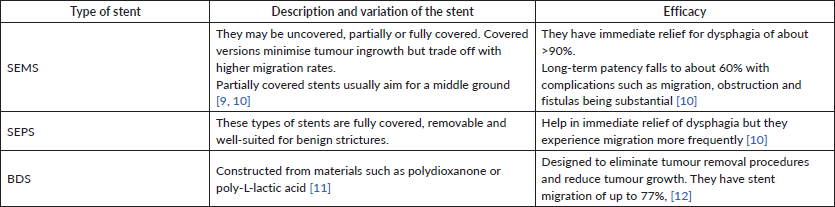
Table 1 shows a summary of the different types of esophageal stents, their descriptions and various efficacies.
Glasgow prognostic score (GPS) as a prognostic tool for esophageal stenting
The GPS is an inflammation-based score that combines the levels of proteins such as C-reactive protein (CRP) and albumin [13, 14]. The GPS was originally used in the routine clinical assessment of colorectal cancers but has, over time, been applied to predict clinical outcomes across several malignancies, including EC and gastric cancers [13, 15]. Patients are categorised into three groups based on their CRP and albumin levels: GPS 0, where patients have normal levels of both CRP and albumin; GPS 1, where patients have an abnormal value of either CRP or albumin; and GPS 2, where patients have abnormalities in both CRP and albumin [16].
Table 1. A summary of the different types of esophageal stents, their descriptions and efficacy.
Several studies have shown that the GPS is a valuable predictor of esophageal stenting clinical outcomes [17]. A cohort study of 209 patients conducted at a tertiary institution in Leeds indicated that higher GPS recovery and survival scores at stent insertion were independently associated with 30-day mortality [18]. The study showed a 30-day mortality of 5% for GPS 0 group, 23% in GPS 2 group and 33% for the GPS 2 group with the hazard ratio for the post-stent mortality being higher in the GPS 2 group (Adjusted hazard ratio = 2.4). Similarly, Vashist et al [19] found that the median survival rate post-stenting dropped from about 21 months in GPS 0 group to about 4 months in GPS 2 group. This information is summarised in Table 2.
Indications of esophageal stenting
Esophageal stenting is indicated for both benign and malignant conditions [12, 19].
Malignant indications for esophageal stents
The malignant indications include EC, malignant dysphagia and tracheoesophageal fistulas (TEFs). Esophageal stents show high immediate success rates when used in the palliation of dysphagia in circumstances where there is unresectable EC or cardiac cancer [19]. self expanding metal stents (SEMSs) have been used in the management of TEFs with a high success rate of over 70% [20].
Benign indications of esophageal stents
Esophageal stents have been used in the management of refractory strictures, leaks and perforations [12] with self-expanding plastic stents (SEPSs) and biodegradable stents (BDSs) being used for post- dilation refractory strictures [20]. In esophageal leaks and perforations, stents have an efficacy of 80%–90% in sealing the defects [21, 22].
Contraindications of esophageal stents
Despite the various uses of esophageal stenting in palliative care, it is contraindicated in circumstances where the risks outweigh the benefits [23]. These include: when life expectancy is very limited (less than <4 weeks), when there are proximal lesions within 2 cm of the proximal esophageal sphincter, when the perforation risks outweigh the benefits, uncorrectable coagulopathy, potential for significant airway compression and recent high-dose CRT within 3–6 weeks.
Table 2. Table summarising the GPS scores and their expected outcomes.

Advantages and disadvantages of esophageal stenting
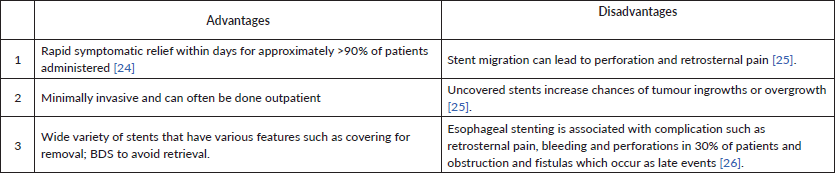
Table 3 shows a summary of the advantages and disadvantages of esophageal stenting.
Clinical outcomes of esophageal stenting
Stent placement in patients suffering from EC is an effective and reliable intervention for dysphagia and serves to improve their nutrition as they undergo neoadjuvant therapy, despite migration occurring in response to therapy [27]. A 1996 study done by Winkelbauer et al [28] assessed the effectiveness of self-expanding uncoated nitinol stents. The mean dysphagia grade improved from 3.5 to 0.6 after the stents were placed, and the patients had a mean survival of 5 months, with mortality being attributed to disease. Three patients were still alive at the end of the study, with the dysphagia greatly improved; grade 1 in one patient and grade 0 in two [28]. A study done by Reijm et al [29] involving 997 patients who received SEMS to assess clinical outcomes over time surprisingly found contradicting information to this. 31% of these patients had recurrence of dysphagia, with evident stent migration. Furthermore, retrosternal pain appeared to be the most common side effect observed in these patients [29]. Sasaki et al [30] found that combining radiotherapy with stent placement resulted in more complications compared to using stents alone, a finding that was statistically insignificant. Furthermore, the complications were not life-threatening, especially with the use of a fully covered self-expanding metallic stent (FCSEMS) [30].
Different types of stents vary in their effectiveness and potential complications. A review by Yang et al [31] found that anti-reflux stents provide better relief from dysphagia, reduce acid reflux and have fewer complications compared to conventional metallic stents. Additionally, brachytherapy has been associated with a higher chance of survival, making it a valuable alternative to traditional self-expanding metallic stents, particularly when combined with radiation therapy [31]. According to Siersema et al [32], three types of covered metal stents – the Flamingo Wallstent, Ultrflex Stent and Z Stent – did not differ in their effectiveness in providing palliation to patients. However, complications resulting from these stents such as tumour growth and stent migration, were non-significant and stent migration can be managed by reducing the diameter of the stent [32]. A systematic review and meta-analysis by Fuccio et al [33] showed no significant differences in success rates between treatment with SEMSs, SEPS and BDS, as well as migration rates.
Esophageal stent placement before neoadjuvant CRT is associated with increased toxicity and a low overall survival rate. A retrospective study by Francis et al [34] demonstrated that esophageal stent placement before CRT was associated with Grade ≥3 acute toxicities (p < 0.01), esophageal toxicity (39% versus 20%, p = 0.05), dehydration (29% versus 13%, p = 0.07) and anorexia (14% versus 5%, p = 0.13) for the stent group and non-stent group, respectively. Furthermore, the overall survival was better for the non-stent group as compared to the stent group (22.0 versus 11.5 months, respectively) [34]. Similarly, a study by Bakheet et al [35] evaluated the effect of prior chemotherapy on SEMS placement using two cohorts: one that received chemotherapy before stent placement and one that did not. Chemotherapy did not affect the clinical success of stent placement, with both groups having a success rate greater than 95% [35]. This was supported by a study conducted in Finland by Helminen et al [36], which showed that esophageal stents were associated with a higher 90-day mortality. In their analysis, the Cox proportional hazards model depicted a 1.29 HR for preoperative stenting against 1.25 HR for no stenting [36].
Table 3. Table summarising advantages and disadvantages of esophageal stenting.
Pre-esophagectomy stenting has been demonstrated to significantly worsen outcomes for patients with EC in terms of 3-year survival and recurrence rates. A comprehensive multicenter study across European hospitals revealed that the use of SEMSs was associated with increased mortality and morbidity compared to control groups that did not receive stents. Specifically, patients with SEMS had a median recurrence time of only 6.5 months, while those in the control group experienced a longer median recurrence time of 9.0 months. Furthermore, the overall survival rate at the 3-year mark was only 25% for patients with stents, contrasting sharply with 44% for those in the control group. These findings underscore the concerning implications of employing stent placements before esophagectomy, highlighting the need for careful consideration of stenting in treatment planning for EC [37]. The adverse oncologic effect observed with the use of pre-operative stents is biologically consistent and supported by a number of cohort and registry studies.
Biologically, the placement of SEMSs can result in recurrent radial stretch and micro-injuries to the esophageal wall, leading to local inflammatory responses, disruption of tissue planes and potential release of tumour cells into peri-esophageal lymphovascular spaces, thus facilitating early disease spread [37]. Moreover, complications such as migration, pressure necrosis and perforation associated with stenting may delay definitive surgical resection or neoadjuvant treatment, leading to suboptimal tumour control.
A Dutch nationwide study by Mariette et al [37] found a significant decrease in R0 resection rates and a trend towards reduced disease-free survival rates with stents, and increased complications with preoperative SEMS before esophagectomy. A recent meta-analysis including more than 1,000 patients demonstrated a relative increase in mortality and recurrence with the use of preoperative stenting, suggesting the hypothesis that the mechanical (and biological) changes associated with the stent may promote tumour progression rather than only palliation [39]. Taken together, these findings indicate that although SEMS can offer temporary dysphagia relief, the use of preoperative stents introduces adverse oncological factors that have a negative impact on disease control and survival, suggesting that rigorous patient selection and consideration of alternative nutritional or bridging measures, if possible, should be prioritised [40].
In a Belfast study by Gray et al [41] out of 53 patients who had SEMS inserted as an intervention for inoperable EC, 23 developed a total of 27 complications, with only one patient surviving by the end of the study. Most of the mortality was as a result of advancement of the underlying malignancy rather than the stent procedure; among the 52 patients who died, 37 succumbed to disease progression, while the rest died from complications such as pneumonia [11], coexisting breast carcinoma [1] and 1 empyema following esophagectomy [1]. The cause of death of one patient was unavailable [41]. Siersema et al [42] divided these complications for metal stent placement into two: procedure-related and stent-related. Procedure-related events occurred in about a third of patients and included aspiration pneumonia, chest pain, perforation, hemorrhage and fever, while those related to stents occurred in about 45% of patients and included bleeding, fistula formation, gastroesophageal reflux, migration of the stents, obstruction of food boluses and tumour overgrowth [42]. A follow-up study on 100 patients observed tumour overgrowth in 33% of the patients, with tumour obstruction being evident at the gastroesophageal junction. Esophageal perforations due to stent invasion into the walls of the esophagus were also observed, with a delayed mediastinal abscess occurring in one of the patients, who later died from sepsis. These findings align with several studies that have linked the use of esophageal stents to an increased risk of sepsis, mediastinal abscess formation [43], tumour perforation [37], bleeding, aspiration and formation of fistulas [44].
Retrosternal pain is a predictable outcome of esophageal stenting that affects the quality of life among EC patients. Multiple studies have pointed out retrosternal pain to be the single most common side effect of esophageal stenting. For instance, in a study conducted by Bektaş et al [45], 56.9% of the patients recruited in the retrospective analysis experienced retrosternal pain post-stenting, which responded well to non-steroidal anti-inflammatory drugs (NSAIDs).
Patient response
Psychosocial, physical and emotional impact
Physical impact
The primary physical benefit reported by patients following esophageal stenting is the rapid and significant relief of dysphagia. As previously mentioned, studies consistently show technical and clinical success rates above 90% for self-expandable metallic stents (SEMSs), with most patients experiencing near-immediate improvement in swallowing and nutritional intake [46]. In a multicenter cohort study, 85% of patients reported marked improvement in food passage following stent insertion [26].
Nonetheless, stents are associated with notable adverse physical effects. Retrosternal chest pain is frequently reported, especially during the initial days post-procedure. Furthermore, complications such as bleeding, reflux, aspiration pneumonia and stent migration can worsen the patient’s physical well-being [47]. Long-term survivors frequently require reinterventions due to tumour overgrowth, ingrowth or stent blockage. In one cohort study of 100 patients with malignant esophageal strictures, 78% of those surviving beyond 120 days experienced recurring dysphagia and required further treatment, with a mean interval of approximately 80 days to reintervention [43].
Psychosocial impact
The psychosocial burden of living with an esophageal stent is often under-discussed in clinical literature, yet it plays a significant role in patient adaptation and well-being. In a prospective study assessing quality of life, patients expressed improved social functioning and reduced isolation following relief from dysphagia [26]. The ability to dine with others and participate in social gatherings around meals, which had previously been avoided due to fears of choking or embarrassment, was considered a significant benefit. However, some patients report feelings of bodily foreignness, fears about stent-related complications and limitations in dietary choices, contributing to anxiety and social withdrawal. A randomised prospective study comparing covered Ultraflex stent placement with single-dose brachytherapy found that, although dysphagia improved more rapidly with stent placement, generic health‑related quality of life (including emotional and social function) deteriorated over time in the stent group, suggesting that stent-related anxiety and lifestyle restrictions may outweigh initial benefits in psychosocial domains [48].
Emotional impact
Emotionally, the relief of dysphagia often elicits a sense of hope, improved dignity and emotional recovery in patients. Stent insertion has been described as a ‘second chance at eating’ by patients previously forced to rely on feeding tubes or pureed diets. Relief of a major symptom can also offer psychological reprieve, especially in palliative care contexts where quality of life is prioritised [26].
However, some emotional responses are less positive. The awareness that stenting serves a palliative rather than curative purpose can provoke sadness, anxiety or existential distress. In a retrospective study of patients undergoing CRT for locally advanced EC, those with esophageal stents experienced significantly higher rates of severe acute toxicity, such as esophagitis, dehydration and anorexia, and had poorer overall survival (median 11.5 versus 22.0 months; hazard ratio 2.3) compared to patients treated without stents. This greater physical burden likely amplifies emotional distress, undermining initial psychosocial benefits [34].
Overall, the patient response to esophageal stenting is multifaceted. While many individuals gain substantial physical and psychosocial benefits, a subset encounters recurrent complications and emotional struggles that demand ongoing support from multidisciplinary care teams.
Functional and nutritional improvement – European Organisation for Research and Treatment of Cancer Quality of Life Questionnaire-Oesophageal Module (EORTC QLQ-OES18)
The EORTC QLQ-OES18 is a validated tool used to assess disease-specific quality-of-life domains in patients with EC. It focuses on dysphagia, nutritional status, reflux, choking, eating difficulties, pain and speech-related functions [49]. Esophageal stenting, primarily employed as a palliative measure, significantly influences these functional domains, often offering measurable improvements in short-term physical functioning and nutritional intake.
Stent placement has consistently been shown to alleviate dysphagia, the most prominent symptom in EC. Dysphagia scores, as captured in QLQ-OES18, tend to improve significantly within days after stent insertion, allowing patients to resume oral feeding and avoid enteral tube dependence [48]. The benefits of stenting extend to specific QLQ-OES18 domains such as ‘eating with others,’ ‘reflux’ and ‘dry mouth,’ reflecting not just physical improvement but also social re-engagement and comfort during meals. Notably, patients report better appetite and weight maintenance, especially when stenting is performed early in the disease course or before severe nutritional compromise [43].
However, the long-term functional benefits of esophageal stents are limited by complications such as tumour ingrowth or overgrowth, stent migration and aspiration. These adverse events can result in recurrent dysphagia and secondary decline in QLQ‑OES18 scores. Importantly, while stenting provides rapid symptomatic relief, long-term dysphagia outcomes and quality of life often worsen compared to brachytherapy. A multicenter randomised trial of 209 patients found that although dysphagia improved more quickly after stent placement, long-term dysphagia relief was superior in the brachytherapy group, which also experienced fewer complications and better generic quality-of-life scores [26, 50].
In addition, nutritional indices such as serum albumin levels, body weight and caloric intake often stabilise or improve temporarily following successful stent placement. Some studies have used nutritional screening tools alongside the EORTC QLQ modules to show a reduction in malnutrition risk post-stenting, particularly when dietary counseling is included in post-intervention care [51].
Ultimately, the EORTC QLQ-OES18 demonstrates that while esophageal stenting is not curative, it is functionally restorative. By reducing dysphagia and facilitating normal eating behaviour, it plays a critical role in improving patient autonomy, energy levels and physical strength during palliative care.
Pain and discomfort – retrosternal pain
The type and design of the stent influence the intensity and duration of retrosternal pain. FCSEMS are often associated with more mucosal pressure and friction compared to partially covered or uncovered stents, potentially increasing pain frequency [51]. Additionally, the placement of esophageal stents was previously considered relatively contraindicated due to elevated risks of complications, including perforation, pulmonary aspiration, migration of the stent into the hypopharynx often intolerable sensation of a foreign body [52].
Management of retrosternal pain post-stenting typically includes short-term administration of analgesics, particularly NSAIDs or mild opioids. Proton pump inhibitors may also reduce associated reflux-related symptoms. However, persistent pain warrants investigation for complications such as stent misplacement, esophageal perforation or pressure necrosis [47].
Patient satisfaction
Patient satisfaction reflects improvements in specific domains of the EORTC QLQ-OES18 questionnaire, such as the ability to eat in public, reduced choking episodes and decreased anxiety around meals [49]. The restoration of these functions often allows patients to re-engage in previously avoided social situations, which reinforces emotional resilience and a sense of normalcy despite the terminal nature of the disease [26].
However, satisfaction may decline over time due to resulting complications and should the need for reintervention arise, can negatively impact the patient’s overall experience and confidence in the efficacy of the intervention [43].
Additionally, psychological adaptation and pre-procedural counseling play a role in shaping satisfaction. Patients who are adequately informed about the purpose, limitations and possible outcomes of stenting are more likely to express positive satisfaction regardless of the presence of minor complications [47]. Conversely, those who expected long-term or curative effects from the procedure may experience disappointment or emotional distress when symptoms recur or new complications emerge.
Comparison to other treatment modalities
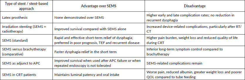
SEMSs have been shown to have fewer incidences of complications, whether early or delayed, compared to latex prostheses. Recurrent dysphagia was, however, not different between metallic stents and latex prostheses. Further analysis showed that treatment with radiotherapy or chemotherapy before SEMS or latex prostheses placement exacerbates device-related complications [53]. Surgical treatment of EC, such as esophageal exclusion, esophageal bypass and fistula resection, is associated with high procedure-related risk, especially in advanced disease. Retrospective studies have shown that the use of esophageal stents for TEF secondary to EC provides better survival outcomes and better infection control in comparison to gastrostomy. Gastrostomy or jejunostomy had been used earlier for the management of TEF before the introduction of stenting. This study has, however, led to the consideration of esophageal stents and a first-line treatment in the management of TEF [54].
Other studies show that irradiation stenting may prolong survival rates in comparison to stenting alone in the treatment of EC [55]. In a randomised controlled trial, stenting provided a rapid and effective relief of dysphagia in the short term, with brachytherapy having more symptomatic relief long-term [56]. Brachytherapy is suitable for patients with unresectable tumours, predicted long-term survival (young, small tumour, no metastases, good performance status) and cancer developing near the upper esophageal sphincter. On the other hand, SEMS is preferred in patients with poor survival outcomes (4–5 months), TEF and recurrent EC after CRT or brachytherapy [57]. Studies comparing the survival outcomes in patients using APC and SEMS have shown no significant differences when APC or SEMS is used alone. The survival rate, however, increases when the two modalities are used together, with APC being the primary palliative modality and SEMS being used secondary to APC failure or when patients cannot tolerate repeated endoscopies [58]. Percutaneous gastrostomy provides better maintenance of nutritional status, as evidenced by higher serum albumin levels and reduced weight loss, as well as prolonged survival rates and fewer complications, including reduced local pain and decreased device dislodgment, compared to SEMS in the treatment of EC [59]. For patients undergoing CRT, the use of esophageal stents is associated with worse pain, decreased albumin and reduced overall quality of life when compared to tube feeding. In further analysis, patients using esophageal stents experience significantly more weight loss, while the patients using nasogastric tubes need narcotics for retrosternal pain relief [60]. Esophageal balloon dilatation is preferably used for benign stenosis, while esophageal stent is approved only for malignant strictures, with both having a 90% success rate [61]. This information is summarised in Table 4.
Emerging trends and future directions
Esophagectomy is one of the minimally invasive methods used in patients diagnosed with EC. This is especially in cases of advanced disease where the tumour has invaded the submucosa. Despite the advantages of esophagectomy in the management of these cases, it has, however, been associated with severe complications such as anastomotic leakage [62]. There are different modalities of esophagectomy that have been used in the management of these patients. Initially, open transthoracic esophagectomies were used until minimally invasive methods were developed. The most recent development has been robot-assisted minimally invasive thoraco-laparoscopic esophagectomies. The robot-assisted method has been found to result in less blood loss and fewer cases of pulmonary and cardiac complications [63].
Table 4. Table comparing esophageal stenting with other treatment modalities.
Conventionally, self-expandable metal and SEPS have been used. However, they are associated with complications such as migration and restenosis. Innovation has led to the devising of biodegradable materials, including synthetic polyester polymers and magnesium-based alloys as stents. Several synthetic polymers have been approved for use as synthetic polymers by the US Food and Drug Administration, including and not limited to polydioxanone, poly-L-lactide, poly (1.3-trimethylene carbonate) and poly (lactic-co-glycolic acid) are being used in research efforts. For these named polymers, their products after degradation are water and carbon dioxide, which means less toxicity to the human body. Of importance to note, is that the BDS are not as stiff and strong as the metal and plastic stents hence have been found to be associated with early displacement and increased susceptibility to acidic environment in the gut leading to a faster degradation [64].
Stent blockage is a challenge commonly faced with the use of self-expanding stents. Drug-eluting stents were a new advancement where the sustained release of docetaxel (DTX) loaded stents was devised. The stents are prepared by dip-coating the dip nitinol stents with DTX-polyurethane [65]. The drug-eluting stents are composed of three sections: the stent body, the stent coating and an anti-proliferative drug portion. The anti-proliferative sections function to localise the drug and reduce the potential systemic cytotoxicity.
As is the case in many industries, technology has been devised to develop 3D printing of stents, which are designed and customised to be tubular, flexible polymer stents with spirals. The stents customised with spirals have been proven to have a higher anti-migration force than the stents that do not have spirals, and they had a significantly reduced migration distance. This has proven that the 3D customisable printing of stents is promising in the management of esophageal malignancies and even benign obstructions [66].
Conclusion
Since the 1970s, esophageal stents have been effective in treating and providing palliative care for EC and other related conditions or side effects that may result from it, such as fistulas, leaks and strictures. Since then, they have undergone significant advancements, starting with rigid prosthetics that were linked to high rates of complications and morbidity. Today, they are made of durable polymers and metal alloy compounds, such as SEPS and self-expanding esophageal metal stents. The clinical results of esophageal stent use are anticipated to improve further with the development of biodegradable, adjunct-embedded and 3D-printed stents. In most cases, palliative brachytherapy and CRT have been shown to provide superior long-term outcomes for dysphagia alleviation than metal and polymer stents.
Conflicts of interest
The author(s) declared no potential conflicts of interest with respect to the research, authorship and/or publication of this article.
Funding
The author(s) received no financial support for the research, authorship and/or publication of this article.
Author contributions
All authors contributed equally towards the preparation of this manuscript.
References
1. Arnal MJD (2015) Esophageal cancer: risk factors, screening and endoscopic treatment in Western and Eastern countries World J Gastroenterol WJG 21(26) 7933–7943 https://doi.org/10.3748/wjg.v21.i26.7933
2. Van Nistelrooij AMJ, Dinjens WNM, and Wagner A, et al (2014) Hereditary factors in esophageal adenocarcinoma Gastrointest Tumors 1(2) 93–98 https://doi.org/10.1159/000362575 PMID: 26675496 PMCID: 4645576
3. Coleman HG, Xie SH, and Lagergren J (2018) The epidemiology of esophageal adenocarcinoma Gastroenterology 154(2) 390–405 https://doi.org/10.1053/j.gastro.2017.07.046
4. He L, He L, and Jin-Hu F, et al (2017) Epidemiology, etiology, and prevention of esophageal squamous cell carcinoma in China Cancer Biol Med 14(1) 33–41 https://doi.org/10.20892/j.issn.2095-3941.2016.0093
5. Ramakrishnaiah VPN, Malage S, and Sreenath GS, et al (2016) Palliation of dysphagia in carcinoma esophagus Clin Med Insights Gastroenterol 9 11–23 https://doi.org/10.4137/CGast.S30303 PMID: 27279758 PMCID: 4896534
6. Guyer DL, Almhanna K, and Mckee KY (2020) Palliative care for patients with esophageal cancer: a narrative review Ann Transl Med 8(17) 1103 https://doi.org/10.21037/atm-20-3676 PMID: 33145322 PMCID: 7575932
7. Mohapatra S, Santharaman A, and Gomez K, et al (2022) Optimal management of dysphagia in patients with inoperable esophageal cancer: current perspectives Cancer Manag Res 14 3281–3291 https://doi.org/10.2147/CMAR.S362666 PMID: 36448034 PMCID: 9701451
8. Vermeulen BD and Siersema PD (2018) Esophageal stenting in clinical practice: an overview Curr Treat Options Gastroenterol 16(2) 260–273 https://doi.org/10.1007/s11938-018-0181-3 PMID: 29557070 PMCID: 5932108
9. Jiménez-Gutiérrez JM, Alonso-Lárraga JO, and Hernández-Guerrero AI, et al (2023) Efficacy and safety of self-expanding metal stents in patients with inoperable esophageal cancer: a real-life study Ther Adv Gastrointest Endosc 16 26317745231200975 https://doi.org/10.1177/26317745231200975 PMID: 37771789 PMCID: 10524049
10. Schembre D (2010) Advances in esophageal stenting: the evolution of fully covered stents for malignant and benign disease Adv Ther 27(7) 413–425 https://doi.org/10.1007/s12325-010-0042-5
11. Shen Y, Yu X, and Cui J, et al (2022) Development of biodegradable polymeric stents for the treatment of cardiovascular diseases Biomolecules 12(9) 1245 https://doi.org/10.3390/biom12091245 PMID: 36139086 PMCID: 9496387
12. Hindy P, Hong J, and Lam-Tsai Y, et al (2022) Comprehensive review of esophageal stents Gastroenterol Hepatol (N Y) 8(8) 526–534
13. Mcmillan DC (2013) The systemic inflammation-based Glasgow Prognostic Score: a decade of experience in patients with cancer Cancer Treat Rev 39(5) 534–540 https://doi.org/10.1016/j.ctrv.2012.08.003
14. Proctor MJ, Morrison DS, and Talwar D, et al (2011) A comparison of inflammation-based prognostic scores in patients with cancer. a Glasgow Inflammation Outcome Study Eur J Cancer 47(17) 2633–2641 https://doi.org/10.1016/j.ejca.2011.03.028 PMID: 21724383
15. Proctor MJ, Horgan PG, and Talwar D, et al (2013) Optimization of the systemic inflammation‐based Glasgow Prognostic Score: a Glasgow Inflammation Outcome Study Cancer 15(12) 2325–2332 https://doi.org/10.1002/cncr.28018
16. Lee SN, Moon D, and Moon KW, et al (2019) The Glasgow prognostic score as a significant predictor of clinical outcomes in patients with acute coronary syndrome J Cardiol 74(2) 130–135 https://doi.org/10.1016/j.jjcc.2019.02.007 PMID: 30878352
17. Wang Y, Li P, and Li J, et al (2019) The prognostic value of pretreatment Glasgow Prognostic Score in patients with esophageal cancer: a meta-analysis Cancer Manag Res 11 8181–8190 https://doi.org/10.2147/CMAR.S203425 PMID: 31564977 PMCID: 6732518
18. Driver RJ, Handforth C, and Radhakrishna G, et al (2018) The Glasgow Prognostic Score at the time of palliative esophageal stent insertion is a predictive factor of 30-day mortality and overall survival J Clin Gastroenterol 52(3) 223 https://doi.org/10.1097/MCG.0000000000000773
19. Vashist YK, Loos J, and Dedow J, et al (2011) Glasgow prognostic score is a predictor of perioperative and long-term outcome in patients with only surgically treated esophageal cancer Ann Surg Oncol 18(4) 1130–2118 https://doi.org/10.1245/s10434-010-1383-7
20. Sharma P and Kozarek R (2010) Role of esophageal stents in benign and malignant diseases Gastroenterol PPC AC 105(2) 258 [https://doi.org/10.1038/ajg.2009.684]
21. D’Cunha J, Rueth NM, and Groth SS, et al (2011) Esophageal stents for anastomotic leaks and perforations J Thorac Cardiovasc Surg 142(1) 39–46 https://doi.org/10.1016/j.jtcvs.2011.04.027
22. Kochman ML, Mcclave SA, and Boyce HW (2005) The refractory and the recurrent esophageal stricture: a definition Gastrointest Endosc 62(3) 474–475 https://doi.org/10.1016/j.gie.2005.04.050 PMID: 16111985
23. Martinez JC, Puc MM, and Quiros RM (2011) Esophageal stenting in the setting of malignancy Int Sch Res Not 2011(1) 719575 [https://doi.org/10.5402/2011/719575]
24. Vermeulen BD and Siersema PD (2018) Esophageal stenting in clinical practice: an overview Curr Treat Options Gastroenterol 16(2) 260–273 [http://doi.org/10.1007/s11938-018-0181-3]
25. Singh P, Singh A, and Singh A, et al (2016) Long term outcome in patients with esophageal stenting for cancer esophagus - our experience at a rural hospital of Punjab, India J Clin Diagn Res 10(12) PC06–PC09 [https://doi.org/10.7860/JCDR/2016/22950.8994]
26. Homs MY, Steyerberg EW, and Eijkenboom WM, et al (2004) Single-dose brachytherapy versus metal stent placement for the palliation of dysphagia from oesophageal cancer: multicentre randomised trial Lancet 364(9444) 1497–1504 https://doi.org/10.1016/S0140-6736(04)17272-3 PMID: 15500894
27. Nagaraja V, Cox MR, and Eslick GD (2014) Safety and efficacy of esophageal stents preceding or during neoadjuvant chemotherapy for esophageal cancer: a systematic review and meta-analysis J Gastrointest Oncol 5(2)
28. Winkelbauer FW, Schöfl R, and Niederle B, et al (1996) Palliative treatment of obstructing esophageal cancer with nitinol stents: value, safety, and long-term results AJR Am J Roentgenol 166(1) 79–84 https://doi.org/10.2214/ajr.166.1.8571911 PMID: 8571911
29. Reijm A, Didden P, and Schelling S, et al (2019) Self-expandable metal stent placement for malignant esophageal strictures - changes in clinical outcomes over time Endoscopy 51(1) 18–29 https://doi.org/10.1055/a-0644-2495
30. Sasaki K, Osako Y, and Urata M, et al (2021) Clinical outcomes of fully covered self-expanding metallic stent placement for palliation of incurable esophageal cancer with or without radiotherapy Anticancer Res 41(1) 385–389 https://doi.org/10.21873/anticanres.14787 PMID: 33419835
31. Yang C, Li J, and Fu W, et al (2014) Interventions for Dysphagia in Oesophageal Cancer (Cochrane Library) [Internet] [https://www.cochranelibrary.com/cdsr/doi/10.1002/14651858.CD005048.pub4/full] Date accessed: 04/01/26
32. Siersema P, Hop W, and Vanblankenstein M, et al (2001) A comparison of 3 types of covered metal stents for the palliation of patients with dysphagia caused by esophagogastric carcinoma: a prospective, randomized study Gastrointest Endosc 54(2) 145–153 https://doi.org/10.1067/mge.2001.116879 PMID: 11474382
33. Fuccio L, Hassan C, and Frazzoni L, et al (2016) Clinical outcomes following stent placement in refractory benign esophageal stricture: a systematic review and meta-analysis Endoscopy 48(2) 141–148 [https://doi.org/10.1055/s-0034-1393331]
34. Francis SR, Orton A, and Thorpe C, et al (2017) Toxicity and outcomes in patients with and without esophageal stents in locally advanced esophageal cancer Int J Radiat Oncol Biol Phys 99(4) 884–894 https://doi.org/10.1016/j.ijrobp.2017.06.2457 PMID: 28864404
35. Bakheet N, Hu HT, and Park JH, et al (2020) Clinical effectiveness and safety of self-expanding metal stent placement following palliative chemotherapy in patients with advanced esophageal cancer Abdom Radiol N Y 45(2) 563–570 https://doi.org/10.1007/s00261-019-02245-3
36. Helminen O, Sihvo E, and Helmiö M, et al (2023) Preoperative esophageal stenting and 5-year survival in patients undergoing esophagectomy for esophageal cancer: a population-based nationwide study from Finland J Gastrointest Surg 27(6) 1078–1088 https://doi.org/10.1007/s11605-023-05643-7 PMID: 36882628 PMCID: 10267252
37. Mariette C, Gronnier C, and Duhamel A, et al (2015) Self-expanding covered metallic stent as a bridge to surgery in esophageal cancer: impact on oncologic outcomes J Am Coll Surg 220(3) 287 https://doi.org/10.1016/j.jamcollsurg.2014.11.028 PMID: 25617915
38. Noordman BJ, Spaander MCW, and Valkema R, et al (2018) Detection of residual disease after neoadjuvant chemoradiotherapy for oesophageal cancer (preSANO): a prospective multicentre, diagnostic cohort study Lancet Oncol 19(7) 965–974 https://doi.org/10.1016/S1470-2045(18)30201-8 PMID: 29861116
39. Halsema EEV (2015) Clinical outcomes of self-expandable stent placement for benign esophageal diseases: a pooled analysis of the literature World J Gastrointest Endosc 7(2) 135–153 https://doi.org/10.4253/wjge.v7.i2.135 PMID: 25685270 PMCID: 4325310
40. Tham JE, Tharian B, and Allen PB, et al (2019) Oesophageal stents for potentially curable oesophageal cancer – a bridge to surgery? Ulster Med J 88(1) 10–14 PMCID: 6342033
41. Gray RT, O’Donnell ME, and Scott RD, et al (2011) Self-expanding metal stent insertion for inoperable esophageal carcinoma in Belfast: an audit of outcomes and literature review Dis Esophagus Off J Int Soc Dis Esophagus 24(8) 569–574 https://doi.org/10.1111/j.1442-2050.2011.01188.x
42. Siersema PD, Marcon N, and Vakil N (2003) Metal stents for tumors of the distal esophagus and gastric cardia Endoscopy 35(1) 79–85 https://doi.org/10.1055/s-2003-36418 PMID: 12510232
43. Christie NA, Buenaventura PO, and Fernando HC, et al (2001) Results of expandable metal stents for malignant esophageal obstruction in 100 patients: short-term and long-term follow-up Ann Thorac Surg 71(6) 1797–1802 https://doi.org/10.1016/S0003-4975(01)02619-4 PMID: 11426750
44. Doosti-Irani A, Mansournia MA, and Rahimi-Foroushani A, et al (2017) Complications of stent placement in patients with esophageal cancer: a systematic review and network meta-analysis PLoS One 12(10) 184784 https://doi.org/10.1371/journal.pone.0184784
45. Bektaş H, Gürbulak B, and Düzköylü Y, et al (2017) Clinical outcomes of upper gastrointestinal stents and review of current literature JSLS 21(4) https://doi.org/10.4293/JSLS.2017.00058
46. Conigliaro R, Battaglia G, and Repici A, et al (2007) Polyflex stents for malignant oesophageal and oesophagogastric stricture: a prospective, multicentric study Eur J Gastroenterol Hepatol 19(3) 195–203 https://doi.org/10.1097/MEG.0b013e328013a418 PMID: 17301645
47. Verschuur EML, Kuipers EJ, and Siersema PD (2007) Esophageal stents for malignant strictures close to the upper esophageal sphincter Gastrointest Endosc 66(6) 1082–1090 https://doi.org/10.1016/j.gie.2007.03.1087 PMID: 17826774
48. Homs MYV, Essink-Bot ML, and Borsboom GJJM, et al (2004) Quality of life after palliative treatment for oesophageal carcinoma – a prospective comparison between stent placement and single dose brachytherapy Eur J Cancer 40(12) 1862–1871 https://doi.org/10.1016/j.ejca.2004.04.021 PMID: 15288288
49. Blazeby JM, Alderson D, and Winstone K, et al (1996) Development of an EORTC questionnaire module to be used in quality of life assessment for patients with oesophageal cancer Eur J Cancer 32(11) 1912–1917 https://doi.org/10.1016/0959-8049(96)00199-2
50. Diamantis G (2011) Quality of life in patients with esophageal stenting for the palliation of malignant dysphagia World J Gastroenterol 17(2) 144–150 [10.3748/wjg.v17.i2.144] https://doi.org/10.3748/wjg.v17.i2.144 PMID: 21245986 PMCID: 3020367
51. Zhang D, Luo T, and Gao F, et al (2025) Comparison of fully covered versus uncovered self-expandable metallic stents in treating inoperable malignant distal biliary obstruction: a retrospective study Ther Adv Gastrointest Endosc 18 26317745251376845 https://doi.org/10.1177/26317745251376845 PMCID: 12480805
52. Shim CS (2012) Esophageal stent for cervical esophagus and esophagogastric junction Clin Endosc 45(3) 235–239 https://doi.org/10.5946/ce.2012.45.3.235 PMID: 22977809 PMCID: 3429743
53. Siersema PD, Hop WCJ, and Dees J, et al (1998) Coated self-expanding metal stents versus latex prostheses for esophagogastric cancer with special reference to prior radiation and chemotherapy: a controlled, prospective study Gastrointest Endosc 47(2) 113–120 https://doi.org/10.1016/S0016-5107(98)70342-6 PMID: 9512274
54. Chen YH, Li SH, and Chiu YC, et al (2012) comparative study of esophageal stent and feeding gastrostomy/jejunostomy for tracheoesophageal fistula caused by esophageal squamous cell carcinoma PLoS One 7(8) e42766 https://doi.org/10.1371/journal.pone.0042766 PMID: 22912737 PMCID: 3418295
55. Tinusz B, Soós A, and Hegyi P, et al (2020) Efficacy and safety of stenting and additional oncological treatment versus stenting alone in unresectable esophageal cancer: a meta-analysis and systematic review Radiother Oncol 147 169–177 https://doi.org/10.1016/j.radonc.2020.05.015 PMID: 32422302
56. Steyerberg EW, Homs MYV, and Stokvis A, et al (2005) Stent placement or brachytherapy for palliation of dysphagia from esophageal cancer: a prognostic model to guide treatment selection Gastrointest Endosc 62(3) 333–340 https://doi.org/10.1016/S0016-5107(05)01587-7 PMID: 16111947
57. Wallace MB (2005) Stent vs. brachytherapy: does life expectancy help you decide? Gastrointest Endosc 62(3) 341–343 https://doi.org/10.1016/j.gie.2005.06.007 PMID: 16111948
58. Sigounas DE, Krystallis C, and Couper G, et al (2017) Argon plasma coagulation compared with stent placement in the palliative treatment of inoperable oesophageal cancer United Eur Gastroenterol J 5(1) 21–31 https://doi.org/10.1177/2050640616650786
59. Wang T, Wen Q, and Zhang Y, et al (2021) Percutaneous gastrostomy compared with esophageal stent placement for the treatment of esophageal cancer with dysphagia J Vasc Interv Radiol 32(8) 1215–1220 https://doi.org/10.1016/j.jvir.2021.04.019 PMID: 33957245
60. Yu FJ, Shih HY, and Wu CY, et al (2018) Enteral nutrition and quality of life in patients undergoing chemoradiotherapy for esophageal carcinoma: a comparison of nasogastric tube, esophageal stent, and ostomy tube feeding Gastrointest Endosc 88(1) 21–31 https://doi.org/10.1016/j.gie.2017.11.030
61. Therasse E, Oliva VL, and Lafontaine E, et al (2003) Balloon dilation and stent placement for esophageal lesions: indications, methods, and results RadioGraphics 23(1) 89–105 https://doi.org/10.1148/rg.231025051 PMID: 12533645
62. Fabbi M, Hagens ERC, and Van Berge Henegouwen MI, et al (2020) Anastomotic leakage after esophagectomy for esophageal cancer: definitions, diagnostics, and treatment Dis Esophagus 39 https://doi.org/10.1093/dote/doaa039 PMID: 32476017 PMCID: 7801633
63. Van Der Sluis PC, Schizas D, and Liakakos T, et al (2020) Minimally invasive esophagectomy Dig Surg 37(2) 93–100 https://doi.org/10.1159/000497456
64. Yang Y, Yang Y, and Hou Z, et al (2023) Comprehensive review of materials, applications, and future innovations in biodegradable esophageal stents Front Bioeng Biotechnol 11 1327517 https://doi.org/10.3389/fbioe.2023.1327517 PMID: 38125305 PMCID: 10731276
65. Fouladian P, Jin Q, and Arafat M, et al (2021) Drug-loaded, polyurethane coated nitinol stents for the controlled release of docetaxel for the treatment of oesophageal cancer Pharmaceuticals 14(4) 311 https://doi.org/10.3390/ph14040311 PMID: 33915787 PMCID: 8067330
66. Lin M, Firoozi N, and Tsai CT, et al (2019) 3D-printed flexible polymer stents for potential applications in inoperable esophageal malignancies Acta Biomater 83 119–129 https://doi.org/10.1016/j.actbio.2018.10.035