Clinical characteristics, treatment patterns and survival outcomes of pancreatic cancer patients treated at a Lakeshore Cancer Center in Lagos: a 10-year review
Agbakwuru Chidiebere1,2, Zainab Adegbite2, Clement Awe2, Toluwanimi Aduloju2, Temabor Akemu2, Joy Olowe2, Adewale Ahmed2, Chineye Iwuji2, Mutiu Jimoh2,3
1Medserve-LUTH Cancer Centre, Lagos University Teaching Hospital, Lagos 101283, Nigeria
2Department of Oncology, Lakeshore Cancer Center, Lagos 100271, Nigeria
3Radiation Oncology Department, College of Medicine, University of Ibadan, Ibadan 200284, Nigeria
Abstract
Background: Pancreatic cancer outcomes in Nigeria are predominantly reported from public hospitals. This study describes clinical characteristics, treatment patterns, and survival outcomes at a private oncology center in Lagos.
Methods: A retrospective study of 42 patients diagnosed with pancreatic cancer and managed at Lakeshore Cancer Center (2014–2024) was conducted. Data were extracted from the institutional cancer registry. Overall survival was estimated using the Kaplan–Meier method.
Results: Most patients were female (57.1%) and aged 60 years or older (64.3%). Common presenting symptoms included abdominal pain (78.6%), weight loss (71.4%), and jaundice (64.3%). Among patients with comorbidities (n = 33), diabetes (57.6%) and hypertension (48.5%) were most frequent. Advanced disease predominated, with 76.2% presenting at Stage IV and only one patient (2.4%) at Stage I. Tumours were mainly located in the pancreatic head (64.3%). Treatment modalities were not mutually exclusive: chemotherapy was administered to 45.2% (n = 19), best supportive care to 35.7% (n = 15), and surgery to 11.9% (n = 5), while 21.4% (n = 9) had no documented treatment. No patient received radiotherapy. Median overall survival was 17 weeks (95% CI: 3.9–24.6), and mean survival was 21.2 weeks (95% CI: 14.0–28.3). Female patients had a longer median survival than male patients (23 versus 17 weeks).
Conclusion: Most patients presented with advanced disease and had poor survival, typically less than 5 months, consistent with reports from public hospitals. Access to private healthcare did not result in earlier diagnosis. Strengthening referral pathways, improving access to diagnostics, and expanding health insurance coverage may improve outcomes.
Keywords: pancreatic cancer, Nigeria, private oncology, survival, late-stage presentation, Lakeshore Cancer Center
Correspondence to: Zainab Adegbite
Email: zainab.lakeshorecc@gmail.com
Published: 24/09/2026
Received: 15/05/2026
Publication costs for this article were supported by ecancer (UK Charity number 1176307).
Copyright: © the authors; licensee ecancermedicalscience. This is an Open Access article distributed under the terms of the Creative Commons Attribution License (http://creativecommons.org/licenses/by/4.0), which permits unrestricted use, distribution, and reproduction in any medium, provided the original work is properly cited.
Introduction
Pancreatic cancer kills nearly as many people as it diagnoses. With a 5-year survival rate below 12% largely unchanged over four decades, pancreatic ductal adenocarcinoma (PDAC) is one of the most lethal solid malignancies in oncology [1,2]. It is projected to become the second leading cause of cancer-related death in Western countries by 2030, surpassing breast and colorectal cancers [3]. PDAC arises from the exocrine ductal epithelium and accounts for over 90% of pancreatic malignancies. It generates a thick fibrotic stroma that physically blocks chemotherapy from reaching cancer cells. Perineural and vascular invasion happen early, often before diagnosis. The tumour microenvironment is hypoxic and packed with immunosuppressive cells, which is why checkpoint inhibitors that work in melanoma and lung cancer have largely failed here [4]. At the molecular level, activating Kirsten Rat Sarcoma Viral Oncogene Homologue mutations occur in approximately 90% of cases, with sequential inactivation of TP53, CDKN2A and SMAD4 driving progression from precursor lesions to invasive carcinoma [4].
This malignancy is typically diagnosed in older adults, with a median age of 65–70 years in high-income countries, although it can occur in younger patients. Pancreatic cancer has one of the lowest survival rates among major cancers, even with multimodality treatment; the overall 5-year survival rate is only around 10% [1].
Globally, over 500,000 new cases of pancreatic cancer are diagnosed each year [5]. Curative treatment options remain limited. Surgical resection offers the only potential for cure, yet only a minority of patients present with disease localised enough to be resectable. For those who undergo successful pancreatic resection followed by adjuvant chemotherapy, outcomes improve considerably, with median survival reaching several years in optimally treated cases; however, such patients represent a small fraction of the total [3].
Clinical presentation and symptoms
Pancreatic cancer often presents with insidious symptoms that overlap with many benign conditions. Common complaints include vague upper abdominal or back pain, weight loss and anorexia. Patients with tumours in the head of the pancreas may develop painless obstructive jaundice due to bile duct compression. The deep retroperitoneal location of the pancreas and the gradual, unobtrusive growth of the tumour account for this lack of specificity. There is no effective routine screening test for asymptomatic individuals at average risk, unlike cancers such as breast or colorectal cancer [15]. Imaging or endoscopic investigations are typically arranged only after significant symptoms have manifested, by which point the disease may have advanced.
Diagnostic challenges and delayed detection
Delayed diagnosis is a persistent challenge in pancreatic cancer. Even after initial presentation, additional delays occur during the diagnostic workup. Arranging specialised imaging (CT or MRI) or endoscopic procedures and obtaining tissue biopsies can take considerable time, particularly in low-resource settings. Evidence suggests that prolonged intervals from symptom onset to diagnosis negatively impact survival [6]. The combination of an asymptomatic early phase and nonspecific presenting features means that most patients are not diagnosed until their disease is advanced.
Stage at diagnosis, treatment options and survival
The stage distribution at diagnosis is heavily skewed towards advanced disease. Contemporary data indicate that only around 15% of patients are diagnosed with truly localised, surgically resectable tumours [3]. The remaining 85% have either regionally advanced unresectable tumours or distant metastatic spread at the time of detection. Over half of patients already have metastases to the liver, peritoneum or other sites when the cancer is first identified. This stage distribution, with most cases being stage III or IV, is observed worldwide and reflects the biologically aggressive yet clinically silent nature of pancreatic cancer. It is the fundamental reason that overall outcomes remain so poor. Late-stage diagnosis translates directly into high mortality.
Pancreatic cancer in Sub-Saharan Africa and Nigeria
Pancreatic cancer in Sub-Saharan Africa shares the global pattern of late presentation, and outcomes in the region are particularly poor. Most patients are diagnosed at an advanced stage after prolonged symptom duration. A multicentre study in Nigeria reported that the majority of pancreatic cancer patients had experienced symptoms for over 6 months before definitive diagnosis, and over 80% had unresectable locally advanced or metastatic disease at presentation [7]. Curative surgical resections were exceedingly rare. Similar findings have been reported elsewhere in Africa; a Kenyan tertiary hospital study found that more than half of patients presented with metastatic disease, and the median age at diagnosis (around 58 years) was notably younger than in Western populations [8].
Structural barriers compound these clinical challenges. In Nigeria and similar settings, healthcare is largely financed out-of-pocket, placing CT scans and chemotherapy beyond the reach of most families [9]. Public hospitals contend with equipment failures, drug shortages and long waiting times. Histological confirmation is frequently delayed or simply unavailable. Studies from Nigeria and South Africa indicate that the majority of patients receive palliative care only, and surgical resection rates remain low [10,7]. Reported survival figures are worse than those from high-income centres, though incomplete follow-up limits direct comparisons [9].
Rationale for the study and study objective
Although less prevalent than other cancers, pancreatic cancer contributes meaningfully to cancer mortality in Nigeria. The country’s healthcare system is marked by disparities in access to specialised diagnostics and treatment, complicating management. Most published data on pancreatic cancer in Nigeria derive from public teaching hospitals, which serve populations with limited financial resources and face well-documented operational constraints.
Private oncology centres play an increasingly important role in delivering specialised cancer care. These facilities often serve patients with distinct socioeconomic backgrounds and potentially earlier access to diagnostic imaging and systemic therapy. However, clinical outcomes in this setting remain poorly characterised. Understanding the demographic profile, stage distribution, treatment patterns and survival of pancreatic cancer patients treated in the private sector may inform service planning and identify opportunities for improvement.
This study aimed to describe the clinical characteristics, treatment patterns and survival outcomes of patients diagnosed with pancreatic cancer at Lakeshore Cancer Center, Lagos, over 10 years (2014–2024).
Methodology
Study design
This was a retrospective study on patients diagnosed and treated with pancreatic cancer at Lakeshore Cancer Center over a period of 10 years, from 2014 to 2024. Lakeshore Cancer Center has an electronic database that captures demographic, clinical and treatment data for all cancer patients.
Study site
Lakeshore Cancer Center is one of the nine cancer centres in Nigeria and the only private cancer centre affiliated with Roswell Park Cancer Center in the United States, the oldest cancer centre in the United States. It is the first comprehensive cancer centre in Nigeria and was established in 2014. Lakeshore Cancer Center provides comprehensive oncology services that include screening, diagnostics, treatment, follow-up and palliative care. The centre screens for the top four cancers in Nigeria, namely breast, prostate, colorectal and cervical cancer. The facility provides an in-house blood laboratory, mammograms, X-rays and CT scans. Whilst some surgical procedures (e.g. mastectomy) are performed in the operating theatre at Lakeshore, patients are referred to partner hospitals for more complex surgeries. Breast cancer surgery is performed by a team of two surgeons who operate at Lakeshore. Colorectal cancer patients who require surgery will have surgery performed at local hospitals and referred to Lakeshore for adjunct therapy. Therefore, Lakeshore provides systemic chemotherapy and immunotherapy treatment to patients who may be referred from other medical facilities, identified via screening or who ‘walk in’ requesting a consultation. However, the centre does not currently have radiotherapy available on-site, and therefore patients requiring radiotherapy treatment are referred to Medsereve-LUTH Cancer Center.
Study population
The study population comprises of all patients diagnosed with pancreatic cancer and registered in the Lakeshore Cancer Center’s hospital-based cancer registry between 2014 and 2024.
Eligibility criteria
Inclusion criteria: patients were eligible for inclusion, if they met all of the following criteria:
a. patients with a confirmed diagnosis of pancreatic cancer, established by histological and/or radiological evidence,
b. diagnosis made and/or managed at the study centre within the specified study period (2014–2024) and
c. availability of complete medical records in the institutional registry, including key demographic, clinical, staging and outcome variables required for analysis.
Exclusion criteria: patients were excluded from the study if they met any of the following criteria:
a. patients with incomplete or missing data for key study variables,
b. patients whose diagnosis of pancreatic cancer was later found to be incorrect or revised following further evaluation (i.e. misdiagnosed cases) and
c. patients with pancreatic lesions subsequently determined to be benign or nonpancreatic malignancies.
Data collection tools
Data were extracted from the hospital-based cancer registry at Lakeshore Cancer Center for patients diagnosed with pancreatic cancer between January 2014 and December 2024. A structured data abstraction form was developed for this study based on the objectives and variables of interest.
Variables collected included: demographics (age at diagnosis, sex and marital status), clinical history (smoking history, alcohol use, family history of cancer and family history of pancreatic cancer), comorbidities (hypertension, diabetes mellitus, hepatitis B, peptic ulcer disease and others), presenting symptoms (jaundice, abdominal pain, weight loss, nausea/vomiting and others), tumour characteristics (anatomical site and stage at diagnosis), treatment received, outcome status and date of last contact or death.
Data analysis
All collected data were entered into a secure database and analysed using the Statistical Package for the Social Sciences (SPSS) software (version 27.0). Descriptive statistics were used to summarise the demographic and clinical characteristics of the study population. Frequencies and percentages were used for categorical variables, while means and standard deviations (medians and interquartile ranges, as appropriate) were used for continuous variables. Survival analysis will be performed using the Kaplan–Meier method to estimate overall survival rates, and log-rank tests will be used to compare survival curves across different groups (e.g. by stage and treatment type). Cox proportional hazards regression will identify factors independently associated with survival. All statistical tests will be two-sided, and a p-value of <0.05 will be considered statistically significant.
Ethics
This study used secondary data. However, ethical approval was obtained from the Health Research Ethics Committee (HREC) of the College of Medicine, University of Lagos, Lagos State, CMUL/HREC/08/25/2097. De-identified data were stored on password-protected laptops during data collection, and all data were stored on secure and password-protected computers.
Results
A total of 44 patients were diagnosed with pancreatic cancer during the 10-year study period at Lakeshore Cancer Center. Of these, 18 (42.9%) were males, and 22 (57.1%) were females, giving a male-to-female ratio of approximately 1:1.4. The majority of patients (64.3%) were >60 years at diagnosis, while 31% were between 41 and 60 years. Only 4.8% were aged 40 years or younger. Most patients were married (78.6%), with smaller proportions being widowed (9.5%), single (7.1%), or divorced (4.8%) (Table 1).
With respect to risk factors, 7.1% of patients reported a history of smoking, while 38.1% reported alcohol use. A family history of cancer was recorded in 21.4% of patients, although only one individual (2.4%) had a family history of pancreatic cancer specifically. Comorbidities such as hypertension, diabetes and peptic ulcer disease were variably documented but not consistently reported across records.
The table below illustrates the distribution of presenting symptoms among patients with pancreatic cancer. Abdominal pain was the most frequently reported symptom, occurring in 81% of patients. This was followed by weight loss (71.4%) and jaundice (64.3%). Nausea and vomiting were less common, reported by 23.8% of patients. With these, the results show that nonspecific gastrointestinal symptoms such as abdominal pain and weight loss were dominant at initial presentation. At the same time, classic obstructive symptoms such as jaundice were also common but not universal.
Presenting symptoms among the 42 patients were predominantly abdominal pain, reported by 33 patients (78.6%), followed by weight loss in 30 patients (71.4%) and jaundice in 27 patients (64.3%). Nausea and vomiting were less common, occurring in 9 patients (21.4%). Comorbid conditions were common, with 34 patients (81.0%) having at least one documented comorbidity at diagnosis. Notably, some patients had more than one comorbid condition. Diabetes mellitus was the most frequent comorbidity, affecting 20 patients (58.8%), followed by hypertension in 16 patients (47.1%) and hepatitis B infection in 2 patients (5.9%) (Table 2).
Most patients were diagnosed with advanced disease. Stage IV cancer was the most common, accounting for 77% (32) of cases. Stage III disease was present in 12% (5) of patients, while Stage II accounted for 9% (4). Only 2% (1) of patients were diagnosed at Stage I, which was an incidental finding. (Table 2) About 17 patients (39%) received active cancer-directed treatment, while 26 patients (61%) did not receive any form of treatment following diagnosis.
Table 1. Patient demographics and risk factor profile (N = 42)
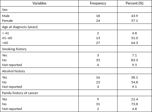
Table 2. Distribution of presenting symptoms and comorbidities (N = 42)
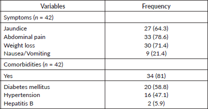
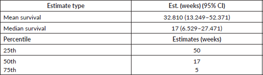
Table 3. Mean duration of survival (weeks)

Table 4. Overall survival estimates (Kaplan–Meier).
Among those who received multiple modalities of treatment, palliative care was the predominant approach, accounting for 16 patients (94.1%). Only 3 patients (17.6%) received chemotherapy, and 1 patient (5.9%) underwent surgical intervention.
The survival analysis in terms of weeks showed poor outcomes among patients with pancreatic cancer in this cohort. Overall survival was assessed among patients with pancreatic cancer and followed for up to approximately 60 weeks. Survival declined early after diagnosis, with cumulative survival falling to about 85%–90% within the first 4–6 weeks, indicating early death events. By around 8–10 weeks, survival decreased further to approximately 65%. At about 17 weeks, survival dropped to 50%, indicating that half of the patients had died by this time. After this point, survival declined more gradually, with a period of relative stability observed between 20 and 45 weeks. Beyond 50 weeks, survival declined again, reaching approximately 10% by the end of follow-up, with no additional deaths recorded thereafter. Median overall survival was 17 weeks, indicating that half of the patients died within approximately 4 months of diagnosis. However, survival varied widely, with 25% of patients dying within about 5 weeks, while a smaller proportion survived for up to 50 weeks, resulting in a higher mean survival estimate (Tables 3 and 4; Figure 1).
Survival differed by disease stage, with earlier stages demonstrating longer survival and advanced disease associated with rapid mortality. Patients diagnosed with Stage II disease showed the most favourable survival pattern. The median survival was 57 weeks, indicating that at least half of these patients survived for more than 1 year following diagnosis. Notably, the 25th percentile survival was also 57 weeks, suggesting that early mortality was uncommon in this group. However, by the 75th percentile, survival declined to 13 weeks, reflecting heterogeneity in outcomes and possible late deaths among a subset of patients (Table 5; Figure 2).
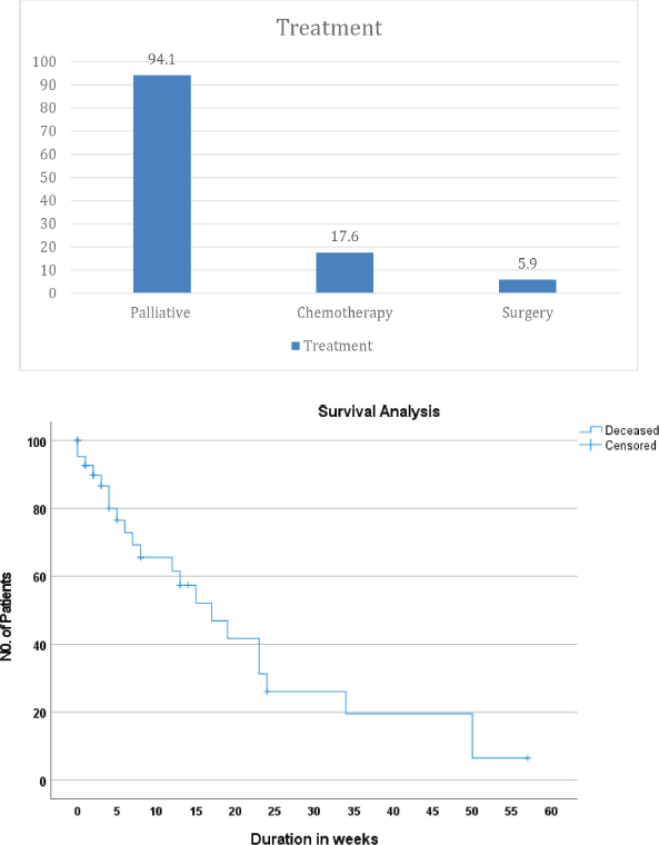
Figure 1. Distribution of treatment modalities and Kaplan–Meier overall survival curve (N = 42).
In contrast, patients with Stage III disease experienced substantially poorer survival. The median survival was 8 weeks, with 25% of patients dying within 17 weeks and 75% dying within 6 weeks, indicating both early and rapid mortality following diagnosis.
Table 5. Survival estimates by disease stage (percentile analysis).

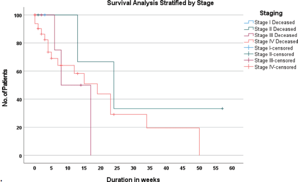
Figure 2: Kaplan–Meier survival curve stratified by disease stage
Patients with Stage IV disease demonstrated the worst outcomes overall. Median survival was 19 weeks, but early mortality was prominent, with 25% of patients dying within 4 weeks of diagnosis. Although a subset of patients survived longer (25th percentile at 50 weeks), the survival curve declined steeply, underscoring the aggressive nature of metastatic disease.
When all stages were considered together, the overall median survival was 17 weeks, with 25% of patients dying within 5 weeks and 75% dying within 50 weeks, highlighting wide variability in survival but an overall predominance of short survival durations.
Survival analysis stratified by sex revealed differences in survival patterns between male and female patients. Kaplan–Meier curves showed that female patients maintained higher cumulative survival probabilities over time, while male patients experienced a steeper and earlier decline in survival, indicating poorer outcomes among males.
Median survival time was 17.0 weeks (95% CI: 2.9–31.0 weeks) for male patients, compared to 23 weeks (95% CI: 0.3–45.7 weeks) for female patients. Similarly, the estimated mean survival time was substantially longer in females (36.5 weeks) than in males (12.8 weeks) (Table 6; Figure 3).
Overall, while female patients appeared to have a survival advantage over male patients in this cohort, sex did not emerge as a statistically significant determinant of survival.
Table 6. Survival estimates by sex.

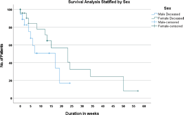
Figure 3: Kaplan–Meier survival curve stratified by sex
Table 7. Survival estimates by age group.

Overall survival was assessed across three age groups: ≤40 years, 41–60 years and >60 years. Patients aged ≤40 years had an average survival of 34 weeks. Because very few deaths occurred in this group, survival estimates remained largely unchanged over time, and the results should be interpreted cautiously. Among patients aged 41–60 years, survival declined more quickly after diagnosis. The average survival was 22.7 weeks, and half of the patients in this age group died by 8 weeks, indicating relatively early mortality. Patients aged >60 years had an average survival of 36.8 weeks, with a median survival of 19 weeks. Although survival declined steadily, some patients in this group lived beyond 40–50 weeks after diagnosis (Table 7; Figure 4).
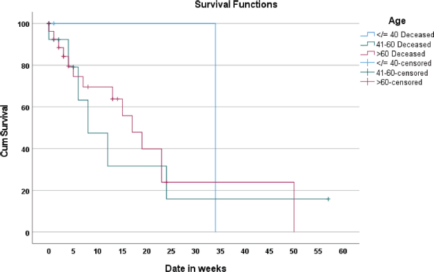
Figure 4. Kaplan–Meier survival curve stratified by age group.
Discussion
This retrospective cohort of 42 patients provides the first long-term report on pancreatic cancer from a private oncology centre in Lagos. Patients were predominantly female and older than 60 years, and most had at least one metabolic comorbidity (hypertension or diabetes) at diagnosis. Classical risk factors such as tobacco smoking were rare, whereas a family history of cancer was documented in one-fifth of cases. Presenting complaints were dominated by nonspecific gastrointestinal symptoms, abdominal pain, weight loss and jaundice, with very few patients reporting nausea or vomiting. Three-quarters of the cohort (77%) presented with stage IV disease, and only one patient (2%) presented with stage I disease, reflecting overwhelming late presentation. The one patient, diagnosed with Stage I pancreatic cancer at presentation, reflects an incidental finding. She was a 74-year-old woman who came to the hospital after having symptoms for only about 3 weeks. Her main symptom was jaundice, along with weight loss, tiredness and mild chest discomfort. Because the jaundice appeared early, doctors arranged imaging tests quickly. A CT scan showed a small tumour in the head of the pancreas (about 1.8 × 1.2 cm) that was blocking the bile duct but had not spread to nearby blood vessels, lymph nodes or other organs. The pancreatic duct was normal, and no other tumours were seen. This early evaluation allowed the cancer to be diagnosed before it had spread, explaining why this patient was identified at Stage I, unlike most other patients in the study who presented with advanced disease.
Symptom type did not discriminate stage, sex, or age in this small series. Survival analysis demonstrated a median overall survival of 19 weeks, with 25% of patients dying within 5 weeks and a small subset surviving almost 1 year; stage-stratified curves showed a survival gradient favouring stage II disease but were not significant because of sparse early-stage cases. Mean survival was longer for women than men (36.5 versus 12.8 weeks), but this difference was not statistically significant. Age-stratified survival curves overlapped substantially, indicating that chronological age did not determine prognosis in a cohort dominated by advanced disease.
The predominance of stage IV disease mirrors reports from public tertiary hospitals in Nigeria and across Sub-Saharan Africa. A study shows that of 178 patients treated between 2013 and 2021 in southeastern Nigeria noted that 87.1% of patients presented more than 6 months after symptom onset and that financial constraints and late referrals were major contributors to diagnostic delays [9]. A multicentre study of gastrointestinal cancers in southwestern Nigeria found that approximately 90% of tumours were diagnosed at advanced stage (III–IV) [11]. Similarly, historical reviews from public hospitals reported uniformly late-stage disease: a 96 patient series from Obafemi Awolowo University Teaching Hospital concluded that most patients were presented with advanced pancreatic cancer and underwent only palliative bypass surgery, while a 126 patient study from Ibadan (1999–2013) found a mean age of 60 years and noted that disease was “increasingly diagnosed in young adults at an advanced stage” [12]. These patterns are not confined to Nigeria; a Kenyan tertiary hospital series reported that >54% of patients had metastatic disease at presentation and a Moroccan hospital reported that only 19.8% of pancreatic cancers were diagnosed at a resectable stage [8,13]. Compared to these reports, the present Lakeshore cohort shows an even higher proportion of stage IV cases (73%), emphasising that private-sector access does not necessarily translate into earlier diagnosis.
The low prevalence of smoking (6.8%) and moderate alcohol use (36.4%) in the Lakeshore cohort contrasts with higher smoking and alcohol rates reported in North African and East African studies; 22.8% smokers and 12.7% drinkers were noted in the Moroccan series [13]. Comorbidity patterns in the present study, particularly the high frequency of diabetes (58.8 %) and hypertension (47.1 %) are consistent with reports from Morocco, where diabetes was present in 38% of patients, and support growing evidence that metabolic disease may be an important driver of pancreatic cancer in African populations [13]. Documentation gaps in smoking and alcohol history, however, limit direct comparisons. The absence of peptic ulcer disease in our cohort suggests that gastro-duodenal ulceration may not be a common comorbid condition in Nigerian pancreatic cancer patients.
Unlike earlier Nigerian studies conducted in public tertiary hospitals, the current series comes from a private cancer centre. Nevertheless, the patient population reflected socioeconomic barriers similar to those documented in public settings. In a cross-sectional survey of cancer care in Nigeria and Romania, it was observed that the Nigerian government spends only about 3% of its gross domestic product on health and that 64%–90% of Nigerian cancer patients pay for care entirely out of pocket [14]. The same survey reported that patients receiving care in private facilities perceived better service but still experienced communication gaps and cost barriers. Similarly, a retrospective review of 548 patients at Lakeshore Cancer Center found that 90% of patients paid out of pocket for their healthcare and 67% did not complete treatment [15]. These findings echo national assessments of Nigeria’s cancer system: Ogunniyi et al [19]. report that the country has only 27 cancer treatment centres for >200 million people and that most cancer patients pay for chemotherapy entirely themselves because the National Health Insurance Scheme does not cover cancer care [11]. In contrast, the Moroccan study was conducted in a public university hospital; although a fifth of patients had resectable disease, delays in diagnosis were attributed to resource constraints and affordability [13]. Collectively, these data suggest that the private sector setting does not obviate upstream barriers such as late referral, lack of insurance coverage and unaffordable diagnostics.
Survival outcomes in the present cohort are markedly worse than those reported in high-income countries. Analyses from high-income jurisdictions indicate that 1-year net survival after pancreatic cancer ranges from 21.1% to 30.9% and 3-year net survival from 6.6% to 10.9%, with more than half of patients in these countries still diagnosed at a distant stage [16]. Cancer Research UK reports that, for localised pancreatic cancer, about 55% of patients survive at least 1 year, and >25% survive 3 years; for regional disease, these figures are approximately 50% and 15%, while for metastatic disease only 10% survive 1 year and 1% survive 3 years (Cancer Research). The ICBP SURVMARK 2 study similarly found that 53.9%–83.3% of pancreatic cancer cases in high-income countries are diagnosed with distant disease [16]. Our median survival of 19 weeks (≈4.5 months) is substantially shorter than these benchmarks, underscoring the severe survival disadvantage faced by Nigerian patients. Even stage-specific comparisons are unfavourable: global data indicate 5-year survival rates of 32% for localised disease, 12% for stage III and 3% for stage IV; in contrast, the stage II subgroup in our cohort had a median survival of 57 weeks and the stage IV subgroup had a median of 19 weeks with a 25th percentile of 4 weeks and 75th percentile of 50 weeks [17]. Survival rates in Asia are similarly higher: a meta-analysis of 39 studies reported a pooled 1-year survival of 27.6% and a 5-year survival of 9.7%, both considerably higher than the outcomes observed in Lagos [18].
The pervasive late presentation observed in both private and public Nigerian series is likely multifactorial. First, pancreatic cancer often manifests with vague abdominal symptoms that are easily attributed to benign conditions, leading to protracted health-seeking delays. Bharadwaj
et al [10]. noted that >87% of Nigerian patients presented after 6 months of symptoms and identified financial constraints and late referral as key drivers of delay. Second, limited diagnostic capacity and long waiting times exacerbate delays; Nigeria has only 27 cancer treatment facilities and each radiotherapy machine serves far more patients than its designed capacity [19]. Third, financial barriers impede timely evaluation and treatment. More than 64%–90% of Nigerian cancer patients pay entirely out of pocket for care, and even at the Lakeshore centre, 90% of patients self-funded treatment [15,14]. Out-of-pocket spending remains high because cancer care is not covered by the national health insurance scheme [19]. Fourth, Nigeria faces a critical shortage of specialists; there are only about 110 practising gastrointestinal surgeons for a population of more than 200 million [10]. In contrast, high-income countries benefit from organised screening, timely access to imaging, universal health coverage and multidisciplinary care pathways, facilitating earlier detection and improved survival [16] Cancer Research).
There may also be biological and socioeconomic factors at play. The high prevalence of diabetes and hypertension in this cohort aligns with the metabolic syndrome hypothesis. It may reflect different etiological pathways compared to smoking-driven pancreatic cancer in Western populations. The absence of stage sex and stage age associations suggests that once disease is established, tumour biology rather than host factors predominates. The female predominance and slightly longer survival among women warrant further investigation but could reflect selection bias in a private facility where women may be more likely to seek care.
These findings have several implications. First, they underscore the urgency of implementing public awareness campaigns to promote earlier presentation of abdominal and systemic symptoms suggestive of pancreatic malignancy. Second, expanding access to diagnostic imaging and specialist evaluation, through public–private partnerships, mobile diagnostic units and subsidised imaging, could shorten referral times. Third, national health insurance reform is imperative. A cross-sectional survey found that only 3% of Nigeria’s GDP is allocated to healthcare and that most patients pay out of pocket [14]. Countries with similar per capita income but broader insurance coverage (e.g. Kenya) report much lower out-of-pocket expenditure [19]. Innovative financing mechanisms, such as the Cancer Health Fund and chemotherapy access partnerships, are promising but require expansion [19]. Fourth, workforce development and training to increase the number of gastrointestinal surgeons, oncologists and endoscopists are critical to reduce diagnostic delays [9]. Fifth, private centres should strengthen linkages with primary care and public hospitals to facilitate early referral and ensure continuity of care; the high proportion of patients who did not complete treatment at Lakeshore (67%) indicates that financial and logistical barriers persist even after diagnosis [15].
Strengths and limitations
The principal strength of this study is that it provides the first longitudinal analysis of pancreatic cancer from a Nigerian private cancer centre, complementing numerous reports from public hospitals. Its detailed description of symptom patterns, stage distribution and survival outcomes fills a gap in the literature on private-sector oncology in Sub-Saharan Africa. The use of Kaplan–Meier methods and stage-stratified percentiles provides a nuanced view of survival heterogeneity. However, several limitations warrant caution. The retrospective design relies on medical records that lacked complete data on risk factors and comorbidities; metabolic and lifestyle histories may therefore be under-reported. The sample size (n = 44) is small, limiting statistical power to detect associations and resulting in wide confidence intervals for stage-specific survival estimates. Differences in patient selection constrain comparison with public hospitals; private centres may serve a wealthier and more health-seeking population, yet late presentation persisted. Survival analysis did not adjust for treatment modality or performance status, which may confound observed differences. Finally, because follow-up was incomplete for some patients, median survival estimates may still overestimate actual survival due to censoring.
Acknowledgments
The authors acknowledge the staff of Lakeshore Cancer Center for their support in data collection.
Conflicts of interest
The authors declare no competing interests.
Authors’ contributions
All authors contributed to the design of the study, data collection, analysis and manuscript preparation. All authors read and approved the final version of the manuscript.
Funding
This study received no external funding.
References
1. Mizrahi JD, Surana R, and Valle JW, et al (2020) Pancreatic cancer Lancet 395(10242) 2008–2020 https://doi.org/10.1016/S0140-6736(20)30974-0
2. Siegel RL, Giaquinto AN, and Jemal A (2024) Cancer statistics, 2024 CA A Cancer J For Clinicians 74(1) 12–49 https://doi.org/10.3322/caac.21820
3. Park W, Chawla A, and O’Reilly EM (2021) Pancreatic cancer: a review JAMA 326(9) 851–862 https://doi.org/10.1001/jama.2021.13027
4. Halbrook CJ, Lyssiotis CA, and Pasca di Magliano M, et al (2023) Pancreatic cancer: advances and challenges Cell 186(8) 1729–1754 [https://doi.org/10.1016/j.cell.2023.02.014]
5. Hu JX, Zhao CF, and Chen WB, et al (2021) Pancreatic cancer: a review of epidemiology, trend, and risk factors World J Gastroenterol 27(27) 4298–4321 https://doi.org/10.3748/wjg.v27.i27.4298 PMID: 34366606 PMCID: 8316912
6. Ponz De Leon Pisani R, Altieri G, and Stasio RC, et al (2025) Gastrointestinal symptoms in the journey of pancreatic cancer patients Expert Rev Anticancer Therapy https://doi.org/10.1080/14737140.2025.2517888
7. Ogbuanya AU, Ugwu NB, and Nwigwe GC, et al (2023) Profile of pancreatic cancers and management characteristics at major hospitals in Southeast Nigeria East Afr Med J 100(11) 6445–6455
8. Muchiri SW, Ogutu EO, and Oyiro PO, et al (2024) Clinicopathologic characteristics and treatment outcomes of pancreatic cancer patients at a tertiary referral hospital in Kenya Ecancermedicalscience 18 1682 https://doi.org/10.3332/ecancer.2024.1682 PMID: 38566769 PMCID: 10984841
9. Bharadwaj HR, Bone M, and Dahiya DS, et al (2025) The current landscape of pancreatic cancer management in Sub-Saharan Africa – a perspective review Health Sci Rep 8(10) e71330 https://doi.org/10.1002/hsr2.71330 PMID: 41064197 PMCID: 12500524
10. Nhleko ML, Edoka I, and Musenge E (2024) Pancreatic cancer mortality in South Africa: a case-control study South Afr Med J 114(1) 27–32 https://doi.org/10.7196/SAMJ.2024.v114i1.1132
11. Onwuka JU, Wuraola FO, and Owoade IA, et al (2024) Delays in presentation, diagnosis, and treatment among gastrointestinal cancer patients in southwest Nigeria JCO Global Oncol 10 e2400060 https://doi.org/10.1200/GO.24.00060
12. Alegbeleye BJ, Afolabi AO, and Alegbeleye BJ (2019) Clinicopathologic pattern and outcome of management of pancreatic carcinoma in Ibadan, Nigeria Sudan J Med Sci 14(4) 172–187 https://doi.org/10.18502/sjms.v14i4.5898
13. Amri F, Koulali H, and Jabi R, et al (2023) Pancreatic cancer: experience from an emerging country in North Africa J Cancer Res Clin Oncol 149(15) 14297–14302 https://doi.org/10.1007/s00432-023-05245-5 PMID: 37561209 PMCID: 11796776
14. Ago BU, Olaitan JO, and Stefan DC, et al (2023) Cancer care in Nigeria and Romania: a cross-sectional survey Ecancermedicalscience 17 1538
15. Fapohunda A, Farouk S, and Ige O, et al (2020) Out-of-pocket expenditure and treatment non-completion at Lakeshore Cancer Center Ecancermedicalscience 14 1105
16. Arnold M, Rutherford MJ, and Bardot A, et al (2019) Progress in cancer survival, mortality, and incidence in seven high-income countries 1995–2014 (ICBP SURVMARK-2) Lancet Oncol 20(11) 1493–1505
17. Rawla P, Sunkara T, and Gaduputi V (2019) Epidemiology of pancreatic cancer: global trends, etiology and risk factors World J Oncol 10(1) 10–27 [https://doi.org/10.14740/wjon1166]
18. Ghaem H, Safari H, and Jahromi HK, et al (2025) Survival rate of pancreatic cancer in Asian countries: A systematic review and meta-analysis Medicine 104(41) e45104
19. Ogunniyi TJ, Fatokun BS, and Isah KO, et al (2025) Current status of cancer diagnosis and treatment in Nigeria Health Sci Rep 8(6) e7087