
Supporting oncology professionals through education
The content on this site is intended for healthcare professionals only
The content on this site is intended for healthcare professionals only
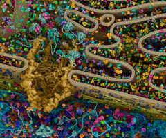
A review in Genes & Diseases explores how a system designed to restore protein balance can influence an inflammatory form of cell death.
The article connects endoplasmic reticulum stress with necroptosis, highlighting potential treatment targets across conditions affecting the heart, kidneys, digestive tract, bones and other tissues, as well as cancer.
Prepared by a team at Hainan Medical University and its First Affiliated Hospital, the review examines what happens when the endoplasmic reticulum, a cellular compartment responsible for processing and folding proteins, becomes overwhelmed.
Accumulating misfolded proteins activate the unfolded protein response, a protective mechanism that attempts to restore function.
When stress is manageable, this response supports survival. If stress persists or becomes excessive, however, its signals can contribute to cell death.
Understanding that transition could help explain how an initially protective process becomes linked to inflammation and tissue damage.
The review focuses on three signalling branches, PERK, IRE1α and ATF6, that connect protein stress with downstream changes in cell behaviour.
These pathways can influence necroptosis, a regulated process in which cells lose membrane integrity and release their contents, potentially triggering inflammatory responses in surrounding tissue.
The classical necroptosis machinery involves RIPK1, RIPK3 and MLKL. Activation of this molecular sequence ultimately enables MLKL to disrupt the cell membrane.
The article examines how stress-response pathways may interact with this machinery, while emphasising that several connecting steps remain unresolved.
These interactions have different implications depending on the disease. Limiting excessive necroptosis could help protect vulnerable tissues from inflammatory injury.
In cancer, encouraging necroptosis in tumour cells may offer a way to overcome resistance to other forms of cell death.
Effective treatment would therefore require careful control of where and when these pathways are altered.
Potential approaches include compounds that adjust endoplasmic reticulum stress or selectively influence signalling branches.
The review also discusses delivery technologies, including extracellular vesicles, as possible ways to improve how such agents reach their targets.
Uncertainties remain, particularly around IRE1α and ATF6. It is not yet clear exactly how each branch controls RIPK1, RIPK3 and MLKL, or whether alternative necroptosis pathways are involved.
Evidence also varies between organs and disease settings, limiting broad conclusions about therapeutic benefit.
Source: Compuscript Ltd
We are an independent charity and are not backed by a large company or society. We raise every penny ourselves to improve the standards of cancer care through education. You can help us continue our work to address inequalities in cancer care by making a donation.
Any donation, however small, contributes directly towards the costs of creating and sharing free oncology education.
Together we can get better outcomes for patients by tackling global inequalities in access to the results of cancer research.
Thank you for your support.