A rare case of clear cell sarcoma of the foot with a cascade of pathological misdiagnosis—the importance of expert sarcoma pathology
Anand Rajendran1, Gaurav Gupta1, Adarsh Barwad2 and Sameer Rastogi3
1Department of Medicine, All India Institute of Medical Sciences, New Delhi 110029, India
2Department of Pathology, All India Institute of Medical Sciences, New Delhi 110029, India
3Department of Medical Oncology, All India Institute of Medical Sciences, New Delhi 110029, India
Abstract
Sarcoma pathology discrepancy is well known owing to the extremely heterogenous and rare nature of this tumour. Through this case, we want to highlight the difficulty that a patient has to undergo in a case of misdiagnosis. A 20-year-old male presented with swelling in the right foot for 4 months, which was initially diagnosed as alveolar rhabdomyosarcoma, subsequently as synovial sarcoma and finally as Ewing’s sarcoma (based upon positive Ewing Sarcoma Breakpoint Region 1 (EWSR1) by fluorescence in situ hybridisation and he underwent neoadjuvant chemotherapy and surgical excision with grafting before he presented to our institute, where the pathologists reviewed the biopsy slides, which were positive for HMB45 and negative for Melan-A suggestive of clear cell sarcoma. The next-generation sequencing suggested EWSR1-ATF1 fusion, which again reinforced the diagnosis. This case throws light on the importance of expert pathology and interpreting molecular results in the right context.
Keywords: sarcoma, clear cell sarcoma, misdiagnosis, EWSR1, diagnostic discrepancy
Correspondence to: Sameer Rastogi
Email: Samdoc_mamc@yahoo.com
Published: 31/10/2022
Received: 17/05/2022
Publication costs for this article were supported by ecancer (UK Charity number 1176307).
Copyright: © the authors; licensee ecancermedicalscience. This is an Open Access article distributed under the terms of the Creative Commons Attribution License (http://creativecommons.org/licenses/by/4.0), which permits unrestricted use, distribution, and reproduction in any medium, provided the original work is properly cited.
Introduction
Soft tissue sarcomas (STSs) are a group of remarkably diverse neoplasms that frequently pose significant diagnostic challenges to surgical pathologists [1]. Pathological discrepancy between various institutes has been reported from 25% to 50% across the globe [2–6]. Molecular testing is now becoming an integral part of diagnosis of sarcomas and thus influencing the management. In a prospective multi-centre observational study (GENSARC) which included 384 patients with sarcoma, diagnosis was modified eventually by molecular methods in 53 patients with the maximum percentage among dedifferentiated liposarcoma patient cohort (23%) [7].
Clear cell sarcoma (CCS) is a rare tumour first described in 1968 and also referred to as malignant melanoma of soft parts [8]. It has characteristics of both STS and malignant melanoma with a predilection for the deep soft tissues of lower extremity and typically affects young adults [9]. This tumour is characterised by the translocation t(12;22)(q13;q12) and consequent fusion of the Ewing Sarcoma Breakpoint Region 1 (EWSR1) and ATF1 genes which is not documented in malignant melanoma [9] and its detection using advanced molecular techniques helps in the confirmation of a diagnosis of CCS. The literature is rife with studies in which CCS has been misdiagnosed as melanoma, metastatic carcinoma, fibrosarcoma, etc. [10–13].
A thorough understanding about the molecular abnormalities of STSs is necessary to avoid misdiagnosis of a tumour, as there are multiple tumours which behave differently with similar morphology and immunopositivity. This case where the diagnosis was altered thrice before coming to the final diagnosis emphasises the need for timely referral to a sarcoma specialist to avoid misdiagnosis and mismanagement.
Case
A 20-year-old male from Kathmandu, Nepal, developed pain on the undersurface of his right foot since January 2021, which was exaggerated by walking. He also noticed a swelling which was better felt than seen (Figure 1) at the site where he was feeling pain and after 3 months since onset of symptoms, an ultrasound of the right foot was done, which showed a suspicious mass under the skin of the sole and an MRI was advised for further characterisation.
An MRI was done (Figure 2) on the 24th of April, which showed a heterogeneously enhancing lobulated mass of size 4.5 × 7.5 × 3.8 cm with occasional non-enhancing areas suggestive of necrosis, involving both subcutaneous tissue and overlying muscles bones and tendons.
The patient underwent a trucut biopsy of the tumour and on histopathological examination, sheets of malignant, round to polygonal cells with intervening fibrous septa were seen with foci of necrosis. The overall impression in the initial report was alveolar rhabdomyosarcoma. Immunostains were not used at this point of time. The patient was administered neo-adjuvant chemotherapy with two cycles of vincristine, cyclophosphamide and dactinomycin on the 12th of May and the 3rd of June 2021 following which, a repeat MRI of foot was done on the 21st of June, which showed no interval changes compared to the previous MRI. Histopathology was again reviewed in another Centre and immunohistochemistry (IHC) markers were done, and the cells were positive for CD99 and TLE-1 and negative for CK, Desmin, S100, CD45, Myo-D1, TdT, NKX2.2 and synaptophysin. The report was subsequently reviewed and a diagnosis of monophasic synovial sarcoma was made. He further underwent wide local excision of the tumour and Ray’s amputation of the 4th and 5th toes of right foot was done with skin and soft tissue grafting on the 27th of June.
In the biopsy specimen, a break-apart fluorescence in situ hybridisation (FISH) for EWSR1 and SS18 rearrangement was done which detected EWSR1 rearrangement (Figures 3 and 4) and hence the diagnosis was changed from synovial sarcoma to Ewing’s sarcoma/PNET.
At our institute, the Pathologists reviewed the slides of the resected specimen. The morphology of the tumour showed oval to spindle shaped cells arranged in nests as well as syncytial pattern. The tumour cells showed moderate pleomorphism with vesicular chromatin and prominent nucleoli with moderate to abundant cytoplasm with epithelioid morphology at places (Figure 5). The tumour cells were immunopositive for HMB45 (Figure 6) and immunonegative for Melan A. Therefore, the EWSR1 rearrangement detected earlier was related to CCS rather than Ewing’s sarcoma/PNET.
We further sent the specimen for next-generation sequencing analysis, which detected EWSR1::ATF gene fusion, which confirmed the diagnosis of CCS. Tumour tissue was analysed using semiconductor-based next-generation sequencing technology (Platform – Thermofisher Ion S5; Mapped fusion reads > 20,000; Median read count 120). High-quality tumour tissue RNA extracted from the submitted specimen was subjected to target enrichment by multiplex polymerase chain reaction (PCR) amplification by Sarcoma Fusion panel. Sequenced data was analysed using a customised in-house pipeline DCGL NGS Bioinformatics v4.2. An FDG PET performed on the 17th of July showed no other focus of disease. Hence, the patient was advised to undergo radiotherapy as there is no role for chemotherapy in CCS in the adjuvant setting. In view of delay in flap healing, radiation therapy was delayed and repeat PET scan after 3 months showed metastatic disease to pelvic lymph nodes and lung metastasis. After which he was started on tab pazopanib but died after 2 months.
Discussion
The above case demonstrates the importance of correctly diagnosing soft tissue tumours in the early phase of presentation itself. CCS has slight female preponderance and is found in 15–35 years age group [12, 14], median age being third decade, similar to our patient. It is most commonly found in the extremities and has a propensity of lymph node metastasis [14].
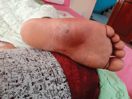
Figure 1. Photograph of the swelling on the plantar aspect of right foot, taken before chemotherapy, 3 months after the onset.
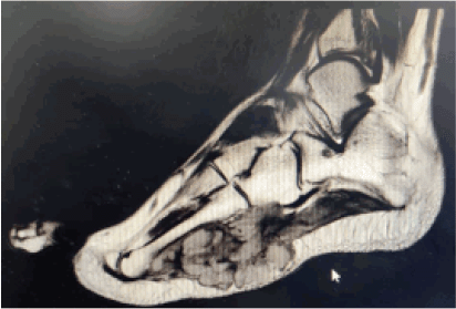
Figure 2. MRI image of right foot (T1W post contrast). Lobulated soft tissue mass with poorly defined margins in the lateral plantar aspect of right foot measuring approx. 4.5 × 7.5 × 3.8 cm (CC × AP × TR). Non- enhancing regions likely representing cystic/necrotic changes. Insinuating between the 4th and 5th metatarsals and involving the intrinsic muscles and abutting the lateral aspect of flexor digitorum longus/brevis muscles and tendons. Also involving the subcutaneous tissue on the plantar aspect of the foot. No definite evidence of signal abnormality or destructive changes in the bones.
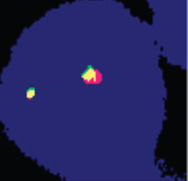
Figure 3. Cell showing two green-orange (fusion) signals indicating negative for SYT gene rearrangement. Test method: FISH. Probe description: Vysis LSI EWSR1 (22q12) Break Apart Rearrangement Probe (CE).
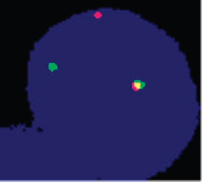
Figure 4. Cell showing one green-orange (yellow) fusion, one orange signal and one green signal, indicating positive for rearrangement of the EWSR1 gene. Test method: FISH. Probe description: Vysis LSI EWSR1 (22q12) Break Apart Rearrangement Probe (CE).
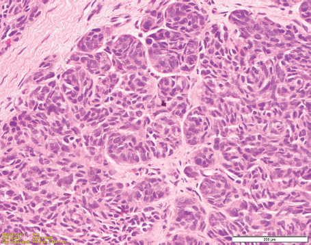
Figure 5. Histopathology photomicrograph of the tumour showing oval to spindle shaped cells arranged in nests as well as syncytial pattern. The tumour cells showed moderate pleomorphism with vesicular chromatin and prominent nucleoli with moderate to abundant cytoplasm with epithelioid morphology at places. (H&E 200×).
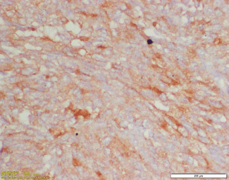
Figure 6. Immunohistochemistry for HMB45 showing diffuse cytoplasmic positivity in the tumour cells.
This case also highlights the pitfalls associated with a delay in referral to a sarcoma speciality clinic in the event of a diagnostic dilemma associated with a STS. Delay in correct diagnosis leads to mistreatment, larger tumour burden, functionally debilitating surgeries and significant distress in the patient which might last longer than treatment duration [15–20]. This study reinforces the need of sarcoma pathologists in developing countries and need for pathology second opinion early in the course of treatment. As per UK guidelines, sarcoma should be reported by sarcoma pathologist who sees significant number of sarcoma in daily practice [21].
A study by Thway et al [22] showed that there were 16.4% major discrepancies (those that could lead to significant change in clinical management) and 11.8% minor discrepancies (those in which the discrepancy was not thought to provoke significant management change) in diagnosis of soft tissue neoplasm after referring to a tertiary sarcoma referral unitClick or tap here to enter text.. The major cause of discrepancies appears attributable to differences in interpretation by the referral and tertiary centre pathologist rather than a lack of or inappropriate use of IHC tests at the referring Centre [22]. This justifies the need for specialist referral for soft tissue tumours in a country like Nepal where a discrepancy can be expected to be higher because of both inexperience and lack of facilities.
EWSR1 gene, located at chromosome22q12, encodes a 656-amino acid nuclear protein that is thought to have roles in meiotic cell division, mitotic spindle formation and stabilisation of microtubules, as well as DNA repair mechanisms and cellular ageing. EWSR1 is a member of TET family of genes which has an RNA binding domain at the carboxy terminus of the gene product which is important in protein-RNA binding, transcription and RNA metabolism [23–25]. EWSR1 has been identified as a translocation partner in a wide range of clinically and pathologically diverse tumours which include the Ewing family of tumours, desmoplastic small round cell tumour, myxoid liposarcomas, extra-skeletal myxoid chondrosarcoma, angiomatoid fibrous histiocytoma, CCS of soft tissue and clear cell sarcoma-like tumours of the gastrointestinal tract, primary pulmonary myxoid sarcoma, myoepithelial tumours of skin, soft tissue and bone and rare examples of low-grade fibro-myxoid sarcoma, sclerosing epithelioid fibrosarcoma and mesothelioma [25].
The errors that accompanied this patient were inappropriate chemotherapy, as CCS is chemo resistant [26, 27] and the patient could have undergone sentinel node biopsy if the diagnosis was clear earlier [28, 29].
The sensitivity to chemotherapy and immunotherapy in this subset has rarely been demonstrated [27, 30]. However, early diagnosis, surgery, sentinel node biopsy and radiation when required are the current treatment of choice for localised CCS [27].
Our case depicts that the molecular markers should be interpreted in the context of meticulous histopathological examination. Also, in Low-middle income countries (LMICs), a well-structured framework is direly needed to improve sarcoma care in the absence of expert pathology, radiology, dedicated sarcoma surgeons and sarcoma medical oncology. The development of pathology services in LMICs faces a number of challenges, including insufficient funds, lack of skilled personnel at all levels (technicians to pathologists), unavailable equipment and unreliable supply chains for consumables [31]. Patients with sarcoma should be referred to tertiary care centres with established sarcoma teams and patient advocacy should be encouraged to educate and empower patients. This also calls for a second opinion for sarcoma pathology, which should be a well-established dogma, unlike other common cancers.
This case shows how a misdiagnosis can change the entire course of the disease rather appallingly. Discrepancy of pathological diagnosis is a well-established entity as far as STSs are concerned. This case adds to the existing knowledge and given the dire need of expertise in pathology in sarcoma, this needs to be re-emphasised with more cases.
Conclusion
This case reveals that expert pathology and appropriate molecular tests for rare sarcomas are essential for appropriate management, along with a second pathology opinion for sarcomas at an expert centre, which should be the norm rather than exception.
Conflicts of interest
No conflicts of interest.
Funding
We did not receive any funding for this manuscript and have no financial interests.
References
1. Lauer S and Gardner JM (2013) Soft tissue sarcomas-new approaches to diagnosis and classification Curr Probl Cancer 37 45–61 https://doi.org/10.1016/j.currproblcancer.2013.03.001 PMID: 23719330
2. Thway K and Fisher C (2009) Histopathological diagnostic discrepancies in soft tissue tumours referred to a specialist centre Sarcoma 2009 741975 https://doi.org/10.1155/2009/741975 PMID: 19503800 PMCID: 2688650
3. Presant CA, Russell WO, and Alexander RW, et al (1986) Soft-tissue and bone sarcoma histopathology peer review: the frequency of disagreement in diagnosis and the need for second pathology opinions. The southeastern cancer study group experience J Clin Oncol 4 1658–1661 https://doi.org/10.1200/JCO.1986.4.11.1658 PMID: 3772418
4. Harris M, Hartley AL, and Blair V, et al (1991) Sarcomas in North West England: I. Histopathological peer review Br J Cancer 64 315–320 https://doi.org/10.1038/bjc.1991.298 PMID: 1892759 PMCID: 1977501
5. Arbiser ZK, Folpe AL, and Weiss SW (2001) Consultative (expert) second opinions in soft tissue pathology. Analysis of problem-prone diagnostic situations Am J Clin Pathol 116 473–476 https://doi.org/10.1309/425H-NW4W-XC9A-005H PMID: 11601130
6. Ray-Coquard I, Montesco MC, and Coindre JM, et al (2012) Sarcoma: concordance between initial diagnosis and centralized expert review in a population-based study within three European regions Ann Oncol 23 2442–2449 https://doi.org/10.1093/annonc/mdr610 PMID: 22331640 PMCID: 3425368
7. Italiano A, Di Mauro L, and Rapp J, et al (2016) Clinical effect of molecular methods in sarcoma diagnosis (GENSARC): a prospective, multicentre, observational study Lancet Oncol 2045 1–7
8. Dim DC, Cooley LD, and Miranda RN (2007) Clear cell sarcoma of tendons and aponeuroses a review Arch Pathol Lab Med 131(1) 152–156 https://doi.org/10.5858/2007-131-152-CCSOTA PMID: 17227118
9. Stenman G, Kindblom LG, and Angervall L (1992) Reciprocal translocation t(12;22)(q13;q13) in clear-cell sarcoma of tendons and aponeuroses Genes Chromosomes Can 4(2) 122–7 https://doi.org/10.1002/gcc.2870040204 PMID: 1373311
10. Jung SJ, Seung NR, and Park EJ, et al (2008) A case of clear cell sarcoma occurring on the abdomen Ann Dermatol 20 45–48 https://doi.org/10.5021/ad.2008.20.1.45 PMID: 27303159 PMCID: 4904049
11. Obiorah IE and Ozdemirli M (2019) Clear cell sarcoma in unusual sites mimicking metastatic melanoma World J Clin Oncol 10 213–221 https://doi.org/10.5306/wjco.v10.i5.213 PMID: 31205866 PMCID: 6556591
12. Juel J and Ibrahim RM (2017) A case of clear cell sarcoma – a rare malignancy Int J Surg Case Rep 36 151–154 https://doi.org/10.1016/j.ijscr.2017.05.034 PMCID: 5459563
13. Arora S, Khanna G, and Rajni, et al (2018) Difficulty in cytological diagnosis of clear cell sarcoma – a clinicopathological correlation J Clin Diagn Res 12 ER01–ER05
14. Chung EB and Enzinger FM (1983) Malignant melanoma of soft parts. A reassessment of clear cell sarcoma Am J Surg Pathol 7 405–413 https://doi.org/10.1097/00000478-198307000-00003 PMID: 6614306
15. Neal RD, Tharmanathan P, and France B, et al (2015) Is increased time to diagnosis and treatment in symptomatic cancer associated with poorer outcomes? Systematic review Br J Cancer 112(1) S92–107 https://doi.org/10.1038/bjc.2015.48 PMID: 25734382 PMCID: 4385982
16. Seinen J, Almquist M, and Styring A, et al (2010) Delays in the management of retroperitoneal sarcomas Sarcoma 2010 702573 https://doi.org/10.1155/2010/702573 PMID: 21048999 PMCID: 2964909
17. Bradford NK, Walker R, and Henney R, et al (2018) Improvements in clinical practice for fertility preservation among young cancer patients: results from bundled interventions J Adolesc Young Adult Oncol 7 37–45 https://doi.org/10.1089/jayao.2017.0042
18. Mesko NW, Mesko JL, and Gaffney LM, et al (2014) Medical malpractice and sarcoma care--a thirty-three year review of case resolutions, inciting factors, and at risk physician specialties surrounding a rare diagnosis J Surg Oncol 110 919–929 https://doi.org/10.1002/jso.23770 PMID: 25155556
19. Meis-kindblom JM (2006) Clear cell sarcoma of tendons and aponeuroses: a historical perspective and tribute to the man behind the ntity Adv Anat Pathol 13 286–292 https://doi.org/10.1097/01.pap.0000213052.92435.1f PMID: 17075294
20. Tang MH, Castle DJ, and Choong PFM (2015) Identifying the prevalence, trajectory, and determinants of psychological distress in extremity sarcoma Sarcoma 2015 745163 https://doi.org/10.1155/2015/745163 PMID: 25767410 PMCID: 4342175
21. Dangoor A, Seddon B, and Gerrand C, et al (2016) UK guidelines for the management of soft tissue sarcomas Clin Sarcoma Res 6 20 https://doi.org/10.1186/s13569-016-0060-4 PMID: 27891213 PMCID: 5109663
22. Thway K, Wang J, and Mubako T, et al (2014) Histopathological diagnostic discrepancies in soft tissue tumours referred to a specialist centre: reassessment in the era of ancillary molecular diagnosis Sarcoma 2014 686902 https://doi.org/10.1155/2014/686902
23. Plougastel B, Zucman J, and Peter M, et al (1993) Genomic structure of the EWS gene and its relationship to EWSR1, a site of tumor-associated chromosome translocation Genomics 18 609–615 https://doi.org/10.1016/S0888-7543(05)80363-5 PMID: 8307570
24. Bertolotti A, Lutz Y, and Heard DJ, et al (1996) hTAF(II)68, a novel RNA/ssDNA-binding protein with homology to the pro-oncoproteins TLS/FUS and EWS is associated with both TFIID and RNA polymerase II EMBO J 15 5022–5031 https://doi.org/10.1002/j.1460-2075.1996.tb00882.x PMID: 8890175 PMCID: 452240
25. Fisher C (2013) The diversity of soft tissue tumours with EWSR1 gene rearrangements: a review Histopathology 64(1) 134–150 https://doi.org/10.1111/his.12269 PMID: 24320889
26. Pasquali S and Gronchi A (2017) Neoadjuvant chemotherapy in soft tissue sarcomas: latest evidence and clinical implications Ther Adv Med Oncol 9 415–429 https://doi.org/10.1177/1758834017705588 PMID: 28607580 PMCID: 5455882
27. Czarnecka AM, Sobczuk P, and Zdzienicki M, et al (2019) Clear cell sarcoma Oncol Clin Pract 14 354–363 https://doi.org/10.5603/OCP.2018.0049
28. de Campos ECR, Adriano Júnior MG, and Winheski MR, et al (2020) Analysis of sentinel lymph node biopsy on clear cell sarcoma treatment J Cancer Ther 11 785–792 https://doi.org/10.4236/jct.2020.1112068
29. van Akkooi ACJ, Verhoef C, and van Geel AN, et al (2006) Sentinel node biopsy for clear cell sarcoma Eur J Surg Oncol 32 996–999 https://doi.org/10.1016/j.ejso.2006.03.044 PMID: 16672185
30. Jones R, Constantinidou A, and Thway K, et al (2010) Chemotherapy in clear cell sarcoma Med Oncol (Northwood, London, England) 28 859–863 https://doi.org/10.1007/s12032-010-9502-7
31. Patel K, Strother RM, and Ndiangui F, et al (2016) Development of immunohistochemistry services for cancer care in western Kenya: implications for low- and middle-income countries Afr J Lab Med 5(1) 187 https://doi.org/10.4102/ajlm.v5i1.187 PMID: 28879100 PMCID: 5436389


