
Supporting oncology professionals through education
The content on this site is intended for healthcare professionals only
The content on this site is intended for healthcare professionals only
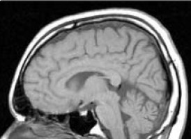
Researchers at the University of Cambridge have demonstrated that an advanced magnetic resonance imaging (MRI) technique can help predict the direction in which glioblastoma, the most aggressive form of primary brain cancer, progresses following surgery. The findings, published in the Journal of Neuro-Oncology, could help inform future approaches to more personalized brain tumor surgery.
Glioblastoma presents a major surgical challenge because cancer cells can infiltrate surrounding brain tissue beyond the tumor boundaries visible on conventional MRI. Even when surgeons completely remove the contrast-enhancing tumor, microscopic disease frequently remains, leading to subsequent progression.
Understanding where this progression is most likely to occur could help surgeons develop more anatomically targeted strategies while minimizing unnecessary removal of healthy brain tissue.
In the new multicenter study, researchers led by Dr. Jawad Fares, at the University of Cambridge, investigated whether preoperative imaging could anticipate the direction of tumor progression in patients with temporal lobe glioblastoma.
The team analyzed imaging and follow-up data from 32 patients who had undergone complete removal of their contrast-enhancing tumors. Three imaging biomarkers were compared: conventional fluid-attenuated inversion recovery (FLAIR), isotropic diffusion (DTI-p), and diffusion anisotropy (DTI-q).
Unlike conventional MRI approaches, DTI-q measures the anisotropic component of water diffusion, providing information about the underlying organization of brain white matter. These structures are particularly relevant because glioblastoma cells can infiltrate along white matter pathways.
DTI-q demonstrated the highest median direction-specific predictive accuracy at 75%, compared with 50% for DTI-p and 44% for FLAIR. It achieved higher classification accuracy across all six anatomical directions, with statistically significant improvements in selected directions.
The findings build on the Cambridge team's previous work investigating diffusion anisotropy as a biomarker of directional glioblastoma progression in the frontal lobe, extending the approach to the anatomically distinct temporal lobe.
Together, these studies contribute to an emerging research direction in neuro-oncology: using advanced imaging to better understand the spatial behavior of brain tumors and inform individualized treatment strategies.
The researchers emphasize that larger independent studies and prospective clinical evaluation are needed before diffusion anisotropy can be incorporated into routine surgical decision-making.
Source: University of Cambridge
We are an independent charity and are not backed by a large company or society. We raise every penny ourselves to improve the standards of cancer care through education. You can help us continue our work to address inequalities in cancer care by making a donation.
Any donation, however small, contributes directly towards the costs of creating and sharing free oncology education.
Together we can get better outcomes for patients by tackling global inequalities in access to the results of cancer research.
Thank you for your support.