Supporting oncology professionals through education
The content on this site is intended for healthcare professionals only
The content on this site is intended for healthcare professionals only
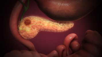
Pancreatic cancer remains one of the most difficult cancers to treat, in large part because tumours do not exist in isolation.
Instead, they are surrounded by a dense and complex network of blood vessels, connective tissue, and immune cells that shape how the disease grows and responds to therapy.
In a recent study led by Faraz Bishehsari, MD, PhD, professor and Atilla Ertan Chair in Gastroenterology Research, researchers developed a “tumour-on-a-chip” system designed to recreate that environment outside the human body, offering a more realistic way to study the disease and evaluate treatments.
“Our goal was to build a model that looks and behaves much more like a real pancreatic tumour than traditional lab models,” said Bishehsari, director of the Gastroenterology Research Centre at McGovern Medical School at UTHealth Houston.
“By recreating the tumour’s environment, we can better understand the disease and test treatments in a patient-specific way.”
Researchers began with pancreatic tumour and blood samples donated by consenting patients and used them to grow three-dimensional organoids that retain key features of the original cancer.
These organoids were then placed into a microfluidic chip alongside blood vessel, stromal, and immune cells to recreate the tumour environment.
Designed to mimic natural conditions, the chip allows fluid to flow like blood, enabling researchers to track how cancer cells interact with surrounding tissues and respond to treatment over time.
Using imaging, molecular analysis, and drug response testing, the team found that the chip-based model closely mirrored the behaviour of human pancreatic tumours.
Notably, the system reproduced interactions between cancer cells and the surrounding scar-like tissue, known as desmoplastic stroma, which is a key factor in why many treatments fail.
“We were able to monitor how cancer cells interacted with neighbouring cells, how the tumour structure evolved, and how it responded to chemotherapy and other therapies,” Bishehsari said.
“By targeting the stromal components in the chip, we found that standard chemotherapy became more effective, which could suggest new strategies for improving treatment response.”
The model also demonstrated potential for studying immune responses, which are often difficult to capture in traditional laboratory systems.
Together, these findings suggest the platform could provide a more accurate and practical way to study pancreatic cancer biology and predict how therapies may perform in patients.
Bishehsari’s team views the work as an important step toward developing next-generation organ-on-a-chip systems that bridge the gap between conventional lab models and human disease.
Moving forward, the team will focus on improving the platform’s scalability, reproducibility, and broader usability.
“This study shows that we can faithfully recreate key features of human pancreatic tumours, including interactions with stromal and immune cells,” Bishehsari said.
“The next step is making these systems more practical so they can be widely used in research and drug development.”
The work reflects a broader shift in cancer research toward more human-relevant models.
Traditional approaches do not always predict how treatments will perform in patients.
By combining patient-derived organoids with organ-on-a-chip technology, researchers are working to close that gap.
“This is a multidisciplinary effort that brings together cancer biology, bioengineering, and clinical science to tackle a very challenging disease,” said Bishehsari, who is also a professor at The University of Texas MD Anderson Cancer Centre UTHealth Houston Graduate School of Biomedical Sciences.
Darbaz Adnan, a PhD student in Bishehsari’s lab, served as first author on the study.
The research was supported by the National Institutes of Health and the National Cancer Institute.
Source: University of Texas Health Science Center at Houston
We are an independent charity and are not backed by a large company or society. We raise every penny ourselves to improve the standards of cancer care through education. You can help us continue our work to address inequalities in cancer care by making a donation.
Any donation, however small, contributes directly towards the costs of creating and sharing free oncology education.
Together we can get better outcomes for patients by tackling global inequalities in access to the results of cancer research.
Thank you for your support.