Last year, Penn State biomedical engineering researchers developed an ultrasound imaging technique to visualise the movement of a type of immune cell called a macrophage, which works to heal wounds and fight infections in the body.
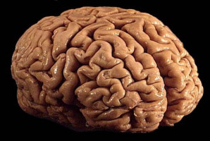
With a five-year, $3.2 million grant from the National Cancer Institute, the team will now apply the technology to monitor the transport of genetically engineered, cancer-fighting macrophages into brain tumours.
Simultaneously, they will use ultrasound to deliver drugs that increase the effectiveness of the macrophages, “supercharging” their potential to attack brain cancer cells.
“Macrophages are being studied now for their ability to localise to a tumour and treat the cancer, using cells that have been genetically modified with a chimaeric antigen receptor to help them specifically recognise malignant cells,” said principal investigator Scott Medina , the William and Wendy Korb Early Career Associate Professor of Biomedical Engineering.
“While our first step was to track where the general immune cells were going in the body, we now want to translate this work to visualise where our bioengineered macrophage therapies are going within the body. Are they moving toward a tumour or somewhere we don't want them to go, where they may cause toxicity?”
Collaborators at the University of Wisconsin, under the leadership of co-principal investigator Igor Slukvin , professor of pathology, will develop the bioengineered macrophages.
Inhye Kim , assistant research professor of biomedical engineering at Penn State, will then use nanoparticle contrast agents paired with diagnostic ultrasound to visualise and track the macrophages, first in a silicone tissue model, then later, in a rodent model.
Kim led the development of the contrast agents, which were published in Small in July 2023.
Once the researchers determine if the macrophages move as expected, they will test whether the cells can better destroy cancer cells with the help of targeted drug delivery.
“We will use ultrasound for dual purposes: to visualise the movement of the macrophages, and to deliver drugs from the nanoparticle contrast agents that will stimulate the macrophages to be more effective in treating cancerous tumours,” Kim said.
“This required us to tweak the nanoparticle design to make it more effective at delivering a variety of drug cargos, including ones that stimulate immune cell function.”
Medina said he looks forward to the work in the grant, which builds upon his group's successes in contrast enhanced ultrasound imaging and drug delivery with the exciting development of now applying it to cancer treatments.
“If we're successful, it may open up other future therapeutic avenues in treating autoimmune disorders, infections and cardiovascular dysfunctions,” he said.
Medina is a leadership fellow within the Huck Institutes of the Life Sciences and is affiliated with the Materials Research Institute.
Julianna Simon , interim director of acoustics and associate professor of biomedical engineering at Penn State, and GirishGirish KirimanjeswaraKirimanjeswara , professor of veterinary and biomedical sciences at Penn State, will collaborate as co-principal investigators on the grant.
Source: Penn State