Supporting oncology professionals through education
The content on this site is intended for healthcare professionals only
The content on this site is intended for healthcare professionals only
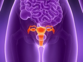
Ovarian cancer is usually diagnosed only after it has reached an advanced stage, with many tumours spread throughout the abdomen.
Most patients undergo surgery to remove as many of these tumours as possible, but because some are so small and widespread, it is difficult to eradicate all of them.
Researchers at MIT, working with surgeons and oncologists at Massachusetts General Hospital (MGH), have now developed a way to improve the accuracy of this surgery, called debulking.
Using a novel fluorescence imaging system, they were able to find and remove tumours as small as 0.3 millimetres - smaller than a poppy seed - during surgery in mice.
Mice that underwent this type of image-guided surgery survived 40 percent longer than those who had tumours removed without the guided system.
"What's nice about this system is that it allows for real-time information about the size, depth, and distribution of tumours," says Angela Belcher, the James Mason Crafts Professor of Biological Engineering and Materials Science at MIT, a member of the Koch Institute for Integrative Cancer Research, and the recently appointed head of MIT's Department of Biological Engineering.
The researchers are now seeking FDA approval for a phase 1 clinical trial to test the imaging system in human patients.
In the future, they hope to adapt the system for monitoring patients at risk for tumour recurrence, and eventually for early diagnosis of ovarian cancer, which is easier to treat if it is caught earlier.
Belcher and Michael Birrer, formerly the director of medical gynaecologic oncology at MGH and now the director of the O'Neal Comprehensive Cancer Center at the University of Alabama at Birmingham, are the senior authors of the study, published online in the journal ACS Nano.
Neelkanth Bardhan, a Mazumdar-Shaw International Oncology Fellow at the Koch Institute, and Lorenzo Ceppi, a researcher at MGH, are the lead authors of the paper.
Other authors include MGH researcher YoungJeong Na, MIT Lincoln Laboratory technical staff members Andrew Siegel and Nandini Rajan, Robert Fruscio of the University of Milan-Bicocca, and Marcela del Carmen, a gynaecologic oncologist at MGH and chief medical officer of the Massachusetts General Physicians Organization.
Because there is no good way to detect early-stage ovarian cancer, it is one of the most difficult types of cancer to treat.
Of 250,000 new cases diagnosed each year worldwide, 75 percent are in an advanced stage.
In the United States, the five-year combined survival rate for all stages of ovarian cancer is 47 percent, only a slight improvement from 38 percent three decades ago, despite the advent of chemotherapeutic drugs such as cisplatin, approved by the FDA in 1978 for ovarian cancer treatment.
In contrast, the five-year combined survival rate for all stages of breast cancer has steadily improved, from around 75 percent in the 1970s to over 90 percent now.
"We desperately need better upfront therapies, including surgery, for these (ovarian cancer) patients," Birrer says.
Belcher and Birrer joined forces to work on this problem through the Bridge Project, a collaboration between the Koch Institute and Dana-Farber/Harvard Cancer Center.
Belcher's lab has been developing a novel type of medical imaging based on light in the near-infrared (NIR) spectrum.
In a paper published in March, she reported that this imaging system could achieve an unprecedented combination of resolution and penetration-depth in living tissue.
In the new study, Belcher, Birrer, and their colleagues worked with researchers at MIT Lincoln Laboratory to adapt NIR imaging to help surgeons locate tumours during ovarian cancer surgery, by providing continuous, real-time imaging of the abdomen, with tumours highlighted by fluorescence.
Previous analyses have shown that survival rates are strongly inversely correlated with the amount of residual tumour mass left behind in the patient during debulking surgery, but many ovarian tumours are so small or hidden that surgeons can't find them.
To make the tumours visible, the researchers designed chemical probes using single-walled carbon nanotubes that emit fluorescent light when illuminated by a laser.
They coated these nanotubes with a peptide that binds to SPARC, a protein that is over-expressed by highly invasive ovarian cancer cells.
This probe binds to the tumours and makes them fluoresce at NIR wavelengths, allowing surgeons to more easily find them with fluorescence imaging.
The researchers tested the image-guided system in mice that had ovarian tumours implanted in a region of the abdominal cavity known as the intraperitoneal space, and showed that surgeons were able to locate and remove tumours as small as 0.3 millimetres.
Ten days after surgery, these mice had no detectable tumours, while mice that had undergone the traditional, non-image-guided surgery, had many residual tumours missed by the surgeon.
By three weeks after the surgery, many of the tumours had grown back in the mice that underwent image-guided surgery, but those mice still had a median survival rate that was 40 percent longer than that of mice that underwent traditional surgery.
No other imaging system would be able to locate tumours that small during a surgical procedure, the researchers say.
"You can't have a patient in a CT machine or an MRI machine and have the surgeon perform this surgical debulking procedure at the same time, and you can't expose the patient to X-ray radiation for multiple hours of the long surgery. This optics-based imaging system allows us to do that in a safe manner," Bardhan says.
For most ovarian cancer patients, tumour debulking surgery is followed by chemotherapy, so the researchers now plan to do another study where they treat the mice with chemotherapy after image-guided surgery, in hopes of preventing the remaining tiny tumours from spreading.
"We know that the amount of tumour removed at the time of surgery for patients with advanced-stage ovarian cancer is directly correlated with their outcome," Birrer says. "This imaging device will now allow the surgeon to go beyond the limits of resecting tumours visible to the naked eye, and should usher in a new age of effective debulking surgery."
Now that they have demonstrated that this concept can be successfully applied to imaging during surgery, the researchers hope to begin adapting the system for use in human patients.
"In principle, it's quite doable," Siegel says. "It's purely the mechanics and the funding at this point, because this mouse experiment serves as the proof of principle and may actually have been more challenging than building a human-scale system."
The researchers also hope to deploy this type of imaging to monitor patients after surgery, and eventually to develop it as a diagnostic tool for screening women at high risk for developing ovarian cancer.
"A major focus for us right now is developing the technology to be able diagnose ovarian cancer early, in stage 1 or stage 2, before the disease becomes disseminated," Belcher says. "That could have a huge impact on survival rates, because survival is related to the stage of detection."
The World Cancer Declaration recognises that to make major reductions in premature deaths, innovative education and training opportunities for healthcare workers in all disciplines of cancer control need to improve significantly.
ecancer plays a critical part in improving access to education for medical professionals.
Every day we help doctors, nurses, patients and their advocates to further their knowledge and improve the quality of care. Please make a donation to support our ongoing work.
Thank you for your support.