Supporting oncology professionals through education
The content on this site is intended for healthcare professionals only
The content on this site is intended for healthcare professionals only
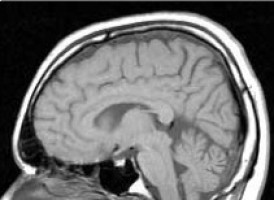
A team of internationally renowned neurologists, bioinformaticians, and pathologists from the United States and India recently published details of the Ivy Glioblastoma Atlas in Science.
The atlas maps out comprehensive, visually-rich information on the anatomic and genetic bases of glioblastoma at the cellular and molecular levels associated with the disease.
It is aimed at helping researchers and physicians improve the diagnosis and treatment of glioblastoma, including finding new drug targets.
While Atlas-based data have been previously available, the new paper in Science represents the first published research outlining the project in detail.
According to , Jill S. Barnholtz-Sloan, PhD, Sally S. Morley Designated Professor in Brain Tumor Research at Case Western Reserve University School of Medicine, there are three aims to the venture.
The first is to make all the relevant data fully transparent and available.
The second is to create a road map of all of the different cells and potential molecular changes that occur in these cells in a glioblastoma.
And the third, is to build a better understanding of how much heterogeneity can be found in these tumours.
"Currently we provide the same standard treatment of surgery, followed by radiation plus chemotherapy, for all glioblastoma patients," says Barnholtz-Sloan, who is also professor and associate director for bioinformatics/translational informatics at CWRU's School of Medicine. "But the tumours are not all the same. They have different molecular changes, which means that we may need to provide separate, tailored treatments to tackle each one. By identifying unique features of these tumours that may benefit from targeted therapies, the Atlas will enable patients to experience the benefits of precision medicine, increasing the chances for better response to treatment and hence better survival."
In virtually all cases of glioblastoma, excised tumours are assessed by highly specialized pathologists, who confirm the presence of malignancy and provide details about the unique characteristics of the tumours they examine.
For this project, Barnholtz-Sloan assisted the team in developing a study design to assess the concordance between features outlined in the tumours by both these human experts and automated machine-learning techniques.
"In many ways it's a belt and suspenders approach," she says. "The gold standard is the pathologist. Machine-driven analysis adds value by bringing further information to the findings of the pathologists. In the article we published, we found that the concordance rates were very high; 85-95 percent."
By way of comparison, Barnholtz-Sloan cites the case of medulloblastoma, a common type of malignant brain tumour mostly seen in children.
In medulloblastoma, researchers have been able to group tumours into four distinct molecularly-defined groups, which have specific drugs that would work best within each subtype.
The new Atlas hopes to replicate this success in glioblastoma.
"As more new information is added to the Atlas and the associated database for clinical and genomic data, we believe that it will continue to be very valuable research and treatment tool, allowing more refined diagnosis and treatment and enabling researchers to generate potentially life-extending hypotheses about causes and treatments," said Barnholtz-Sloan.
Source: Case Western Reserve University
We are an independent charity and are not backed by a large company or society. We raise every penny ourselves to improve the standards of cancer care through education. You can help us continue our work to address inequalities in cancer care by making a donation.
Any donation, however small, contributes directly towards the costs of creating and sharing free oncology education.
Together we can get better outcomes for patients by tackling global inequalities in access to the results of cancer research.
Thank you for your support.