Multimodality management of leiomyosarcoma of the cervix
Vijaya Kumar Jayaram1, Parikshith J1, Geeta Sowmya Narayanan1, Richa Tiwari1, Veena R2, Prathima S3, MS Ganesh4, Chattakonda Sai Snehith1 and Esther Praisy1
1Department of Radiation Oncology, Vydehi Institute of Medical Sciences and Research Centre, Bengaluru 560066, India
2Department of Pathology, HCG Oncology Centre, Bengaluru 560013, India
3Department of Pathology, Vydehi Institute of Medical Sciences and Research Centre, Bengaluru 560066, India
4Department of Surgical Oncology and Oncology, Vydehi Institute of Medical Sciences and Research Centre, Bengaluru 560066, India
Correspondence to: Vijaya Kumar Jayaram. Email: vijaybangalore3@gmail.com
Abstract
We present the case of a young female patient who presented to the outpatient department with a history of bleeding per vagina, diagnosed with leiomyosarcoma of the cervix; the patient underwent total abdominal hysterectomy with pelvic lymph node dissection. In this article, we mainly discuss multimodality therapy in the management of an unusual variety of tumour in the uterine cervix.
Keywords: leiomyosarcoma, young age, multimodality management
Copyright: © the authors; licensee ecancermedicalscience. This is an Open Access article distributed under the terms of the Creative Commons Attribution License (http://creativecommons.org/licenses/by/3.0), which permits unrestricted use, distribution, and reproduction in any medium, provided the original work is properly cited.
Published: 30/04/2018; Received: 10/01/2018
Introduction
Leiomyosarcoma is one of the rare histopathological variants of tumours of the cervix comprising nearly 1% of overall tumours of the cervix. Diagnosis is made on the basis of histopathological findings and immunohistochemical analysis. The treatment of choice in such patients is a triple therapy comprising surgery, chemotherapy and radiation therapy [1].
Case study
A twenty-year-old female patient presented with a history of increased blood flow during her menstrual cycles for the past 3 years. She was symptomatically treated. However, as her symptoms did not subside, she sought further treatment elsewhere, where magnetic resonance imaging (MRI) of the pelvis in August 2017 showed a T1W hypointense, T2W and SPAIR hyper-intense polypoidal mass arising from the cervix measuring 6.2 × 6.9 × 7.2 cm with mild diffusion restriction and the mass was filling the vaginal fornices (Figure 1(A–C)).
A provisional diagnosis of a cervical polyp was made and she underwent a hysteroscopic polypectomy on 8 September 2017. The post-operative histopathology report was in favour of a high grade or poorly differentiated malignancy with the possibility of pleomorphic spindle cell sarcoma/carcinosarcoma (Figure 3 and 4).
Fluoro-deoxy-glucose positron emission tomography - computed tomography (FDG-PET CT) done on 9 October 2017 showed an exophytic mass measuring 7.0 × 5.5 × 6.3 cm3 arising from the uterine cervix, located predominantly on the proximal vagina with relatively well-defined margins and no evidence of significant regional lymphadenopathy (Figure 2(A–C)).
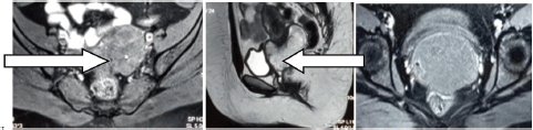
Figure 1(A–C). MRI of the pelvis; T2W and SPAIR hyper-intense polypoidal mass arising from the cervix measuring 6.2 × 6.9 × 7.2 cm3.
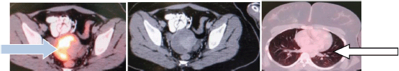
Figure 2(A–C). FDG PET CT a 7.0 × 5.5 × 6.3 cm3 large exophytic mass arising from uterine cervix and normal lung study done as a part of metastatic work up (physiological uptake in lung).
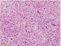
Figure 3. High-grade spindle cell areas with many bizarre giant cells.
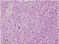
Figure 4. High-grade spindle cell areas with many mitoses.
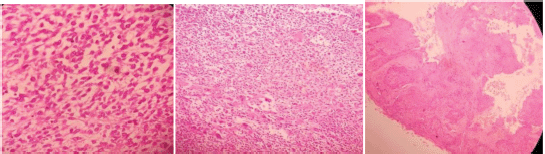
Figure 5 (A–C). High-grade tumour comprising sheets of plump spindle cells with eosinophilic cytoplasm and enlarged pleomorphic hyperchromatic nuclei.
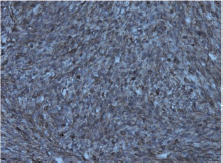
Figure 6. Photomicrograph showing tumour cells showing SMA positivity (200 ×).
The hysteroscopic polypectomy slide was reviewed, suggestive of poorly differentiated malignant tumour with high grade sarcomatous content.
The patient presented to our hospital in mid-October 2017 with a history of bleeding per vagina and yellowish discharge which on examination revealed a polypoidal fungating necrotic mass measuring 4.0 × 6.0 cm2 involving predominantly the anterior lip of the cervix, OS (opening into uterus) was not visualised and bilateral fornices free. She underwent radical hysterectomy type III and bilateral salphingo-oophorectomy with regional pelvic lymph node dissection and omentectomy on 28 October 2017. The post-operative histopathological examination report showed undifferentiated endocervical sarcoma of cervix with all resected lymph nodes, margins and omentum free of tumour (Figure 5(A–C)).
The slide review of the same procedure by an onco-pathologist revealed pleomorphic leiomyosarcoma of the cervix and the immunohistochemistry was positive for vimentin, smooth muscle actin with Ki 67 of 80% (Figure 6 and 7).
The patient and her family members were counselled about the condition and treatment options, regarding the role of adjuvant radiotherapy in view of the high degree of proliferative activity and were planned for 50 Gy in 25 fractions followed by adjuvant chemotherapy. The patient has completed adjuvant external beam radiation therapy (EBRT) (Figure 8) and intravaginal brachytherapy with a dose of 6 Gy in three sessions and the patient has received a third cycle of gemcitabine (900 mg/m2) and docetaxel (75 mg/m2) given every 21st day until the end of March 2018. The patient tolerated the adjuvant treatment without any issue.
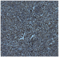
Figure 7. Photomicrograph showing tumour cells showing Vimentin positivity (20 ×).
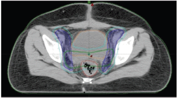
Figure 8. Contoured volumes, cyan-PTV volume, green-CTV tumour volume, blue-CTV nodal volume, red-rectum, orange-urinary bladder.
Discussion and review of the literature
The incidence of leiomyosacroma is less than 1% of all uterine malignancies [1]. Leiomyosarcoma represents 0.21% among all invasive tumours of the uterine cervix [2].
Regarding the epidemiology of the tumour, it commonly presents during the 4th–6th decades of life. It is rarely seen in a young age group similar to the patient in our case report [1]. Whitcombe et al [3] reported a case in which the patient age was 18 years old and was associated with pregnancy. There are four case reports in which leiomyosarcoma of the uterine cervix was associated with pregnancy [3]. Bansal et al [2] analysed the Surveillance Epidemiology and End Results database analysis of tumours of cervix including both carcinoma and sarcoma from 1988 to 2005. A total of 33,074 patients were diagnosed with tumours of carcinoma cervix. Amongst 33,074 patients, 323 patients had sarcoma. Leiomyosarcoma was seen in 67 patients. In this analysis, patients with sarcomas belong to a younger age group, have larger tumours in terms of size and are at a more advanced stage at the time of presentation [2].
Leiomyosarcoma presented with nonpuerperal inversion of the uterus was treated in a 49-year-old nulliparous woman which is an extremely rare event and rare mode of presentation [4]. In contrast to carcinoma cervix, involvement of lymph nodes and parametrium is a very rare event. In view of the rare instances of the involvement of lymph nodes, routine pelvic lymphadenectomy is not recommended, as it does not have any impact on 5-year disease-specific survival. In our patient, pelvic lymphadenectomy was performed in view of the young age of the patient [3].
Ovarian involvement in sarcomas of the cervix, in particular to leiomyosarcoma of the cervix, is a very rare mode of presentation. In view of the rarity of ovarian involvement, the role of oopherectomy in sarcomas of the uterine cervix is a debatable issue. As similar to the other histological variants of sarcoma with some exceptions, the mode of spread to distant sites is through the blood. Lung is the most common site of metastasis [5].
Casanova et al [6] reported a case of localised leiomyosarcoma of the cervix in a 63-year-old woman who presented with lung metastasis after 1 year of diagnosis. There are various prognostic factors including tumour stage, grade, mitotic index, tumour size, age and menopausal status. Amongst these factors, stage, grade and mitotic index were more significant prognostic factors [6].
Diagnosis of leiomyosarcoma of the cervix is based on histopathological findings. Microscopic study shows spindle-shaped cells can be seen which may be both hypercellular and hypocellular in nature. The presence of tumour cell necrosis, mitotic index of 10/10 high power field and atypia of cells ranging from moderate to diffuse are histopathological diagnostic criteria [6]. Various morphological varieties of leiomyosarcoma of the cervix myxoid type, epithelioid type, conventional type and certain variants contain abundant xanthomatous cells [7].
There are no well-established risk factors in the aetiology of leiomyosarcoma of the cervix, various risk factors postulated in the aetiology are radiation exposure to the pelvis in childhood; tamoxifen which is an hormonal drug used in breast carcinoma is postulated in the aetiology without any proper explanation for the associated risk; Epstein–Barr virus infection in immunocomprised status is also recognised as an etiological factor without any proper explanation. Patients with abnormal hereditary retinoblastoma gene are also at an increased risk [8].
Signs and symptoms are bleeding per vagina, discharge per vagina, pain and fullness in the pelvic and abdominal region. Most of the tumours are bulky at the time of presentation ranging from 10 to 12 cm in size. Various pressure symptoms such as increased frequency of micturition and sometimes urinary retention can also be seen. The patient is presented with h/o weight loss due to decreased appetite.
Since it is a rare disease, there is no definitive guideline for management of leiomyosarcoma of the cervix. Most recommended treatment is multimodality comprising surgery, total abdominal hysterectomy with bilateral salpingo-oophorectomy in patients having disease confined to the cervix, followed by post-operative radiation therapy to reduce the incidence of local recurrence. In view of the low incidence of lymph node involvement in leiomyosarcoma of the cervix, the role of prophylactic pelvic lymph node dissection along with primary surgery is debatable, unless there is gross pelvic lymph node involvement [1].
The role of chemotherapy has been extrapolated from sarcomas of the uterine corpus; most commonly used drugs including doxorubicin and ifosfamide. This regimen has an overall response rate of 30% in advanced and metastatic forms of disease [1]. Other chemotherapy drugs used are cyclophosphamide, gemcitabine and docetaxel used with response rates of approximately 30% [9].
Radiation therapy is usually given in the adjuvant setting to prevent loco regional recurrence. The pattern of radiation therapy delivered is external beam radiation therapy dose consisting of 50 Gy in 25 fractions over 5 weeks with 2 Gy per fraction followed by brachytherapy consisting of 2–3 sessions of brachytherapy which can also be delivered during EBRT also to reduce the total duration of treatment which has an impact on locoregional control rates [10, 11]. The prognosis of leiomyosarcoma of the cervix is inferior to its other common counterparts such as squamous cell carcinoma and adenocarcinoma [2].
The main treatment modality of this rare histopathological variant involving the cervix is multimodality management consisting of surgery, chemotherapy and radiation therapy. Multimodality management increases the survival rates in such patients [12].
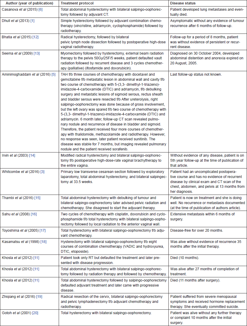
Table 1. Literature review of leiomyosarcoma case reports along with treatment modalities and follow-up status.
Conclusion
Leiomyosarcoma of the cervix must be treated with a multimodality management protocol in view of the rarity of the disease, due to the lack of guidelines for its management. Surgery is the main modality of treatment. Radiation therapy in the form of external beam irradiation and intravaginal brachytherapy improves the loco-regional control rate via decreasing the loco-regional recurrence. The role of chemotherapy and drugs to be used are extrapolated from sarcomas of other anatomical areas and sarcoma of the uterine corpus.
Conflicts of interest
None
References
1. Dhull AK, CA, and Kaushal V, et al (2013) The uncovered story of leiomyosarcoma of the cervix: a rare case report and review of literature BMJ Case Rep 2013 https://doi.org/10.1136/bcr-2013-008616
2. Bansal S, Lewin SN, and Burke WM, et al (2010) Sarcoma of the cervix: natural history and outcomes Gynecol Oncol 118(2) 134–138 https://doi.org/10.1016/j.ygyno.2010.04.021 PMID: 20541244
3. Whitcombe DD, Valente PT, and Acosta OM, et al (2016) Leiomyosarcoma of the uterine cervix associated with pregnancy: a case report and review of literature Gynecol Oncol Rep 17 45–48 https://doi.org/10.1016/j.gore.2016.05.012 PMID: 27355001 PMCID: 4909816
4. Buyukkurt S, Vardar MA, and Zeren H, et al (2007) Non-puerperal inversion of the uterus caused by leiomyosarcoma: a case report and clinical management J Obstet Gynaecol Res 33(3) 402–406 https://doi.org/10.1111/j.1447-0756.2007.00546.x PMID: 17578377
5. Aminimoghaddam S, Arabian S, and Haghighi S, et al (2016) Cervical leiomyosarcoma: a case report J Obstet Gynecol Cancer Res 1(1) e7523 https://doi.org/10.17795/jogcr-7523
6. Casanova J, Huang KG, and Thepsuwan J, et al (2015) Localized leiomyosarcoma of the uterine cervix with rapid lung metastases Gynecol Minimally Invasive Therapy 4 95–97 https://doi.org/10.1016/j.gmit.2014.05.010
7. Fadare O (2006) Uncommon sarcomas of the uterine cervix: a review of selected entities Diagn Pathol 1 30 https://doi.org/10.1186/1746-1596-1-30 PMID: 16981999 PMCID: 1584249
8. [http://www.dovemed.com/diseases-conditions/leiomyosarcoma-uterine-cervix/] Date accessed: 09/10/16
9. Sutton G, Blessing JA, and Malfetano JH (1996) Ifosfamide and doxorubicin in the treatment of advanced leiomyosarcomas of the uterus: a Gynecologic Oncology Group study Gynecol Oncol 62(2) 226–229 https://doi.org/10.1006/gyno.1996.0220 PMID: 8751554
10. Mittal S, Verma YP, and Kumar T (2017) Primary leiomyosarcoma of cervix: now it counts! Int J Curr Res 9(4)
11. Khosla D, Gupta R, and Srinivasan R, et al (2012) Sarcomas of uterine cervix: clinicopathological features, treatment, and outcome Int J Gynecol Cancer 22(6) 1026–1030 https://doi.org/10.1097/IGC.0b013e31825a97f6 PMID: 22740005
12. Bhatia V, Taksande R, and Natekar A, et al (2015) Cervical leiomyosarcoma: a rare entity South Afr J Gynaecol Oncol 7(2) 64–66 https://doi.org/10.1080/20742835.2015.1030887
13. Seema G, Mary A, and MK M (2009) Leiomyosarcoma of the cervix J Obstet Gynecol India 59(4) 364–366
14. Irvin W, Presley A, and Andersen W, et al (2003) Leiomyosarcoma of the cervix Gynecol Oncol 91(3) 636–642 https://doi.org/10.1016/j.ygyno.2003.08.037 PMID: 14675691
15. Thambi R, Subitha K, and Pothen L, et al (2016) Leiomyosarcoma of uterine cervix–a case report National J Lab Med 5(2) 72–73
16. Sahu L, Bupathy A, and Badhe BA (2008) Leiomyosarcoma of the uterine cervix in a young woman J Obstet Gynaecol Res 34(2) 717–720 https://doi.org/10.1111/j.1447-0756.2008.00914.x PMID: 18840189
17. Toyoshima M, Okamura C, and Niikura H, et al (2005) Epithelioid leiomyosarcoma of the uterine cervix: a case report and review of the literature Gynecol Oncol 97(3) 957–960 https://doi.org/10.1016/j.ygyno.2005.02.028 PMID: 15890394
18. Kasamatsu T, Shiromizu K, and Takahashi M, et al (1998) Leiomyosarcoma of the uterine cervix Gynecol Oncol 69(2) 169–171 https://doi.org/10.1006/gyno.1998.4986 PMID: 9600826
19. Zhiqiang L, Bin S, and Min F, et al (2016) Leiomyosarcoma of cervical stump following subtotal hysterectomy: a case report and review of literature Eur J Gynaecol Oncol 37(1) 148–151 PMID: 27048131
20. Gotoh T, Kikuchi Y, and Takano M, et al (2001) Epithelioid leiomyosarcoma of the uterine cervix Gynecol Oncol 82(2) 400–405 https://doi.org/10.1006/gyno.2001.6288 PMID: 11531304


