Supporting oncology professionals through education
The content on this site is intended for healthcare professionals only
The content on this site is intended for healthcare professionals only
Scientists at Oregon Health & Science University have uncovered a previously unknown system of internal “trade winds” that help cells rapidly move essential proteins to the front of the cell, reshaping how researchers understand cell migration, cancer spread and wound healing.
The discovery, published today in Nature Communications, reshapes what researchers thought they knew about how cells direct proteins to the right place at the right time.
For decades, biology textbooks have taught that free‑floating proteins inside cells move mainly by diffusion, drifting randomly until they happen to reach their destination.
But the new study shows that cells don’t leave this to chance.
Instead, they create targeted streams of fluid that push essential proteins toward the cell’s leading edge, where movement and repair begin.
Accidental discovery to major breakthrough
The study’s co-corresponding authors Catherine (Cathy) Galbraith, Ph.D., and James (Jim) Galbraith, Ph.D., both associate professors in the OHSU Biomedical Engineering Department and Discovery Engine Investigators in the OHSU Knight Cancer Institute, trace the discovery to an unexpected observation years ago during the neurobiology course at the Marine Biological Laboratory in Massachusetts.
“It actually started out as an unexpected finding,” Cathy said.
“We were just conducting an experiment with students in class.”
They used a laser to make proteins invisible in a strip across the back of a living cell — a standard technique for tracking how materials move inside cells — when something unexpected appeared: a second small, dark line showed up at the front edge that the cell extends as it moves.
“We kind of did it for fun and then realised this gave us a way of measuring something that wasn’t able to be measured before,” she said.
That unexpected dark line turned out to be a wave of soluble actin — one of the key proteins that helps cells move — being pushed rapidly to the cell’s leading edge.
Until now, scientists assumed actin mostly arrived there by diffusion, drifting through the cell randomly.
But the Galbraiths’ experiments revealed something else entirely.
“We realised the cartoon models in textbooks were missing a huge piece,” Jim said.
“There had to be some kind of flow in the cell pushing things forward. Cells really do ‘go with the flow.’”
Understanding cancer cell migration
Cathy and Jim were recruited to OHSU in 2013 from the National Institutes of Health, where they had collaborated with Nobel Laureate Eric Betzig, Ph.D., at the nearby Howard Hughes Medical Institute’s Janelia Research Campus, on the development of live-cell super-resolution microscopy.
Using custom imaging assays, the scientists discovered that cells actively create directional fluid flows, which the team compares to atmospheric rivers.
These currents push actin and other proteins forward much faster than diffusion could.
“We found that the cell can actually squeeze at the back and target where it sends that material,” Jim said.
“If you squeeze half a sponge, the water only goes on that half. That’s basically what the cell is doing.”
This internal flow is nonspecific, meaning it sweeps up many types of proteins at once.
The result is a fast, efficient delivery system that fuels protrusion, adhesion and rapid shape changes, all critical processes for cell movement, immune response and tissue repair.
The published findings confirm that these flows occur within a specialised compartment at the cell’s front, separated from the rest of the cell by an actin‑myosin condensate barrier that acts like a physical wall and targets the flows to advancing regions along the cell edge.
To visualise the currents, the team developed an inverse version of a standard fluorescence technique.
Instead of using a laser to bleach light away, they activated fluorescent molecules at a single point and watched how they moved.
The team nicknamed one of the key experiments FLOP, or Fluorescence Leaving the Original Point.
“It wasn’t a flop at all,” Cathy said.
“It was the opposite. It is anything but a flop, because it worked.”The team’s discovery may help explain why certain cancer cells move so aggressively.
“We know these highly invasive cells have this really cool mechanism to push proteins really fast, really rapidly where they need them at the front of the cell,” Jim said.
“All cells have basically the same components inside, much like a Porsche and a Volkswagen have many of the same parts, but when those parts are assembled into the final machine, they behave and function very differently.”
By understanding those differences, researchers hope to identify new ways to interrupt how cancer cells migrate.
“If you can understand the differences, you can target future therapies based on how cancer cells and normal cells work differently,” he said.
Collaboration for key findings
The project brought together engineering, physics, microscopy and cell biology, with key contributions from collaborators at Janelia Research Campus in Virginia, including experts in fluorescence correlation spectroscopy and 3D super‑resolution imaging.
“The instrumentation we needed doesn’t exist in most places,” Cathy said.
“Janelia had a one‑of‑a‑kind setup that let us test and confirm what we were seeing.”
Much of the project relied on advanced imaging techniques developed at Janelia — among them iPALM, an interferometric method for resolving nanometer-scale structures that the Galbraiths contributed to developing.
“iPALM allowed us to physically see the compartments,” Jim said.
“There’s no other light‑based technique that could do that.”
The researchers say the study reveals a “pseudo‑organelle,” or a functional compartment that isn’t surrounded by a membrane but still shapes how the cell behaves.
“Just as small shifts in the jet stream can change the weather, small changes in these cellular winds could change how diseases begin or progress,” Cathy said.
The team believes the work opens new directions for cancer research, drug delivery, tissue repair and synthetic biology.
“All you had to do was look,” Cathy said.
“The flows were there all along. Now we know how cells use them.”
In addition to the Galbraiths, coauthors on this study are Brian English, Ph.D., of Janelia Research Campus, and Ulrike Boehm, Ph.D., formerly with Janelia and now with Carl Zeiss AG of Germany.
Source: Oregon Health & Science University
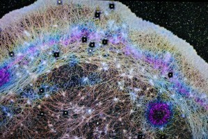
Image: Actin proteins inside a cell - A 3D single-molecule super-resolution microscopy (iPALM) image showing individual actin protein molecules inside a cell, each rendered as a single dot and captured at extraordinary detail — roughly 10,000 times finer than a human hair. Colors indicate depth within the cell, from blue at the bottom to magenta at the top. The blue and magenta dots cluster into curved structures that form a wall-like barrier separating the region of active fluid flow from the rest of the cell interior. Credit: OHSU/Christine Torres Hicks
We are an independent charity and are not backed by a large company or society. We raise every penny ourselves to improve the standards of cancer care through education. You can help us continue our work to address inequalities in cancer care by making a donation.
Any donation, however small, contributes directly towards the costs of creating and sharing free oncology education.
Together we can get better outcomes for patients by tackling global inequalities in access to the results of cancer research.
Thank you for your support.