Clinical adoption of radiobiological models and artificial intelligence in radiotherapy treatment planning: a cross-sectional practice survey
Kalyan Mondal1,2a, Abhijit Mandal3b and Anuj Vijay1c
1Department of Physics, Institute of Applied Science & Humanities, GLA University, Mathura 281406, Uttar Pradesh, India
2Department of Radiation Oncology, North Bengal Medical College & Hospital, Darjeeling 734012, West Bengal, India
3Department of Radiotherapy and Radiation Medicine, Institute of Medical Sciences, Banaras Hindu University, Varanasi 221005, Uttar Pradesh, India
ahttps://orcid.org/0000-0001-7685-7391
bhttps://orcid.org/0000-0001-5626-1072
chttps://orcid.org/0000-0001-6610-3844
Abstract
Introduction: Radiobiological models optimising tumour control probability (TCP) and normal tissue complication probability (NTCP) underpin effective radiotherapy treatment planning; however, real-world adoption – particularly in low- and middle-income country (LMIC) settings and for plan evaluation – remains poorly characterised alongside emerging artificial intelligence and machine learning (AI/ML) applications.
Methods: This cross-sectional survey is a pre-specified component of a mixed-methods systematic review registered with PROSPERO (CRD420251146188); the survey itself is not separately registered. A 40-item anonymous instrument was distributed via the MEDPHYS mailing list and professional society channels. Forty-two self-selected professionals from 10 countries (India 71.4%) responded. Analysis was descriptive, with Wilson 95% confidence intervals; proportions are reported on the full sample (N = 42), with the number of non-respondents stated for every item. Reporting follows CHERRIES guidelines for internet e-surveys.
Results: Confirmed local calibration of radiobiological model parameters was reported by 14.3% (95% CI 6.7%–27.8%) of respondents; 40.5% (95% CI 27.0%–55.5%) reported no calibration and a further 40.5% did not know their institution’s calibration status. Biologically effective dose and equivalent dose in 2 Gy fractions were widely recognised (>70%), while formal TCP and NTCP models were reported substantially less often (<35%). Radiobiological models were used for plan evaluation by 38.1% of respondents; dedicated software was uncommon (commercial 11.9%; open-source 2.4%). Routine clinical use of AI/ML was reported by 19.0% (95% CI 10.0%–33.3%), with auto-contouring the most frequently reported application (50.0%). Publication activity in this domain was low: 83.3% (95% CI 69.4%–91.7%) reported no peer-reviewed publications. The most frequently cited barriers were limited software access (45.2%), inadequate infrastructure (40.5%) and absence of clinical validation guidelines (31.0%).
Conclusion: In this small, self-selected sample, these exploratory findings describe a multi-layered implementation gap spanning model calibration, plan evaluation, training and AI/ML adoption. They are hypothesis-generating rather than representative of national or global practice and indicate priorities for larger, geographically balanced studies and for the accompanying systematic review evidence synthesis (PROSPERO: CRD420251146188).
Keywords: surveys and questionnaires, artificial intelligence, radiobiology, radiotherapy, treatment planning, low- and middle-income countries, TCP, NTCP, plan evaluation
Correspondence to: Kalyan Mondal
Email: kalyanmondal.res@gmail.com
Published: 22/10/2026
Received: 22/05/2026
Publication costs for this article were supported by ecancer (UK Charity number 1176307).
Copyright: © the authors; licensee ecancermedicalscience. This is an Open Access article distributed under the terms of the Creative Commons Attribution License (http://creativecommons.org/licenses/by/4.0), which permits unrestricted use, distribution, and reproduction in any medium, provided the original work is properly cited.
Introduction
Radiotherapy is central to cancer management, with approximately 50% of patients receiving radiation during their treatment course [1]. Low- and middle-income countries (LMICs) account for nearly 50% of global cancer deaths yet access only 5% of global radiotherapy resources [2,3]. Effective treatment planning requires maximising tumour control probability (TCP) while minimising normal tissue complication probability (NTCP) – a balance that radiobiological models have refined over five decades, from the foundational linear-quadratic (LQ) model through biologically effective dose (BED), equivalent dose in 2 Gy fractions (EQD2), generalised equivalent uniform dose (gEUD) and formal TCP/NTCP frameworks [4–9]. A clinically consequential yet underexplored application of these models is biological plan evaluation, wherein radiobiological indices complement conventional dose–volume metrics to inform plan selection and clinical decision-making [10]. For example, when two plans are dosimetrically comparable, NTCP estimates for parallel organs such as lung or parotid can identify the plan with the lower predicted complication risk; explicit TCP–NTCP trade-off analysis has also been used to individualise dose escalation and to select patients for proton therapy under model-based protocols [11,12].
Despite extensive model development, formal TCP/NTCP implementation in routine clinical practice remains limited across diverse healthcare systems; reviews from North America, Europe and Asia consistently report biologically based plan evaluation at a minority of centres, with physical dose–volume metrics remaining the clinical standard [13–16]. The Netherlands’ national NTCP-based proton therapy selection protocol represents a notable exception, demonstrating feasibility when supported by national infrastructure and validated model sets [11,12]. Concurrently, artificial intelligence and machine learning (AI/ML) are transforming radiotherapy workflows across automated contouring, knowledge-based planning, dose prediction and outcome modelling [17–19], yet the 2024 AAPM–ESTRO guideline confirmed that clinical adoption remains limited relative to technological development [20]. Recent surveys indicate increasing AI use among European radiologists (48% in 2024), with significant disparities persisting between high-income countries (HICs) and LMICs [21,22].
In India, an estimated 1.46 million annual cancer cases coincide with persistent radiotherapy infrastructure deficits and workforce challenges [2,23,24]. While IMRT/VMAT adoption has improved substantially (~88% nationally [25]), integration of radiobiological optimisation and AI/ML-based plan evaluation into routine practice remains largely undocumented. Resource-constrained settings may derive disproportionate benefit from these tools: biological optimisation can help extract maximal therapeutic value from limited fractionation and machine capacity and standardise planning quality across heterogeneous centres, while AI-based automation can partially offset workforce shortages [22,26]. To our knowledge and based on a structured literature search of MEDLINE and PubMed (1975–2025) using combinations of the terms ‘radiobiological model’, ‘TCP’, ‘NTCP’, ‘plan evaluation’, ‘artificial intelligence/machine learning’ and ‘survey’, no prior study has jointly assessed classical radiobiological model adoption and AI/ML implementation with specific attention to plan evaluation practices and LMIC settings; existing surveys have addressed these domains separately [21,22,27].
This survey was designed as a pre-specified component of a mixed-methods systematic review registered with PROSPERO (CRD420251146188), enabling direct comparison between published evidence and clinical practice upon review completion. Specific objectives were as follows: (i) to characterise radiobiological model adoption and calibration practices; (ii) to assess AI/ML implementation patterns; (iii) to characterise radiobiological model use for plan evaluation; (iv) to identify barriers and training needs; and (v) to describe respondents’ publication activity in this domain. The theme of this study is illustrated in Figure 1.
Methods
Study design and registration
This cross-sectional observational study was approved by the Institutional Review Board of GLA University, Mathura, India (September 23, 2025). It is a pre-specified component of a mixed-methods systematic review prospectively registered with PROSPERO on September 15, 2025 (CRD420251146188). The registered protocol names the survey component in its title and its secondary review question and pre-specifies the target population (radiation oncology departments worldwide, with medical physicists, radiation oncologists and department heads as respondents), the survey domains and a descriptive analysis plan stratified by region and centre type. Because PROSPERO registers systematic reviews and does not accept registrations of stand-alone cross-sectional surveys, the survey reported here is not itself separately registered, and no claim of independent registration is made. The registered protocol further specified a minimum of 20 responding centres per subcontinental region for quantitative regional synthesis, with regions below that threshold to be described narratively only; this pre-specified threshold governs how geography is reported here (Statistical Analysis). Informed electronic consent was obtained from all participants. The study was conducted in accordance with the Declaration of Helsinki and is reported according to the CHERRIES guidelines [28]. Upon systematic review completion, survey findings will be compared with the published literature synthesis.

Figure 1. Conceptual overview of the study theme: the roles of classical radiobiological models and artificial intelligence/machine learning (AI/ML) across the radiotherapy treatment-planning workflow, with radiobiology-based plan evaluation as the focus. Top (blue): classical radiobiological models – dose‑equivalence models (linear-quadratic, LQ; modified‑LQ; universal survival curve, USC; biologically effective dose, BED; equivalent dose in 2 Gy fractions, EQD2) and outcome models (tumour control probability, TCP; normal tissue complication probability, NTCP; generalised equivalent uniform dose, gEUD). Middle (grey): the clinical workflow – imaging, segmentation, plan optimisation, plan evaluation, patient-specific quality assurance (QA), treatment delivery and clinical outcome and toxicity; plan optimisation and plan evaluation each combine a conventional dosimetric component (dose–volume histogram, DVH; conformity index, CI; homogeneity index, HI; Dx; Vx) and a radiobiological component (TCP/NTCP/gEUD). Bottom (orange): AI/ML methods – deep learning (convolutional neural network, CNN; U‑Net), machine learning (random forest, RF; support vector machine, SVM), knowledge‑based planning (RapidPlan) and multi-criteria optimisation (MCO)– and their applications across the workflow (synthetic CT, image registration/fusion, auto-contouring, automated planning, dose prediction, AI-assisted plan evaluation, clinical decision support, QA prediction, adaptive radiotherapy, motion management, toxicity prediction, outcome prediction and patient stratification). Radiobiological models inform plan optimisation and, in particular, plan evaluation (green, biological–AI convergence), where radiobiological indices complement conventional dosimetric evaluation, while AI/ML methods act as data‑driven counterparts across the same stages. Population-specific clinical-outcome data are fed back (dashed) to locally calibrate radiobiological model parameters (e.g. α/β, TD50, n, m) and to continuously train and validate AI models, supporting biologically informed, AI‑augmented personalised radiotherapy. Created with BioRender.com.
Survey instrument
The 40-item questionnaire comprised six domains: (1) demographics and institutional characteristics; (2) clinical infrastructure; (3) radiobiological model usage, calibration and documentation; (4) AI/ML implementation; (5) plan evaluation tools and barriers and (6) training needs and research activity. Question types included single-select, multiple-select, Likert scales and ranking items. The instrument was developed iteratively by the investigator team with expertise in medical physics, radiation oncology, radiobiology and AI/ML and pilot-tested with five colleagues (three medical physicists and two radiation oncologists) across Indian radiation oncology departments prior to full distribution. The full instrument is provided in Supplementary Table S1. To reduce interpretive variability, key response options were anchored with explicit descriptors: AI/ML status used the categories ‘routine clinical use (fully integrated)’, ‘limited clinical implementation (pilot)’, ‘planning to implement’ and ‘not using’, and frequency of radiobiological model use was banded by percentage of cases (>75%, 50%–75%, 25%–50%, <25%, never). A four-step data quality assurance (QA) protocol assessed completeness, internal consistency, plausibility and discordance. Item-level completeness was 100% for demographics and clinical infrastructure and ranged from 88.1% to 97.6% across the radiobiological-model, plan-evaluation, training and publication items; it was 81.0% for implementation methods, 76.2% for the barriers item and 64.3%–66.7% for the items answered only by respondents currently using AI/ML. The number of respondents who did not answer each item is stated in the corresponding table. Full QA results are provided in Supplementary Table S2.
Survey distribution
A multi-channel distribution strategy was employed: the global MEDPHYS mailing list (>4,000 subscribers) with an initial invitation (November 4, 2025) and reminder (January 13, 2026); direct email to >300 Indian radiation oncology professionals; dissemination via AMPI, EFOMP and AFOMP; and LinkedIn, Facebook and WhatsApp professional groups. The survey was open October 28, 2025–February 20, 2026 (117 days); all 42 responses were received within the first 83 days of the active window.
Participation was open to practising radiation oncology professionals (medical physicists, radiation oncologists, dosimetrists and radiotherapy researchers) involved in treatment planning or evaluation; a pre-survey consent item screened participants, and non-consenting respondents exited automatically. Duplicate submissions were limited by the platform’s one-response-per-account setting and screened during quality assurance by cross-checking demographic, institutional and infrastructure responses; no duplicate patterns were detected. Because the instrument was disseminated through overlapping open channels with unavoidable audience overlap, the number of eligible individuals who viewed the invitation cannot be established; consequently, CHERRIES view, participation and completion rates and a true response rate cannot be calculated. Instrument validation comprised iterative content review by the multidisciplinary investigator team and pilot testing with five colleagues; formal psychometric validation was not undertaken, consistent with the study’s exploratory objective.
Statistical analysis
The analysis is descriptive throughout. Categorical variables are reported as frequencies and percentages with Wilson score 95% confidence intervals. No inferential testing between geographical subgroups was performed. The registered protocol required a minimum of 20 responding centres per subcontinental region before a region could enter quantitative synthesis, and specified that regions falling below that threshold would be described narratively only; no region other than South Asia reached this threshold in the present sample (respondents practising outside India, n = 12 across nine countries). Country of practice is therefore reported descriptively and no India-versus-international comparison is presented. All proportions are calculated on the full sample (N = 42); every table reports the number of respondents who did not answer that item, so single-select items sum to 42, while multiple-select items may sum to more than 100%. Exploratory stratification by profession and institution type is reported descriptively, without significance testing, in Supplementary Table S3. Analyses were performed in Python 3.12 with pandas and NumPy, and all tables and figures were regenerated directly from the de-identified response file deposited at Zenodo.
Results
Participant demographics and geographic distribution
Forty-two valid responses were received from 10 countries across five continental regions (Fig. 2). India contributed 71.4% (n = 30), followed by the United States (n = 3, 7.1%), Germany (n = 2, 4.8%), and one respondent each from seven other countries; no region other than South Asia reached the 20-centre threshold pre-specified in the registered protocol for regional quantitative synthesis. Medical physicists comprised 78.6% (n = 33), radiation oncologists 19.0% (n = 8) and research scientists 2.4% (n = 1). Experience was distributed across career stages, with 2–5 years the most common group (26.2%, 11/42; two respondents did not state their experience). Full demographic and institutional characteristics are presented in Table 1; geographic and professional details are provided in Supplementary Figure S3.
Clinical infrastructure
Advanced EBRT capabilities were widely available: VMAT (88.1%), IMRT (85.7%), IGRT-CBCT (81.0%), 3DCRT (78.6%), SBRT (59.5%) and SRT (54.8%; Fig. 3A). Eclipse™ was the most frequently reported treatment planning system (26 of 52 selections, 50.0%), followed by Monaco™ (23.1%), Telecobalt-based systems (7.7%) and Precision™ (5.8%); one respondent did not answer this item (Fig. 3B). Brachytherapy was available at 83.3% of centres. These findings are broadly consistent with published analyses of India’s installed radiotherapy fleet [25,29].
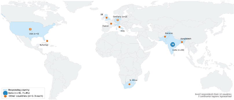
Figure 2. Global distribution of survey respondents (N = 42; 10 countries, 5 continental regions). Bubble size is proportional to the square root of respondent count. India (n = 30, 71.4%) is shown in blue; all other contributing countries in orange. Shaded country borders indicate responding nations.
Table 1. Demographic and institutional characteristics of survey respondents (N = 42).

Radiobiological model adoption and calibration practices
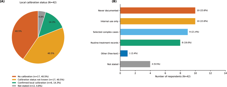
Local parameter calibration to the institution’s own patient population was confirmed by 14.3% (6/42; 95% CI 6.7%–27.8%) of respondents. An equal number reported no calibration (40.5%, 17/42; 95% CI 27.0%–55.5%) and did not know their institution’s calibration status (40.5%, 17/42; 95% CI 27.0%–55.5%); two respondents did not answer the item (Table 2; Supplementary Fig. S2A). These categories are reported separately throughout: uncertainty about calibration status is not evidence that calibration is absent, and the proportion of respondents unable to state whether their own institution calibrates its model parameters is itself a finding. Parameter sources were predominantly published literature values (52.4%), which are largely Western-derived.
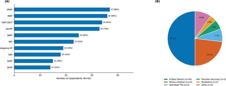
Figure 3. Clinical infrastructure at responding institutions. (A) Availability of external beam radiotherapy (EBRT) modalities (multiple selections permitted); bars show the number of respondents reporting each modality (N = 42). (B) Distribution of treatment planning systems (TPS) in use (multiple selections permitted; n = 52 TPS selections from the 41 respondents who answered this item, shown as pie slices). CBCT = cone-beam computed tomography; IGRT = image-guided radiotherapy; IMRT = intensity-modulated radiotherapy; SBRT = stereotactic body radiotherapy; SRT = stereotactic radiotherapy; VMAT = volumetric modulated arc therapy.
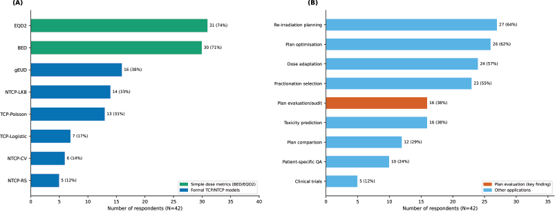
Figure 4. Radiobiological model adoption (N = 42; multiple selections permitted in both panels; percentages calculated on the full sample). (A) Specific radiobiological models or dose metrics recognised and used (answered by 37 respondents): green bars denote simple dose metrics (BED, biologically effective dose; EQD2, equivalent dose in 2 Gy fractions); blue bars denote formal models (gEUD, generalised equivalent uniform dose; TCP, tumour control probability; NTCP, normal tissue complication probability; LKB, Lyman–Kutcher–Burman). (B) Clinical contexts in which radiobiological models are applied (answered by 39 respondents); the red bar highlights plan evaluation/audit (key finding, 38.1%) and blue bars denote other applications.
Table 2. Radiobiological model adoption, calibration practices and plan evaluation type (N = 42).

Formal clinical use of radiobiological models was generally infrequent: 9.5% used them in routine practice (>75% of cases), 33.3% rarely (<25%) and 16.7% never; one respondent described a non-standard pattern of use in free text, and two did not answer the item. BED and EQD2 were the most widely recognised models (71.4% and 73.8%, respectively), consistent with their integration into fractionation guidelines [5,30,31]. Formal TCP/NTCP models were reported substantially less often: gEUD (38.1%), NTCP-LKB (33.3%), TCP-Poisson (31.0%), TCP-Logistic (16.7%), NTCP-Critical Volume (14.3%) and NTCP-Relative Seriality (11.9%; Fig. 4A); five respondents did not answer this item.
The most common contexts for radiobiological model use were re-irradiation planning (64.3%), plan optimisation (61.9%) and dose prescription adaptation (57.1%; Fig. 4B); three respondents did not answer this item. Notably, 38.1% (16/42) used radiobiological models for plan evaluation or audit – fewer than for re-irradiation planning, plan optimisation or dose adaptation. Documentation practices were variable: 19.0% routinely included radiobiological calculations in treatment planning records (Supplementary Fig. S2B). In exploratory stratification (descriptive only, given small strata), confirmed local calibration occurred exclusively at academic/teaching institutions (6/22) and at no non-academic centre (0/19), while all respondents reporting routine AI/ML clinical use were medical physicists (8/33; 0/8 radiation oncologists); these unadjusted patterns are hypothesis-generating (Supplementary Table S3).
AI/ML implementation
The AI/ML landscape reflected a field in early transition: 19.0% (8/42; 95% CI 10.0%–33.3%) reported routine clinical use, 33.3% had no plans to implement, 16.7% were planning implementation within 1 year and 9.5% were in limited pilot testing; a further 14.3% described other statuses in free text (research use only, testing or validation or implementation planned beyond 1 year), and 7.1% did not answer the item (Fig. 5A).
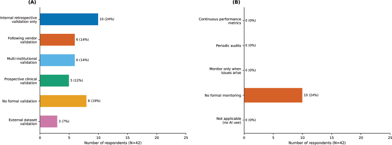
Auto-contouring/segmentation was the most widely reported AI application (50.0%), followed by plan optimisation (28.6%) and automated planning (26.2%; Fig. 5B); this item was answered by the 28 respondents reporting any AI/ML activity, and percentages are calculated on the full sample. AI-based plan evaluation (16.7%) and toxicity/outcome prediction (11.9%) – representing the convergence of radiobiological models and AI/ML – were among the least frequently reported applications, despite favourable predictive performance in several published cohorts [32–34]. AI/ML validation and monitoring practices were limited relative to recommended standards [20]: 11.9% reported prospective clinical validation and 23.8% reported no formal monitoring programme (Supplementary Fig. S1); these items were answered by 27 and 28 respondents, respectively.
Barriers, training needs and plan evaluation tools
The most frequently reported barriers overall were limited software access (45.2%), inadequate infrastructure (40.5%) and lack of clinical validation or guidelines (31.0%; Table 3; Fig. 6A). This item was answered by 32 of 42 respondents (76.2%), the lowest completeness of any item; percentages are calculated on the full sample, so the frequency of each barrier among respondents who did answer is correspondingly higher than the values shown.
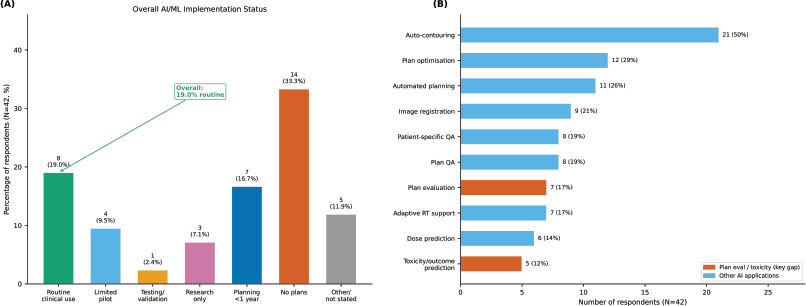
Figure 5. Artificial intelligence/machine learning (AI/ML) implementation (N = 42; percentages calculated on the full sample). (A) Overall distribution of AI/ML implementation status across all respondents; each colour represents one implementation stage, the final bar combines free-text statuses with unanswered responses and the overall routine clinical use rate (19.0%) is annotated. (B) AI/ML applications currently used or under evaluation (multiple selections permitted; answered by 28 respondents); red bars highlight AI-aided plan evaluation and toxicity/outcome prediction, the lowest-adoption applications despite high potential convergence with radiobiological modelling.
Table 3. Barriers to adoption of radiobiological models and AI/ML (N = 42).

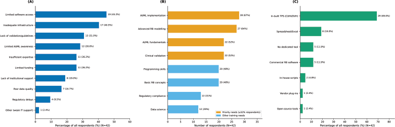
Figure 6. Barriers, training needs and plan evaluation tools (N = 42; multiple selections permitted in all panels; percentages calculated on the full sample). (A) Barriers to adopting radiobiological models and AI/ML (answered by 32 respondents). (B) Training needs ranked by respondents (answered by 38 respondents); orange bars indicate priority needs (≥52% endorsement). (C) Plan evaluation tools used (answered by 40 respondents). CI = conformity index; HI = homogeneity index; DVH = dose–volume histogram; RB = radiobiological.
A training deficit was reported: 38.1% of respondents (16/42; 95% CI 25.0%–53.2%) had received no formal training in radiobiological modelling or AI/ML; five respondents did not answer this item. Top-ranked training needs were AI/ML implementation (66.7%), advanced radiobiological modelling (64.3%) and, jointly, AI/ML fundamentals and clinical validation methods (52.4% each; Fig. 6B); four respondents did not answer this item.
Plan evaluation tool use was dominated by built-in TPS modules (69.0%), followed by spreadsheet-based calculations (19.0%; Table 4; Fig. 6C); two respondents did not answer this item. Dedicated radiobiological software was reported by 11.9% (commercial: BioSuite [35], dose–volume histogram (DVH) Analyser [36] and PlanIQ [37]) and 2.4% (open-source: CERR [38], DREES [39], HART [40], RADBIOMOD [41] and RBMODELv1 [42]); 85.7% reported using neither. Publication activity in this domain was low: 83.3% (35/42; 95% CI 69.4%–91.7%) reported no peer-reviewed publications on radiobiological modelling or AI/ML in radiotherapy, and three respondents did not answer the item. This measures the research output of the respondents rather than the quality of their clinical practice; no directly comparable published estimate was identified.
The key observations of this study are given in Table 5.
Table 4. Plan evaluation tools used (N = 42).

Table 5. Key findings and suggested priorities for future research.

Discussion
The radiobiological model paradox and calibration practice
The finding that BED/EQD2 are widely recognised (>70%) while formal TCP/NTCP models remain less used (<35%) reflects a persistent paradox: despite five decades of model development, the tools most capable of informing individualised treatment decisions have the lowest clinical penetration [14,15]. This is consistent with Yorke’s characterisation of achieving high TCP with low NTCP as still ‘challenging’ due to insufficient integration of dosimetric and clinical correlates into decision workflows [13], and with broader assessments of formal TCP/NTCP use as ‘still limited’ in most clinical settings [14,15]. The Netherlands’ NTCP-based proton therapy selection protocol [11,12] demonstrates feasibility when national infrastructure, validated model sets and institutional research capacity are in place – prerequisites that most centres in this small sample did not report. These findings (Table 5) are summarised in the conceptual framework (Fig. 7), which is descriptive and hypothesis-generating rather than a validated model; it annotates each pipeline stage with the corresponding survey-derived observation.
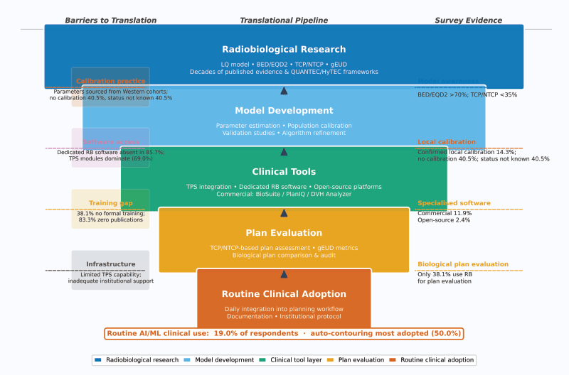
Figure 7. Conceptual framework depicting the translational pipeline from radiobiological research to routine clinical adoption. Five pipeline stages (coloured funnel boxes) are annotated by barriers (left) and survey-derived descriptive evidence (right). The bottom bar summarises overall routine AI/ML use across all respondents. Stage annotations are illustrative and derived from this exploratory survey of 42 self-selected respondents; the framework is not a validated model.
Calibration practice warrants separate consideration. Confirmed local calibration was uncommon (14.3%), and an equal number of respondents reported no calibration (40.5%) as were unable to state their institution’s calibration status (40.5%). These two groups are distinct and are not combined here. Where calibration is confirmed absent, radiobiological plan evaluation may carry uncertainty of unquantified magnitude, the clinical impact of which cannot be determined from these data and warrants formal investigation. Where calibration status is simply unknown to the respondent, the finding speaks to awareness and documentation rather than to the underlying practice: an institution may calibrate without that being visible to the individual preparing or checking the plan. That four in ten respondents could not answer this question about their own department is, on its own terms, a notable observation and a candidate subject for future study. The QUANTEC initiative established dose–volume–outcome relationships predominantly from Western cohorts [30], and subsequent validation studies have demonstrated that model performance varies significantly across populations and treatment techniques [4]. For diverse patient populations – where genetic polymorphisms, comorbidity patterns, nutritional status and treatment delivery variations may differ from derivation cohorts [43] – application of uncalibrated parameters creates systematic uncertainty in predicted TCP/NTCP values [44,45]. Low reported publication activity (83.3% with no publications) may be relevant here because locally generated outcome data are what the iterative calibration cycle requires. Publication output is, however, a measure of research activity rather than of clinical practice or competence and should be read only as an indicator of limited local outcome-data generation.
The plan evaluation gap
In this sample, 38.1% of respondents used radiobiological models for plan evaluation, and dedicated evaluation software was reported by 14.3% – consistent with a specific translational gap, although the sample is too small and too geographically skewed to establish its extent. Physical DVH metrics remained dominant (73.8%), and the scarcity of purpose-built radiobiological evaluation tools in these clinical environments is consistent with the divide described between published evaluation methodology and clinical implementation [16,46]. Recent validation studies further underscore that NTCP model performance varies substantially across cohorts [47], reinforcing the case for population-specific calibration as a prerequisite for valid biological plan evaluation. Two questions follow for future research: whether integrating validated radiobiological evaluation tools within commercial TPS platforms changes evaluation practice, and whether institutional pipelines for population-specific outcome data collection can support parameter calibration – the latter being an objective of the accompanying systematic review. Candidate topics for that work include multicentre collaborations to pool population-specific outcome data, prospective validation studies preceding clinical use, structured training embedded in professional-society programmes and accessible, ideally open-source, radiobiological evaluation tools within commercial TPS platforms. These are research priorities suggested by an exploratory sample, not recommendations for practice.
AI/ML adoption and the unexplored convergence
The overall 19.0% routine use rate, with auto-contouring as the most frequently reported application (50.0%), is consistent with the documented AI maturity hierarchy in radiotherapy [18–20], corroborated by a systematic overview of AI-based applications that identified auto-contouring as the most widely implemented modality with substantial geographic disparities [48], and with broader surveys reporting LMIC–HIC adoption disparities [21,22]. The present sample contains too few respondents outside South Asia to support regional comparison, so this survey cannot test whether such disparities are present in its own data, and no comparison between Indian and other respondents is reported. The barriers most frequently endorsed overall – limited software access, inadequate infrastructure and absence of clinical validation guidance – span both infrastructure and evidence-translation categories, and are consistent with those described by Petragallo et al [27] and by a systematic review of AI implementation barriers across healthcare settings [49].
The convergence of radiobiological modelling with AI/ML for personalised plan evaluation – where machine learning approaches have demonstrated favourable predictive performance relative to classical NTCP models [32,34,50] – was rarely reported in this sample: AI-based toxicity prediction (11.9%) and AI-aided plan evaluation (16.7%) were among the least frequently reported applications. Whether this reflects a general pattern cannot be determined from 42 self-selected respondents, but it identifies a question worth addressing in future implementation research. Adoption is also shaped by factors this instrument did not measure – integration into existing clinical workflows, the complexity and opacity of some models, absence of reimbursement pathways and medico-legal and regulatory uncertainty around model-guided decisions.
Contributions and limitations
This study contributes several observations. Based on the structured search underpinning our registered systematic review, we are not aware of a previous survey that reports, within a single instrument, publication activity in this domain (83.3% with no publications), local calibration practice (14.3% confirmed calibration; 40.5% no calibration; 40.5% status not known) and radiobiological model use specifically for plan evaluation (38.1%). A substantial training deficit was also reported (38.1% with no formal training). As a pre-specified component of a registered systematic review, the data provide an empirical complement to the published evidence base for integration upon review completion. These observations describe the respondents to this survey and are offered as a starting point for larger studies rather than as an estimate of practice in any country, region or income group.
Several limitations must be acknowledged, and they bound the interpretation of every finding above. The sample size (N = 42) is small, though comparable to published surveys in specialised radiotherapy communities [22,27], and supports exploratory description only. The sample is a self‑selected convenience sample recruited through open channels; it is not statistically representative of national or global practice, and no finding here should be read as an estimate of practice in any country, region or income group. The geographic distribution is heavily weighted towards India (71.4%); the 12 respondents practising in nine other countries are too few, and too heterogeneous in health system and resource level, to constitute a comparator group, and the 20-centre-per-region threshold pre-specified in the registered protocol was not met outside South Asia. Country of practice is therefore reported descriptively only, and no India-versus-international comparison is presented. Although institution type and patient load were captured, staffing levels and a full technology inventory were not; these unmeasured institutional factors may influence adoption and warrant more granular future surveys. Approximate engagement from the MEDPHYS listserv (~1%) is consistent with norms for unsolicited online professional surveys [51,52]; because the instrument was disseminated through overlapping open channels, the true eligible denominator – and therefore a genuine response rate – cannot be calculated, so the magnitude of non-response and self-selection bias cannot be quantified. Self-reported data carry inherent social desirability bias, which may inflate reported adoption. Item non-response varied by domain, from none for demographics and infrastructure to 23.8% for the barriers item; because percentages are calculated on the full sample throughout, reported proportions are conservative for items with missing responses. The calibration question did not allow respondents to distinguish between active institutional calibration unknown to the individual and confirmed absence of any calibration process, which is why ‘not sure’ responses are reported as a separate category throughout rather than combined with ‘no’. Internal consistency analysis identified 9.5% response discordance, indicating some measurement imprecision. Within these limits, concordance of key infrastructure findings with published benchmarks (Supplementary Table S4), high completeness for the core items, and transparent QA reporting support the internal validity of the descriptive findings.
Conclusion
Among 42 self-selected respondents, this exploratory survey describes a multi-layered implementation gap in radiotherapy treatment planning – spanning model calibration, biological plan evaluation, training and AI/ML adoption. The sample is small and predominantly Indian, so these observations are hypothesis-generating and are not offered as a description of national or global practice. They indicate priorities for future research: confirming the calibration and plan-evaluation findings in larger, geographically balanced samples; establishing why calibration status is so frequently unknown to practitioners and evaluating whether accessible evaluation software and structured training alter practice. The dataset also provides the practice-side input for integration with the accompanying systematic review evidence synthesis (PROSPERO: CRD420251146188).
List of abbreviations
AAPM, American Association of Physicists in Medicine; AI/ML, Artificial intelligence/machine learning; API, Application Programming Interface; BED, Biologically effective dose; CBCT, Cone-beam computed tomography; CI, Conformity index (statistics: confidence interval, stated where used); CI, Confidence interval (statistical); CNN, Convolutional neural network; DVH, Dose–volume histogram; EBRT, External beam radiotherapy; EQD2, Equivalent dose in 2 Gy fractions; ESAPI, Eclipse Scripting Application Programming Interface; ESTRO, European Society for Radiotherapy and Oncology; gEUD, Generalised equivalent uniform dose; HDR, High dose rate; HI, Homogeneity index; IGRT, Image-guided radiotherapy; IMRT, Intensity-modulated radiotherapy; KBP, Knowledge-based planning; LDR, Low dose rate; LKB, Lyman–Kutcher–Burman; LMIC, Low- and middle-income country; LQ, Linear-quadratic; NTCP, Normal tissue complication probability; PDR, Pulsed dose rate; QA, Quality assurance; RB, Radiobiological; SBRT, Stereotactic body radiotherapy; SRS, Stereotactic radiosurgery; SRT, Stereotactic radiotherapy; SVM, Support vector machine; U-Net, a convolutional network architecture for biomedical image segmentation; VMAT, Volumetric modulated arc therapy; 2DRT, Two-dimensional conventional radiotherapy.
Acknowledgments
The authors gratefully acknowledge all survey respondents. The authors thank Dr Brenda Byrne (EFOMP Secretary General), Prof. Hasin Anupama Azhari (AFOMP Vice President) and Professor G.A. Zakaria for facilitating dissemination. The authors acknowledge the administrators of the MEDPHYS mailing list at Wayne State University. Special thanks to Dr Ganeshkumar Patel, Dr Shantanu Sapru, Dr Samrat Dutta, Mr Kalpak Chakrabarty, Mr Suman Mitra, Mrs Munmun Chatterjee, Ms Muskaan and Mrs Arti Ghuerai for distribution assistance and feedback.
Conflicts of interest
One of the authors (Abhijit Mandal) is a co-developer of the software RBMODELv1 [42]. The other authors declare no competing interests.
Funding
This research received no specific funding from any public, commercial, or not-for-profit funding agency.
Author contributions
KM: Conceptualisation, methodology, survey design, data collection, formal analysis and manuscript writing. AM: Conceptualisation, survey design, supervision, analysis review and manuscript editing. AV: Survey design, Supervision, resources, approval and manuscript review.
Ethics approval and consent to participate
This study was approved by the Institutional Review Board (IRB) of GLA University, Mathura, India (approval date: 23 September 2025). Informed consent was obtained electronically from all participants prior to survey participation. The study was conducted in accordance with the Declaration of Helsinki.
PROSPERO Registration
The parent mixed-methods systematic review is prospectively registered with PROSPERO (CRD420251146188; registered 15 September 2025): https://www.crd.york.ac.uk/PROSPERO/view/CRD420251146188. The cross-sectional survey reported here is a pre-specified component of that registered protocol. PROSPERO registers systematic reviews and does not accept registrations of stand-alone surveys, so the survey is not separately registered.
Data availability
AI Disclosure
AI-assisted tools (Claude Sonnet 4.6, Anthropic PBC, 2026) were used for language editing and manuscript formatting. All scientific content, data analysis, interpretation and conclusions are the responsibility of the authors.
References
1. Delaney G, Jacob S, and Featherstone C, et al (2005) The role of radiotherapy in cancer treatment: estimating optimal utilization from a review of evidence-based clinical guidelines Cancer 104 1129–1137 https://doi.org/10.1002/cncr.21324
2. Bray F, Laversanne M, and Sung H, et al (2024) Global cancer statistics 2022: GLOBOCAN estimates of incidence and mortality worldwide for 36 cancers in 185 countries CA Cancer J Clin 74 229–263 [https://doi.org/10.3322/caac.21834] PMID: 38572751
3. Starling MTM, Abdihamid O, and Vanderpuye V, et al (2025) Access to radical and palliative radiotherapy in low- and middle-income countries: challenges, progress, and future directions Curr Opin Support Palliat Care 19 256–262 https://doi.org/10.1097/SPC.0000000000000783 PMID: 41128500
4. Gulliford S and El Naqa I (2022) Modelling of Radiotherapy Response (TCP/NTCP) Machine and Deep Learning in Oncology, Medical Physics and Radiology eds El Naqa I and Murphy MJ (Cham: Springer International Publishing) pp 399–437 https://doi.org/10.1007/978-3-030-83047-2_17
5. Fowler JF (1989) The linear-quadratic formula and progress in fractionated radiotherapy Br J Radiol 62 679–694 https://doi.org/10.1259/0007-1285-62-740-679 PMID: 2670032
6. McMahon SJ (2018) The linear quadratic model: usage, interpretation and challenges Phys Med Biol 64 01TR01 https://doi.org/10.1088/1361-6560/aaf26a
7. Lyman JT (1985) Complication probability as assessed from dose-volume histograms Radiat Res 104 S13–S19 https://doi.org/10.2307/3576626
8. Källman P, Ågren A, and Brahme A (1992) Tumour and normal tissue responses to fractionated non-uniform dose delivery Int J Radiat Biol 62 249–262 https://doi.org/10.1080/09553009214552071 PMID: 1355519
9. Wang CKC (2010) The progress of radiobiological models in modern radiotherapy with emphasis on the uncertainty issue Mutation Res/Reviews Mutation Res 704 175–181 https://doi.org/10.1016/j.mrrev.2010.02.001 PMID: 20178860
10. Nahum A, and Onjukka E (2021) Radiobiological evaluation and optimisation of treatment plans Handbook of Radiotherapy Physics 2nd ed, Boca Raton, FL: CRC Press https://doi.org/10.1201/9780429201493-51
11. Langendijk JA, Lambin P, and De Ruysscher D, et al (2013) Selection of patients for radiotherapy with protons aiming at reduction of side effects: the model-based approach Radiotherapy Oncol 107 267–273 https://doi.org/10.1016/j.radonc.2013.05.007
12. Langendijk JA, Hoebers FJP, and De Jong MA, et al (2021) National Protocol for Model-Based Selection for Proton Therapy in Head and Neck Cancer Int J Part Therapy 8 354–365 https://doi.org/10.14338/IJPT-20-00089.1
13. Yorke E (2023) Modeling clinical outcomes in radiotherapy: NTCP, TCP and the ‘TECs.’ Med Phys 50 122–124 https://doi.org/10.1002/mp.16274
14. Jones B and Dale RG (2018) The evolution of practical radiobiological modelling Br J Radiol 92 20180097 https://doi.org/10.1259/bjr.20180097
15. Chaikh A, Thariat J, and Thureau S, et al (2020) Construction des modèles radiobiologiques de type TCP (tumor control probability) et NTCP (normal tissue complication probability) : de la dose à la prédiction des effets cliniques Cancer/Radiothérapie 24 247–257 https://doi.org/10.1016/j.canrad.2019.12.004
16. Allen Li X, Alber M, and Deasy JO, et al (2012) The use and QA of biologically related models for treatment planning: Short report of the TG-166 of the therapy physics committee of the AAPM: TG-166 report Med Phys 39 1386–409 https://doi.org/10.1118/1.3685447 PMID: 22380372
17. Deasy JO (2024) Data science opportunities to improve radiotherapy planning and clinical decision making Semin Radiat Oncol 34 379–394 https://doi.org/10.1016/j.semradonc.2024.07.012 PMID: 39271273 PMCID: 11698470
18. Duke K, and Papanikolaou N (2025) Artificial intelligence in radiation therapy: from imaging to delivery—a comprehensive review BJR|Artificial Intelligence 2 ubaf012 https://doi.org/10.1093/bjrai/ubaf012
19. Huynh E, Hosny A, and Guthier C, et al (2020) Artificial intelligence in radiation oncology Nat Rev Clin Oncol 17 771–781 https://doi.org/10.1038/s41571-020-0417-8 PMID: 32843739
20. Hurkmans C, Bibault JE, and Brock KK, et al (2024) A joint ESTRO and AAPM guideline for development, clinical validation and reporting of artificial intelligence models in radiation therapy Radiother Oncol 197 110345 https://doi.org/10.1016/j.radonc.2024.110345 PMID: 38838989
21. Zanardo M, Visser JJ, and Colarieti A, et al (2024) Impact of AI on radiology: a EuroAIM/EuSoMII 2024 survey among members of the European Society of Radiology Insights Imag 15 240 https://doi.org/10.1186/s13244-024-01801-w
22. McGinnis GJ, Ning MS, and Beadle BM, et al (2022) Barriers and facilitators of implementing automated radiotherapy planning: a multisite survey of low- and middle-income Country radiation oncology providers JCO Global Oncol 8 e2100431 https://doi.org/10.1200/GO.21.00431
23. Gupta N, Chugh Y, and Prinja S (2023) Bridging the cancer care gap and inequities in radiation treatment in India: a narrative review Cancer Res Stat Treat 6 554 [https://doi.org/10.4103/crst.crst_295_23]
24. Datta NR, Samiei M, and Bodis S (2014) Radiation therapy infrastructure and human resources in low- and middle-income countries: present status and projections for 2020 Int J Radiat Oncol Biol Phys 89 448–457 https://doi.org/10.1016/j.ijrobp.2014.03.002 PMID: 24751411
25. Krishnatry R, Mathew A, and Das S, et al (2024) Cross-Sectional National Survey of Practice Patterns in Radiotherapy for Rectal Cancer: a Snapshot of India JCO Glob Oncol 10 e2400410 https://doi.org/10.1200/GO-24-00410 PMID: 39571114
26. Court LE, Aggarwal A, and Jhingran A, et al (2024) Artificial Intelligence–Based Radiotherapy Contouring and Planning to Improve Global Access to Cancer Care JCO Glob Oncol 10 e2300376 https://doi.org/10.1200/GO.23.00376
27. Petragallo R, Bardach N, and Ramirez E, et al (2022) Barriers and facilitators to clinical implementation of radiotherapy treatment planning automation: a survey study of medical dosimetrists J Appl Clin Med Phys 23 e13568 https://doi.org/10.1002/acm2.13568 PMID: 35239234 PMCID: 9121037
28. Eysenbach G (2004) Improving the quality of Web surveys: the checklist for reporting results of internet E-Surveys (CHERRIES) J Med Internet Res 6 e34 https://doi.org/10.2196/jmir.6.3.e34 PMID: 15471760 PMCID: 1550605
29. Chauhan RS, Munshi A, and Pradhan A (2024) An analysis of radiotherapy machine requirements in India: impact of the pandemic and regional disparities J Med Phys 49 370–378 https://doi.org/10.4103/jmp.jmp_20_24 PMID: 39526149 PMCID: 11548067
30. Marks LB, Yorke ED, and Jackson A, et al (2010) Use of normal tissue complication probability models in the clinic Int J Radiat Oncology*Biology*Physics 76 S10–S19 https://doi.org/10.1016/j.ijrobp.2009.07.1754
31. Grimm J, Marks LB, and Jackson A, et al (2021) High dose per fraction, hypofractionated treatment effects in the clinic (HyTEC): an overview Int J Radiat Oncol Biol Phys 110 1–10 https://doi.org/10.1016/j.ijrobp.2020.10.039 PMID: 33864823 PMCID: 9447432
32. Isaksson LJ, Pepa M, and Zaffaroni M, et al (2020) Machine learning-based models for prediction of toxicity outcomes in radiotherapy Front Oncol 10 790 https://doi.org/10.3389/fonc.2020.00790 PMID: 32582539 PMCID: 7289968
33. Valdes G, Simone CB, and Chen J, et al (2017) Clinical decision support of radiotherapy treatment planning: a data-driven machine learning strategy for patient-specific dosimetric decision making Radiotherapy Oncol 125 392–397 https://doi.org/10.1016/j.radonc.2017.10.014
34. Cui S, Hope A, and Dilling TJ, et al (2022) Artificial intelligence for outcome modeling in radiotherapy Seminars Radiat Oncol 32 351–364 https://doi.org/10.1016/j.semradonc.2022.06.005
35. Uzan J and Nahum AE (2012) Radiobiologically guided optimisation of the prescription dose and fractionation scheme in radiotherapy using BioSuite Br J Radiol 85 1279–1286 https://doi.org/10.1259/bjr/20476567 PMID: 22457318 PMCID: 3487060
36. Kim JS, Yoon M, and Park SY, et al (2009) A dose volume histogram analyzer program for external beam radiotherapy J Korean Soc For Therapeutic Radiol Oncol 27(4) 240 https://doi.org/10.3857/jkstro.2009.27.4.240
37. Perumal B, Sundaresan HE, and Ranganathan V, et al (2019) Evaluation of plan quality improvements in PlanIQ-guided autoplanning Rep Pract Oncol Radiother 24 533–543 https://doi.org/10.1016/j.rpor.2019.08.003 PMID: 31641339 PMCID: 6796777
38. Deasy JO, Blanco AI, and Clark VH (2003) CERR: a computational environment for radiotherapy research Med Phys 30 979–985 https://doi.org/10.1118/1.1568978 PMID: 12773007
39. El Naqa I, Suneja G, and Lindsay PE, et al (2006) Dose response explorer: an integrated open-source tool for exploring and modelling radiotherapy dose–volume outcome relationships Phys Med Biol 51 5719–5735 https://doi.org/10.1088/0031-9155/51/22/001 PMID: 17068361
40. Pyakuryal A, Myint WK, and Gopalakrishnan M, et al (2010) A computational tool for the efficient analysis of dose‐volume histograms for radiation therapy treatment plans J Appl Clin Med Phys 11 137–157 https://doi.org/10.1120/jacmp.v11i1.3013
41. Chang JH, Gehrke C, and Prabhakar R, et al (2016) RADBIOMOD: a simple program for utilising biological modelling in radiotherapy plan evaluation Phys Medica Eur J Med Phys 32 248–254 [https://doi.org/10.1016/j.ejmp.2015.10.091]
42. Patel G, Mandal A, and Bharati A, et al (2022) Development and validation of an indigenous, radiobiological model-based tumor control probability and normal tissue complication probability estimation software for routine plan evaluation in clinics J Cancer Res Therapeutics 18 1697 https://doi.org/10.4103/jcrt.JCRT_330_20
43. Miyamoto T, Akutsu SN, and Tauchi H, et al (2018) Exploration of genetic basis underlying individual differences in radiosensitivity within human populations using genome editing technology J Radiat Res 59 ii75–ii82 https://doi.org/10.1093/jrr/rry007 PMID: 29528422 PMCID: 5941146
44. Bentzen SM, Dörr W, and Gahbauer R, et al (2012) Bioeffect modeling and equieffective dose concepts in radiation oncology – Terminology, quantities and units Radiotherapy Oncol 105 266–268 https://doi.org/10.1016/j.radonc.2012.10.006
45. Van Leeuwen CM, Oei AL, and Crezee J, et al (2018) The alfa and beta of tumours: a review of parameters of the linear-quadratic model, derived from clinical radiotherapy studies Radiat Oncol 13 96 https://doi.org/10.1186/s13014-018-1040-z PMID: 29769103 PMCID: 5956964
46. Warkentin B, Stavrev P, Stavreva N, Field C, Fallone BG (2004) A TCP‐NTCP estimation module using DVHs and known radiobiological models and parameter sets J Appl Clin Med Phys 5(1) 50–63 https://doi.org/10.1120/jacmp.v5i1.1970
47. De Vette SPM, Van Rijn-Dekker MI, and Van Den Bosch L, et al (2025) Evaluation of a comprehensive set of normal tissue complication probability models for patients with head and neck cancer in an international cohort Oral Oncol 163 107224 https://doi.org/10.1016/j.oraloncology.2025.107224 PMID: 40023984 PMCID: 11950982
48. Vandewinckele L, Claessens M, and Dinkla A, et al (2020) Overview of artificial intelligence-based applications in radiotherapy: recommendations for implementation and quality assurance Radiother Oncol 153 55–66 https://doi.org/10.1016/j.radonc.2020.09.008 PMID: 32920005
49. Ahmed MI, Spooner B, and Isherwood J, et al (2023) A systematic review of the barriers to the implementation of artificial intelligence in healthcare Cureus 15 e46454 [https://doi.org/10.7759/cureus.46454] PMID: 37927664 PMCID: 10623210
50. Samant P, De Ruysscher D, and Hoebers F, et al (2023) Machine learning for normal tissue complication probability prediction: predictive power with versatility and easy implementation Clin Transl Radiat Oncol 39 100595 [https://doi.org/10.1016/j.ctro.2023.100595] PMID: 36880063 PMCID: 9984444
51. Cunningham CT, Quan H, and Hemmelgarn B, et al (2015) Exploring physician specialist response rates to web-based surveys BMC Med Res Methodology 15 32 https://doi.org/10.1186/s12874-015-0016-z
52. Baruch Y and Holtom BC (2008) Survey response rate levels and trends in organizational research Hum Relations 61 1139–1160 https://doi.org/10.1177/0018726708094863
Supplementary materials
The following 40-item questionnaire was hosted on Google Forms (Google LLC, Mountain View, CA, USA), 28 October 2025 – 20 February 2026. Items are organised by domain. Estimated completion time: 10 minutes (approximately). De-identified aggregate responses are publicly available at the link provided in the main text.
Supplementary Table S1. Full survey instrument.

Supplementary Table S2. Data quality assurance results.

Supplementary Table S3. Exploratory stratification by profession and institution type.

Panel B – by institution type.

Supplementary Table S4. External validation of key survey findings.

Supplementary Figure S1. AI/ML validation and monitoring practices (N = 42; multiple selections permitted; percentages calculated on the full sample). (A) Validation approaches used for AI/ML systems (answered by 27 respondents). (B) Post-implementation monitoring practices (answered by 28 respondents); 11.9% reported prospective clinical validation, and 23.8% reported no formal monitoring programme.
Supplementary Figure S2. Radiobiological model calibration and documentation practices (N = 42). (A) Local parameter calibration status, shown as four mutually exclusive categories on the full sample: confirmed local calibration (Yes, n = 6, 14.3%), no calibration (No, n = 17, 40.5%), calibration status not known (‘Not sure’, n = 17, 40.5%) and not stated (n = 2, 4.8%). The ‘not sure’ responses are reported separately from ‘no’ throughout, because uncertainty about institutional practice is not evidence that calibration is absent. (B) Documentation of radiobiological calculations in clinical records (N = 42), including free-text and unanswered responses.
Supplementary Figure S3. Demographic detail of survey respondents (N = 42). (A) Country of practice (India in blue, all other countries in sky blue). (B) Professional role distribution. (C) Years of experience in current role; two respondents did not state their experience and percentages are calculated on the full sample.